. 2016 Oct 11;113(41):11555-11560.
doi: 10.1073/pnas.1607187113. Epub 2016 Sep 28.

Early somatic mosaicism is a rare cause of long-QT syndrome

Affiliations

Early somatic mosaicism is a rare cause of long-QT syndrome

James Rush Priest et al. Proc Natl Acad Sci U S A. .

Abstract

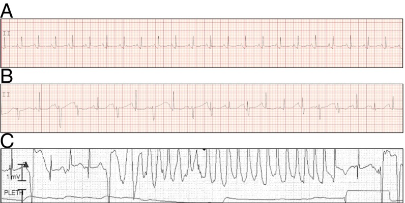
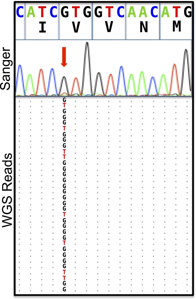
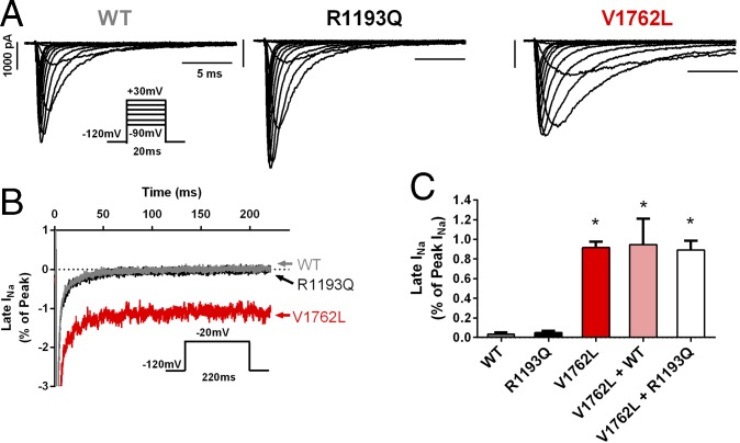
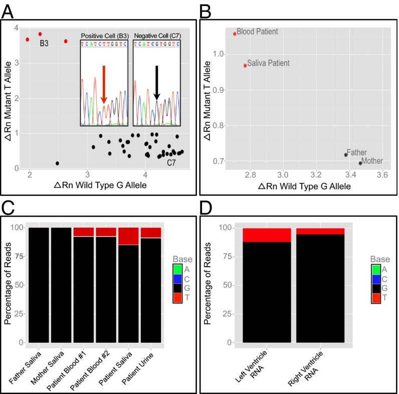
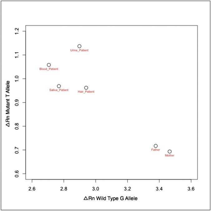
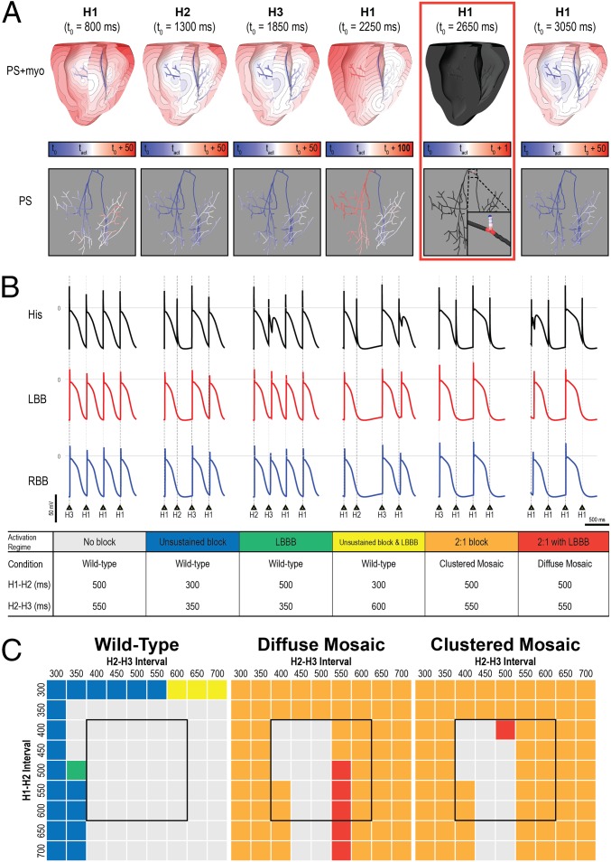
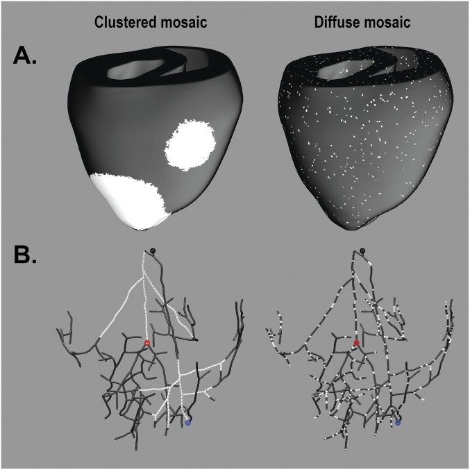
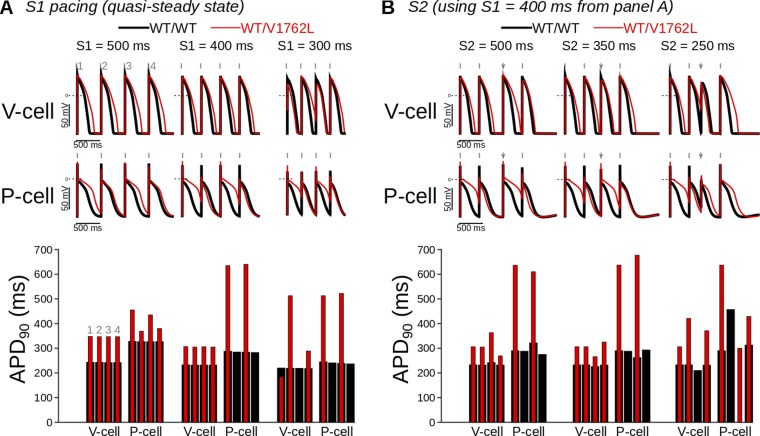
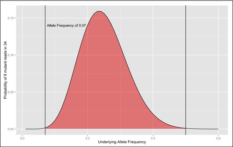
Somatic mosaicism, the occurrence and propagation of genetic variation in cell lineages after fertilization, is increasingly recognized to play a causal role in a variety of human diseases. We investigated the case of life-threatening arrhythmia in a 10-day-old infant with long QT syndrome (LQTS). Rapid genome sequencing suggested a variant in the sodium channel NaV1.5 encoded by SCN5A, NM_000335:c.5284G > T predicting p.(V1762L), but read depth was insufficient to be diagnostic. Exome sequencing of the trio confirmed read ratios inconsistent with Mendelian inheritance only in the proband. Genotyping of single circulating leukocytes demonstrated the mutation in the genomes of 8% of patient cells, and RNA sequencing of cardiac tissue from the infant confirmed the expression of the mutant allele at mosaic ratios. Heterologous expression of the mutant channel revealed significantly delayed sodium current with a dominant negative effect. To investigate the mechanism by which mosaicism might cause arrhythmia, we built a finite element simulation model incorporating Purkinje fiber activation. This model confirmed the pathogenic consequences of cardiac cellular mosaicism and, under the presenting conditions of this case, recapitulated 2:1 AV block and arrhythmia. To investigate the extent to which mosaicism might explain undiagnosed arrhythmia, we studied 7,500 affected probands undergoing commercial gene-panel testing. Four individuals with pathogenic variants arising from early somatic mutation events were found. Here we establish cardiac mosaicism as a causal mechanism for LQTS and present methods by which the general phenomenon, likely to be relevant for all genetic diseases, can be detected through single-cell analysis and next-generation sequencing.

Keywords: arrhythmia; computational modeling; genomics; mosaicism; single cell.


Conflict of interest statement

At the time of this work K.M.K., S.R., and L.B. were employed by Gilead Sciences. M.J.C., S.T.K.G., S.B., J.W., and R.C. were employed by Personalis, Inc. J.W. and E.A.A. are founders of Personalis, Inc. which offers clinical genetic testing but does not offer Clinical Laboratory Improvement Amendments (CLIA)-certified rapid-turnaround whole-genome sequencing. N.A.T. is a co-founder of CardioSolv LLC.

Figures

Fig. 1.
Fig. 1.
Representative electrocardiograms from DOL 1 show a prolonged QT interval and 2:1 atrioventricular block. (A) A rhythm strip from lead II of a 12-lead ECG before treatment shows a prolonged QTc of 542 ms estimated by Bazett’s formula. (B) A rhythm strip showing 2:1 atrioventricular block secondary to prolongation of the QT interval. (C) A rhythm strip demonstrating polymorphic ventricular tachycardia.
Fig. 2.
Fig. 2.
Mosaicism in the SCN5A locus is suggested by rapid genome sequencing and confirmed by augmented deep-exome sequencing. Alignments of next-generation sequencing reads from the SCN5A gene on chromosome 3 derived from rapid genome sequencing, with a cartoon alignment of the reverse complement of next-generation sequencing reads covering base pairs 38, 592, 565–38, 592, and 580 and a Sanger electropherogram suggestive of mosaicism indicated by the red arrow. Coordinates are from the hg19 assembly of the human genome. The allelic balance by genome sequencing (26 G, 8 T) is unlikely to arise from a 50:50 balance at a heterozygous locus (one-sided binomial test, P = 0.001), and the allelic balance observed by exome sequencing (210 G, 17 T) effectively rules out a variant with Mendelian inheritance at this position.
Fig. 3.
Fig. 3.
p.(V1762L) mutant channel exhibits defective INa inactivation consistent with a LQT3 phenotype. (A) Representative whole-cell INa traces recorded from cells expressing WT (Left), the common variant R1193Q (Center), or the mutant V1762L (Right). Note the delayed INa inactivation in the V1762L channel. (B) Extended voltage-step protocols reveal a late (sustained) INa conducted by V1762L (red trace), which was absent in both the WT and R1193Q channels. The dotted line indicates zero current. (C) The average late INa conducted by V1762L was significantly greater than that in WT (0.92 ± 0.06% of peak INa, n = 6, versus 0.03 ± 0.02% of peak INa, n = 6, respectively; *P < 0.001). No late INa was seen with the R1193Q mutation. The V1762L construct exerted a strongly dominant effect when cotransfected with either the WT channel (V1762L + WT) or R1193Q mutant (V1762L + R1193Q), maintaining a profoundly abnormal late INa.
Fig. 4.
Fig. 4.
Single-cell genotyping and targeted resequencing suggest that a mutation event causing mosaicism occurred before gastrulation. (A) qPCR genotyping of amplified DNA from 36 individual peripheral blood mononuclear cells identifies a subpopulation of three cells (red) that contain the T allele encoding the p.(V1762L) variant. A positive cell (B3) and negative cell (C7) are identified for the purposes of Sanger sequencing of cloned PCR products, respectively confirming the presence and absence of the mutant T allele (Inset). For each axis, the ΔRn value represents the magnitude of the signal generated from annealing of the allele-specific fluorescent probe to either the mutant or the WT allele relative to a signal generated from a passive reference dye during PCR amplification. (B) qPCR genotyping of DNA from proband blood, proband saliva, and parental saliva demonstrates the presence of the T allele only in proband samples. (C) Resequencing of PCR amplicons from four separate proband and both parental samples demonstrates that the frequency of the mutation is similar in proband blood (7.9%), urine (9.1%), and saliva (14.8%) samples, but the mutation is absent in both parental samples. (D) RNA sequencing of patient heart samples from the left and right ventricles identifies SCN5A reads containing the mutant allele.
Fig. S1.
Fig. S1.
TaqMan genotyping of DNA from proband tissues originating from all germ layers and both parental samples suggests the presence of the mutant T allele only in the proband samples.
Fig. 5.
Fig. 5.
Mosaic expression of the V1762L mutation in a 3D computational model of fetal ventricles and the Purkinje system leads to 2:1 block and LBBB. (A) Activation maps for six consecutive His bundle stimuli (delivered at t0 shown for each map) for the diffuse mosaic model; cutaway (Upper) and Purkinje-only (Lower) views are shown. Activation time scales are relative to t0 and vary for the different beats. Panel 4 shows an LBBB excitation sequence; excitation of the left side of the Purkinje system was caused by retrograde conduction from ventricular tissue. Panel 5 (red box) shows a conduction block in the His bundle, which initiated the 2:1 block activation regime. (B) Representative action potential sequences from the His bundle and the left/right bundle branches (LBB and RBB) for the six distinct activation regimes observed in response to different pacing protocol configurations: no blocked sinus beats; single blocked beat (i.e., no 2:1 block), without or with LBBB; LBBB with no blocked beats; and 2:1 block without or with LBBB. In cases with LBBB, action potential onset in the LBB was delayed (rightward shift), and late excitation of the His bundle caused by retrograde conduction was observed as a bump during repolarization. Fig. S2B shows the exact Purkinje locations from which action potential traces were extracted. (C) Summary of outcomes for all 243 unique simulations (81 pacing sequences in three models); the color of each entry corresponds to one of the activation regimes in B. Black boxes highlight the range of coupling intervals associated with the normal resting sinus rate for human infants.
Fig. S2.
Fig. S2.
Visualization of diffuse and clustered mosaic models. (A) Images of clustered (Left) and diffuse (Right) distribution of V1762L-positive cells (white) in the ventricular myocardial walls (black). (B) As in A, but for the Purkinje system. Points in B indicate locations from which action potential traces were extracted in Fig. 5 for the His-bundle (His; black), LBB (red), and RBB (blue) activations.
Fig. S3.
Fig. S3.
Simulated pacing-induced single-cell ventricular myocyte and Purkinje action potential traces (WT vs. V1762L-positive) highlight mutant contributions to irregular excitation and repolarization. WT and mutant action potentials are in black and red, respectively. Shown are action potential traces (Upper) and corresponding action potential durations (Lower) (calculated at 90% repolarization time). (A) Single-cell pacing (S) at the cycle lengths of 500, 400, and 300 ms resulted in 1:1 capture in ventricular cells (V-cell) but not in Purkinje cells (P-cell), where long action potentials were followed by skipped beats. (B) Cell responses to pacing at S1 = 400 ms (gray tick marks) followed for a premature or delayed S2 stimulus (S2 = 500, 350, and 250 ms; gray arrows). Among mutant cases (i.e., model configurations including the V1762L mutation), Purkinje cell responses showed inconsistent stimulus capture, whereas ventricular cells responded to all stimuli except those at the shortest coupling interval tested.
Fig. S4.
Fig. S4.
The exact probability of sampling 8 of 34 mutant reads varied by underlying allele frequency. The red area represents allele frequencies for which the probability of sampling 8 of 34 reads is greater than the probability arising from a heterozygous 50% allele ratio (P = 0.00106) by a one-sided binomial test. The maximal probability is an underlying mosaic allele frequency of ∼25% (P = 0.1564).
Fig. S5.
Fig. S5.
Cellular viability impacts DNA yield after single-cell isolation and whole-genome amplification. By staining category, dark lines represent the mean concentration, the boxes represent the first and third quartiles, whiskers represent the full range of included values, and round data points represent outliers not included in the calculations. Note that no individual cells stained uniquely for the apoptosis signal. The DNA detected in the empty wells may represent amplification of cell-free DNA or nonspecific amplification/polymerization of primer sequences from the WGA reaction.
Fig. S6.
Fig. S6.
Primer and probe sequences for TaqMan (A) and PCR (B) experiments.
Fig. S7.
Fig. S7.
TaqMan CNV genotyping at a locus 279 bp distant from the p.V1762L variant does not reveal a microduplication of exon 28. The location of the probe on hg19 chromosome 3 is 38,592,297, whereas the p.V1762L variant occurs at chr3 38,592,576. SE measurements for each sample are denoted by the vertical orange lines.

References

    1. Biesecker LG, Spinner NB. A genomic view of mosaicism and human disease. Nat Rev Genet. 2013;14(5):307–320. - PubMed
    1. Acuna-Hidalgo R, et al. Post-zygotic point mutations are an underrecognized source of de novo genomic variation. Am J Hum Genet. 2015;97(1):67–74. - PMC - PubMed
    1. Thibodeau IL, et al. Paradigm of genetic mosaicism and lone atrial fibrillation: Physiological characterization of a connexin 43-deletion mutant identified from atrial tissue. Circulation. 2010;122(3):236–244. - PubMed
    1. Ruark E, et al. Breast and Ovarian Cancer Susceptibility Collaboration Wellcome Trust Case Control Consortium Mosaic PPM1D mutations are associated with predisposition to breast and ovarian cancer. Nature. 2013;493(7432):406–410. - PMC - PubMed
    1. Jamuar SS, et al. Somatic mutations in cerebral cortical malformations. N Engl J Med. 2014;371(8):733–743. - PMC - PubMed

Publication types

MeSH terms

Substances