. 2016 Sep 29:6:34051.
doi: 10.1038/srep34051.

Rescue of Early bace-1 and Global DNA Demethylation by S-Adenosylmethionine Reduces Amyloid Pathology and Improves Cognition in an Alzheimer's Model

Affiliations

Rescue of Early bace-1 and Global DNA Demethylation by S-Adenosylmethionine Reduces Amyloid Pathology and Improves Cognition in an Alzheimer's Model

Sonia Do Carmo et al. Sci Rep. .

Abstract

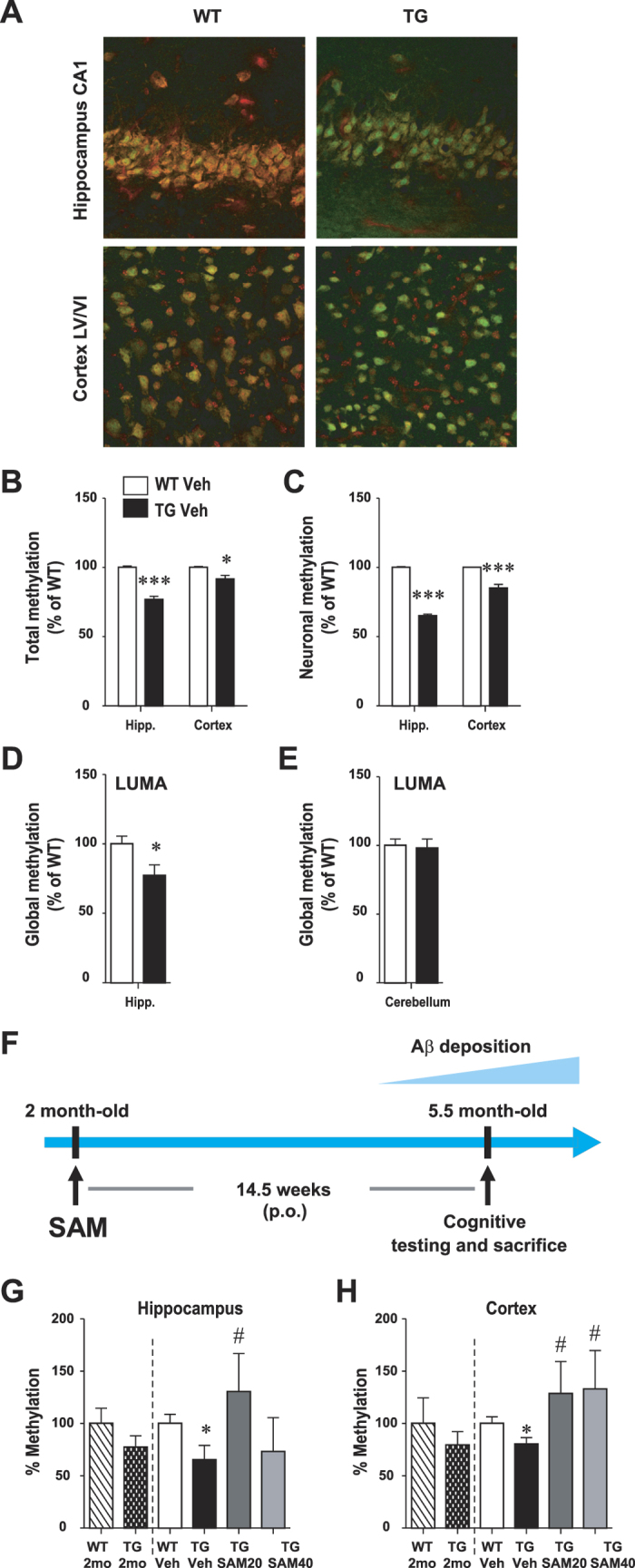
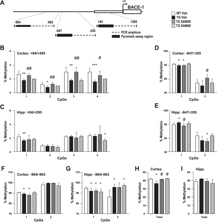
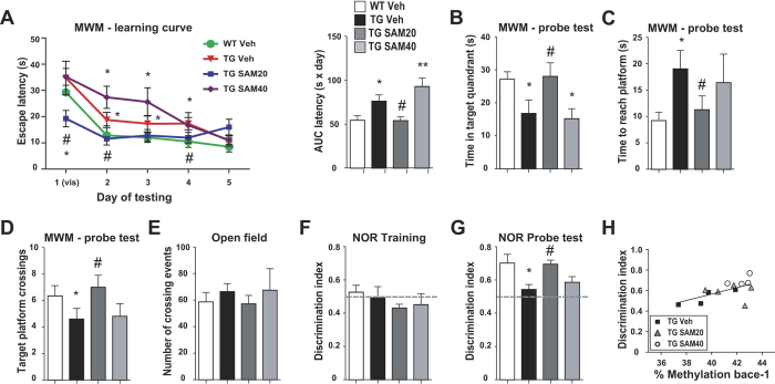
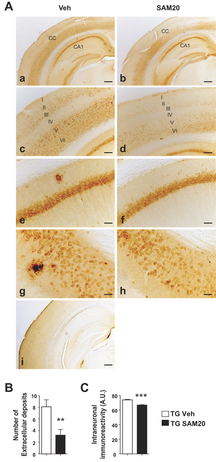
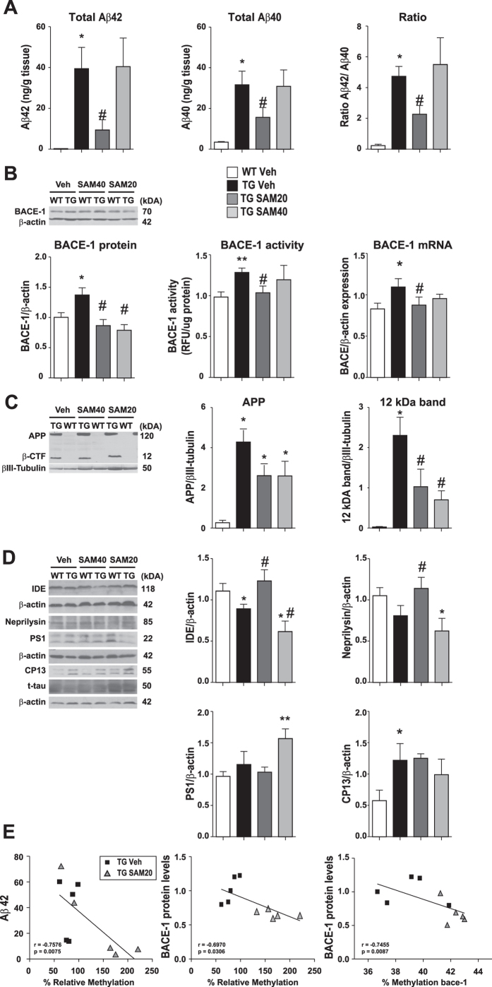
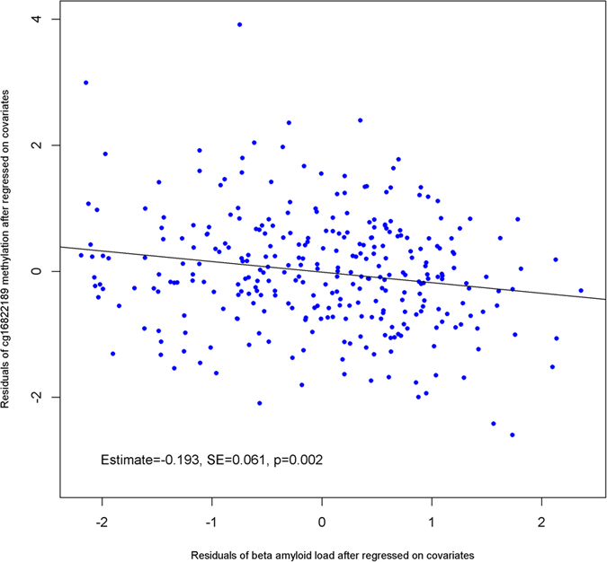
General DNA hypomethylation is associated with Alzheimer's disease (AD), but it is unclear when DNA hypomethylation starts or plays a role in AD pathology or whether DNA re-methylation would rescue early amyloid-related cognitive impairments. In an APP transgenic mouse model of AD-like amyloid pathology we found that early intraneuronal amyloid beta build-up is sufficient to unleash a global and beta-site amyloid precursor protein cleaving enzyme 1 (bace-1) DNA demethylation in AD-vulnerable brain regions. S-adenosylmethionine administration at these early stages abolished this hypomethylation, diminished the amyloid pathology and restored cognitive capabilities. To assess a possible human significance of findings, we examined the methylation at 12 CpGs sites in the bace-1 promoter, using genome-wide DNA methylation data from 740 postmortem human brains. Thus, we found significant associations of bace-1 promoter methylation with β-amyloid load among persons with AD dementia, and PHFtau tangle density. Our results support a plausible causal role for the earliest amyloid beta accumulation to provoke DNA hypomethylation, influencing AD pathological outcomes.


Figures

Figure 1
Figure 1. Global hippocampal and cortical DNA hypomethylation and restoration of methylation levels with SAM administration in McGill-Thy1-APP Tg mice.
(AC) 5-methylcytosine (5-mC) immunofluorescence. Representative images of the hippocampus (CA1 layer) and cerebral cortex (A) in 5 month-old animals showing 5-mC immunoreactivity (red labeling) and neuronal nuclei (NeuN, green labeling). Scale bar: 50 μm. (B,C) Quantification of the total (B) and neuronal (C) 5-mC relative fluorescence (red) in hippocampus and cortex (WT, n = 3; Tg, n = 3). Methylation is significantly decreased in Tg animals, particularly in the hippocampal neurons. (D,E) Global methylation levels in the hippocampus (D) and cerebellum (E) was measured using LUMA. Results are shown as methylation % relative to mean of WT, mean ± SEM. Student’s t-test, *P < 0.05, ***P < 0.001. (F) Experimental design of chronic SAM administration. (G,H) Global methylation was measured by ELISA in the hippocampus (G) and cortex (H) at 2 months of age, time-point where SAM administration started, and at the end of the treatment. All values are presented as relative percentage compared to WT at 2 months of age (WT 2 mo). Data are expressed as mean ± SEM and analyzed with One-Way ANOVA, followed by Bonferroni post-hoc test; *p < 0.05 vs WT Veh; #p < 0.05 vs Tg Veh.
Figure 2
Figure 2. Effect of SAM administration on bace-1 promoter methylation.
(A) Methylation was examined in 3 regions of the bace-1 promoter. Regions amplified by PCR and analysed by pyrosequencing are indicated. Methylation was analysed in region +94/+295 (B,C), −647/−355 (D,E), −904/−663 (F,G) of bace-1 promoter in the cortex (B,D,F) and hippocampus (C,E,G). Values are given as mean percentage of methylation at individual CpG (B–G). (H,I) Cumulative values of all bace-1 promoter regions tested were also calculated. Data are presented as mean ± SEM.*p < 0.05, **p < 0.01, ***p < 0.001 vs WT Veh; #p < 0.05, ##p < 0.01 vs Tg Veh.
Figure 3
Figure 3. SAM chronic administration reversed the cognitive deficits of McGill-Thy1-APP mice.
(AD) Deficits in the Morris water maze. (A) Latency to locate the hidden platform during the learning phase of the test. Animals were given 4 trials per day for 5 days, with the first 2 trials of day 1 involving a visible platform. Mean latency is calculated as area under the curve (AUC, right panel). (B–D) Memory recall was assessed 24 h after completion of the learning phase and time spent in the target quadrant (B), time to first reach the platform (C) and number of crosses over the annulus that contained the platform (D) were analysed. (E) Mice were tested for spontaneous activity in open-field. (F,G) Deficits in Novel Object Recognition test. (F) No preference for either of the 2 objects was recorded during the training phase of the task, when 2 identical objects were presented. (G) During the probe test, carried 24 h after training, WT mice showed a clear preference for the new object, while the vehicle-treated Tg mice failed to discriminate between the familiar and novel object. Treatment with SAM20 reduced this deficit. The 0.5 discrimination ratio, equivalent to chance is indicated by a dashed line. Data are expressed as mean ± SEM and analyzed with One-Way ANOVA, followed by Bonferroni post hoc test; *p < 0.05 vs WT Veh; #p < 0.05 vs Tg Veh.
Figure 4
Figure 4. Effect of SAM administration on AD-like amyloid pathology.
(A) Intense intraneuronal Aβ immunoreactivity is observed in Tg mice throughout the neocortex (a–d), hippocampus (a–f) and subiculum (g,h), as revealed with the anti-Aβ McSA1 monoclonal antibody. Note the absence of McSA1-immunoreactivity in non-transgenic animals (i). Scale bar: a-b, i = 500 μm; c-d = 100 μm; e-h = 50 μm. (B) Quantification of the total number of extracellular Aβ-amyloid plaques. (C) Quantification of the intensity of intraneuronal McSA1 immunoreactivity in cortical lamina V, presented as arbitrary units (A.U.). Data are expressed as mean ± SEM and analyzed with Student’s t-test. *p < 0.05, **p < 0.01, ***p < 0.001.
Figure 5
Figure 5. SAM chronic administration impacts the APP processing cascade.
Biochemical analyses were performed on cortical homogenates. (A) Abundance of human Aβ42 (left) and Aβ40 (middle) peptides was determined by ELISA from guanidine hydrochloride-homogenized tissue. The ratio Aβ42/Aβ40 was also calculated (right). Signals detected in WT animals are considered as assay background. (B) The levels of BACE-1 protein, activity and mRNA were determined by western blot, enzymatic assay and qRT-PCR respectively. (C) The expression of human APP and the major C-terminal cleavage product of APP (β-CTF) were assessed by western blot (left) using 6E10 antibody. (D) The levels of PS1 and of the major Aβ-degrading enzymes IDE and neprilysin were analyzed by western blot. The levels of tau phosphorylated at Ser-202 (CP13) were also determined. Representative blot images of at least three independent experiments are shown. β-actin or βIII-tubulin were used as internal control for quantification and all values are presented as relative levels compared to WT Vehicle. Data are expressed as mean ± SEM and analyzed with One-Way ANOVA, followed by Bonferroni post hoc test; *p < 0.05 vs WT Veh; #p < 0.05 vs Tg Veh. (E) Correlations between the cortical levels of Aβ42 (left) and BACE-1 (center) and the levels of global DNA methylation as detected by ELISA. Correlation between BACE-1 protein levels and bace-1 promoter methylation (right). Only Tg Vehicle and Tg SAM20 were included in the analysis. Spearman’s rank correlation coefficient (ρ) and level of significance (p) are indicated within each graph. Each point represents data from a single animal.
Figure 6
Figure 6. Association of the methylation at cg16822189 located in the bace-1 promoter with β-amyloid load among persons with AD dementia.

References

    1. Cuello A. C. Overview of the Alzheimer’s disease pathology and potential therapeutic targets. In Pharmacological mechanisms in Alzheimer’s therapeutics Cuello A. C., ed. (Springer, New York), pp. 1–27 (2007).
    1. Querfurth H. W. & LaFerla F. M. Alzheimer’s Disease. N Engl J Med 362, 329–344 (2010). - PubMed
    1. LaFerla F. M., Green K. N. & Oddo S. Intracellular amyloid-beta in Alzheimer’s disease. Nat Rev Neurosci 8, 499–509 (2007). - PubMed
    1. Han J. et al.. Effect of 5-aza-2-deoxycytidine microinjecting into hippocampus and prelimbic cortex on acquisition and retrieval of cocaine-induced place preference in C57BL/6 mice. Eur J Pharmacol 642, 93–98 (2010). - PubMed
    1. Levenson J. M. et al.. Evidence that DNA (cytosine-5) methyltransferase regulates synaptic plasticity in the hippocampus. J Biol Chem 281, 15763–15773 (2006). - PubMed

LinkOut - more resources