. 2016 Nov 15;473(22):4243-4254.
doi: 10.1042/BCJ20160674. Epub 2016 Sep 28.

Regulation of divalent metal transporter-1 by serine phosphorylation

Affiliations

Regulation of divalent metal transporter-1 by serine phosphorylation

Young Ah Seo et al. Biochem J. .

Abstract

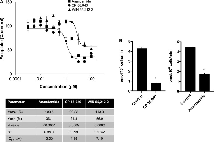
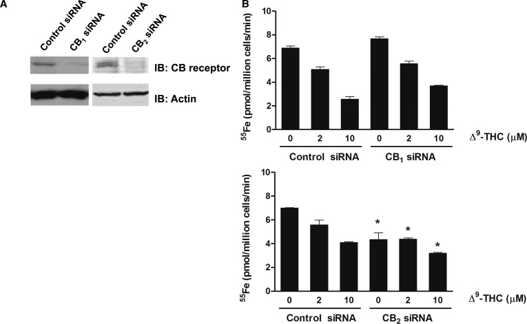
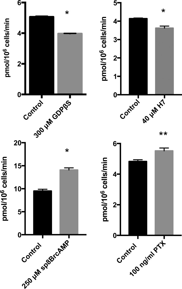
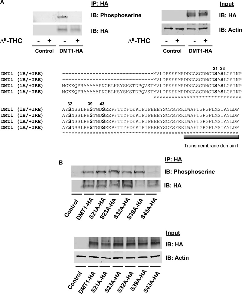
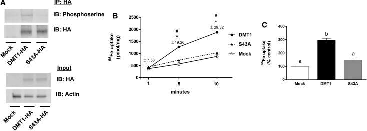
Divalent metal transporter-1 (DMT1) mediates dietary iron uptake across the intestinal mucosa and facilitates peripheral delivery of iron released by transferrin in the endosome. Here, we report that classical cannabinoids (Δ9-tetrahydrocannabinol, Δ9-THC), nonclassical cannabinoids (CP 55,940), aminoalkylindoles (WIN 55,212-2) and endocannabinoids (anandamide) reduce 55Fe and 54Mn uptake by HEK293T(DMT1) cells stably expressing the transporter. siRNA knockdown of cannabinoid receptor type 2 (CB2) abrogated inhibition. CB2 is a G-protein (GTP-binding protein)-coupled receptor that negatively regulates signal transduction cascades involving serine/threonine kinases. Immunoprecipitation experiments showed that DMT1 is serine-phosphorylated under basal conditions, but that treatment with Δ9-THC reduced phosphorylation. Site-directed mutation of predicted DMT1 phosphosites further showed that substitution of serine with alanine at N-terminal position 43 (S43A) abolished basal phosphorylation. Concordantly, both the rate and extent of 55Fe uptake in cells expressing DMT1(S43A) was reduced compared with those expressing wild-type DMT1. Among kinase inhibitors that affected DMT1-mediated iron uptake, staurosporine also reduced DMT1 phosphorylation confirming a role for serine phosphorylation in iron transport regulation. These combined data indicate that phosphorylation at serine 43 of DMT1 promotes transport activity, whereas dephosphorylation is associated with loss of iron uptake. Since anti-inflammatory actions mediated through CB2 would be associated with reduced DMT1 phosphorylation, we postulate that this pathway provides a means to reduce oxidative stress by limiting iron uptake.

Keywords: DMT1; Slc11a2; cannabinoid receptor; iron transport; Δ9-tetrahydrocannabinol.


Figures

Figure 1.
Figure 1.. Dose–response studies of DMT1 inhibition by cannabinoids.
HEK293T(DMT1) cells were incubated at 37°C for 20 min with 0.001–100 μM of the indicated compounds as described in the Experimental Procedures section (n = 6). Shown are curves fit by regression analysis with symbols indicated in (A). (B) 54Mn uptake in the absence and presence of 50 µM anandamide or CP55,940. Shown are means (±SEM) for control cells incubated with vehicle (DMSO) and indicated drug (n = 3); results from one experiment with similar results obtained on at least one other occasion (*P < 0.0001). Dose response curves were best fit by a Hill four-parameter sigmoidal model with values obtained for Ymax, Ymin, and IC50 shown in the inserted table. The P-values and R2 for each of the curve fits are also provided.
Figure 2.
Figure 2.. Loss of CB2 receptor abrogates Δ9-THC inhibition of iron transport.
(A) A representative immunoblot of CB1 or CB2 receptor levels in cells transfected with control, CB1 or CB2 receptor-specific siRNA. Equal loading was verified by immunoblotting with antiactin antibody. (B) Inhibition of iron uptake by CB1 or CB2 receptor-attenuated cells incubated with 2 or 10 μM of Δ9-THC. Results shown are means ± SEM from at least two independent experiments (n = 5). *Two-way ANOVA followed by Holm–Sidak's multiple comparisons test showed that CB2 knockdown reduced iron transport in the absence of Δ9-THC (P = 0.0003), at 2 µM Δ9-THC (P = 0.0114) and at 10 µM Δ9-THC (P = 0.0216) compared with controls.
Figure 3.
Figure 3.. Inhibitors and activators of DMT1-mediated iron uptake.
Results of transport assays performed as described in Figure 1 are shown for indicated agents compared with vehicle control (0.5% DMSO, v/v, bottom panel) or water alone (top panel). Results are from single independent experiments' means ± SEM determined for biological triplicates with similar results obtained on at least two separate occasions. For pertussis toxin (PTX) experiments, cells were treated with 100 ng/ml toxin added to culture medium for at least 24 h prior to assays (*P < 0.01; **P < 0.05).
Figure 4.
Figure 4.. Serine phosphorylation of DMT1 is blocked by Δ9-THC.
(A) HEK293T cells were transiently transfected with an empty vector or DMT1-HA. Cells were preincubated with a phosphatase inhibitor in PBS++ at 37°C for 15 min and then spun down, resuspended in uptake buffer containing a phosphatase inhibitor with 50 μM Δ9-THC or control vehicle (0.5% DMSO, v/v) at 37°C for 20 min. After immunoprecipitation with anti-HA, samples were immunoblotted to determine serine phosphorylation (IP: HA and IB: Phosphoserine) compared with immunoprecipitated DMT1-HA protein (IP: HA and IB: HA). Total cell lysates from the same samples (Input) were used to detect DMT1-HA protein (IB: HA) and actin was used as a loading control (IB: Actin). Amino acid alignment between mouse DMT1 isoforms (1B/+IRE, 1B/−IRE, 1A/+IRE and 1A/−IRE) was generated with ClustalW. The predicted phosphorylation sites are located in a conserved region (marked with bold letters) before the first predicted transmembrane domain (marked with a bold, gray line under amino acid sequence). (B) HEK293 cells were transfected to express DMT1-HA and serine mutants (S21A-HA, S23A-HA, S32A-HA, S39A-HA and S43A-HA). DMT1 was immunoprecipitated from the cell lysates with anti-HA antibody (IP: HA) and immunoblotted with antiphosphoserine antibody (IP: HA and IB: Phosphoserine). Blots were then stripped and reprobed with anti-HA (IP: HA and IB: HA) as a loading control. Total cell lysates from the same samples (Input) were used to detect total abundance of DMT1-HA protein and actin.
Figure 5.
Figure 5.. DMT1 phosphorylation at serine 43.
(A) HEK293T cells were transfected to express DMT1-HA or S43A-HA, and immunoprecipitation and immunoblot analysis were performed as described in Figure 3. (B) Time course of iron uptake by HEK293T cells transfected to express HA-DMT1 (closed circles) or HA-DMT1(S43A) (closed triangles) compared with mock-transfected control (empty circles) cells (n = 3). Similar results were obtained in at least two independent experiments. *P < 0.05 vs. control; #P < 0.05 vs. S43A. SEM values are provided since errors bars are too small to visualize. (C) The extent of iron uptake over a 20-min incubation period was determined for HEK293T cells transfected to express DMT1-HA (filled bar), S43A-HA (gray bar) or mock-transfected (open bar). Results shown are means ± SEM (n = 3) with similar results obtained in at least two independent experiments (n = 3). Means with different letters are significantly different by one-way ANOVA followed by Tukey's multiple comparisons test (P < 0.0001).
Figure 6.
Figure 6.. Staurosporine inhibits DMT1 transport and reduces serine phosphorylation.
(A) Iron uptake by HEK293T(DMT1) cells was assayed after incubation with indicated concentrations of staurosporine in phosphate-buffered saline with glucose (PBSG)++ at 37°C for 20 min. The histogram represents three experiments done in triplicate and results shown are mean values ± SEM for inhibition, *P < 0.001. (B) Serine phosphorylation of DMT1 was determined by immunoprecipitation as described in Figure 3, except after a preincubation step with a phosphatase inhibitor in PBS++ at 37°C for 15 min, followed by assays containing a phosphatase inhibitor with 20 μM staurosporine or vehicle (0.5% DMSO, v/v) at 37°C for 20 min. Immunoprecipitation was performed using an anti-HA antibody (IP: HA) and immunoblotting was performed (IP: HA and IB: Phosphoserine) in comparison with immunoprecipitated DMT1-HA protein (IP: HA and IB: HA). Total cell lysates from the same samples (Input) were used to detect DMT1-HA protein (IB: HA) and actin was used as a loading control (IB: Actin). (C) Mean ratio of DMT1-HA: Actin ± SD from untreated cells (−) and cells treated with staurosporine (+) determined by densitometry of two independent experiments as described in B.

References

    1. Gunshin H., Fujiwara Y., Custodio A.O., Direnzo C., Robine S. and Andrews N.C. (2005) Slc11a2 is required for intestinal iron absorption and erythropoiesis but dispensable in placenta and liver. J. Clin. Invest. 115, 1258–1266 doi: 10.1172/JCI24356 - DOI - PMC - PubMed
    1. Fleming M.D., Romano M.A., Su M.A., Garrick L.M., Garrick M.D. and Andrews N.C. (1998) Nramp2 is mutated in the anemic Belgrade (b) rat: evidence of a role for Nramp2 in endosomal iron transport. Proc. Natl Acad. Sci. USA 95, 1148–1153 doi: 10.1073/pnas.95.3.1148 - DOI - PMC - PubMed
    1. Shawki A., Knight P.B., Maliken B.D., Niespodzany E.J. and Mackenzie B. (2012) Chapter five — H+-coupled divalent metal-ion transporter-1: functional properties, physiological roles and therapeutics. Curr. Top. Membr. 70, 169–214 doi: 10.1016/B978-0-12-394316-3.00005-3 - DOI - PMC - PubMed
    1. Gunshin H., Mackenzie B., Berger U.V., Gunshin Y., Romero M.F., Boron W.F. et al. (1997) Cloning and characterization of a mammalian proton-coupled metal-ion transporter. Nature 388, 482–488 doi: 10.1038/41343 - DOI - PubMed
    1. Hubert N. and Hentze M.W. (2002) Previously uncharacterized isoforms of divalent metal transporter (DMT)-1: implications for regulation and cellular function. Proc. Natl Acad. Sci. USA 99, 12345–12350 doi: 10.1073/pnas.192423399 - DOI - PMC - PubMed

Publication types

MeSH terms

Substances