. 2016 Sep 29:6:34260.
doi: 10.1038/srep34260.

Quantitative lateral flow strip assays as User-Friendly Tools To Detect Biomarker Profiles For Leprosy

Affiliations

Quantitative lateral flow strip assays as User-Friendly Tools To Detect Biomarker Profiles For Leprosy

Anouk van Hooij et al. Sci Rep. .

Abstract

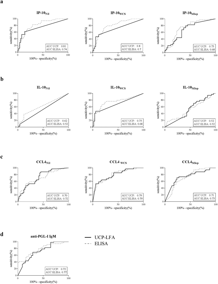
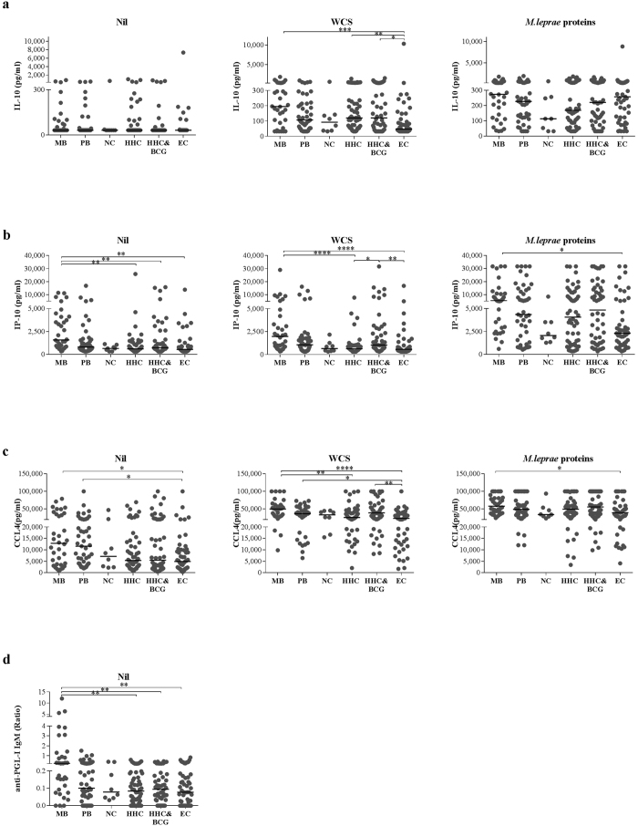
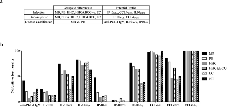
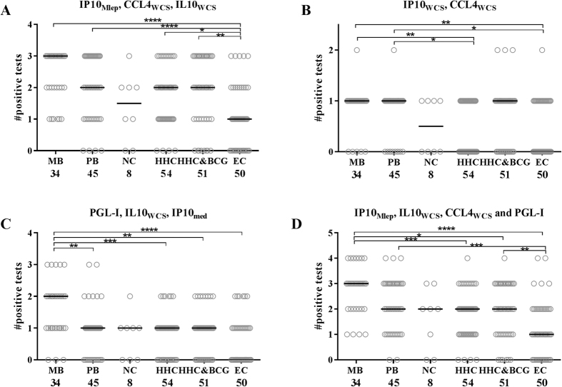
Leprosy is a debilitating, infectious disease caused by Mycobacterium leprae. Despite the availability of multidrug therapy, transmission is unremitting. Thus, early identification of M. leprae infection is essential to reduce transmission. The immune response to M. leprae is determined by host genetics, resulting in paucibacillary (PB) and multibacillary (MB) leprosy associated with dominant cellular or humoral immunity, respectively. This spectral pathology of leprosy compels detection of immunity to M. leprae to be based on multiple, diverse biomarkers. In this study we have applied quantitative user friendly lateral flow assays (LFAs) for four immune markers (anti-PGL-I antibodies, IL-10, CCL4 and IP-10) for whole blood samples from a longitudinal BCG vaccination field-trial in Bangladesh. Different biomarker profiles, in contrast to single markers, distinguished M. leprae infected from non-infected test groups, patients from household contacts (HHC) and endemic controls (EC), or MB from PB patients. The test protocol presented in this study merging detection of innate, adaptive cellular as well as humoral immunity, thus provides a convenient tool to measure specific biomarker profiles for M. leprae infection and leprosy utilizing a field-friendly technology.


Figures

Figure 1
Figure 1. Discriminatory capacity of ELISA and UCP-LFA.
To compare the ability of ELISA (dotted line) and UCP-LFA (solid line) to discriminate between individuals with or without disease ROC curves were computed using data of MB patients and EC. Areas under the curve (AUCs) were compared for all 10 conditions tested, shown in the lower right corner of each graph. (a) ROC curves for IP-10 stimulated and unstimulated samples based on concentration in pg/ml, showing an improved AUC for the UCP-LFA for IP-10Nil and IP-10WCS. (b) ROC curves for IL-10 stimulated and unstimulated samples based on concentration in pg/ml, showing an improved (IL-10Nil) or equal AUC for UCP-LFA. (c) ROC curves for CCL4 stimulated and unstimulated samples based on concentration in pg/ml, showing comparable values for UCP-LFA and ELISA. (d) ROC curves for anti-PGL-I IgM in unstimulated samples based on ratio, showing comparable AUCs for ELISA and UCP-LFA.
Figure 2
Figure 2. Identification of M. leprae specific IL-10, IP-10, CCL4 and anti-PGL-I IgM antibodies by UCP-LFA.
(a) IL-10 concentrations (pg/ml) measured per group per stimulus show that MB patients, HHC and BCG-vaccinated HHC significantly differ from EC upon WCS stimulation. (b) IP-10 concentrations (pg/ml) measured per group per stimulus show that MB patients significantly differ from EC in both stimulated and unstimulated samples, from HHC in unstimulated and WCS stimulated samples and from BCG vaccinated HHC in unstimulated samples. BCG vaccinated HHC significantly differ from HHC and EC upon WCS stimulation. (c) CCL4 concentrations (pg/ml) measured per group, per stimulus show that MB patients significantly differ from EC in both stimulated and unstimulated samples and from HHC in WCS stimulated samples. PB patients significantly differ from EC in unstimulated and WCS stimulated samples and BCG vaccinated HHC significantly differ from EC in WCS stimulated samples. (d) anti-PGL-I IgM ratio measured per groups shows that MB patients have significantly higher levels of anti-PGL-I IgM compared to HHC, BCG vaccinated HHC and EC. P-values: *p ≤ 0.05, **p ≤ 0.01, ***p ≤ 0.001, ****p ≤ 0.0001.
Figure 3
Figure 3. Positive test results per analyte/stimulus combination used to construct potential biomarker profiles.
The groups that should be differentiated to indicate M. leprae infection, disease per se and disease classification are shown. The potential profiles indicated are based on the percentage of positive individuals of these particular groups. The cut-off for positivity was based on values for NEC (Supplementary Table S3) per analyte/stimulus combination the percentage of individuals with a positive test result per group is shown. Based on these data the optimal analyte/stimulus combination to differentiate either infected from non-infected groups, patients and non-patients groups or MB and PB patients were selected to construct the potential profiles described.
Figure 4
Figure 4. Potential of biomarker profiles to indicate M. leprae infection, disease per se and disease classification.
The amount of positive test results per group is shown. (a) IP-10Mlep, CCL4WCS and IL-10WCS significantly differed in MB/PB patients and (BCG-vaccinated) HHC from EC, showing more positive test results in the groups that are exposed to M. leprae and thereby indicating M. leprae infection. (b) CCL4WCS and IP-10WCS enabled the distinction between patients and HHC, thereby indicating the pathogenic immune responses to M. leprae in patients. (c) Anti-PGL-I IgM, IL-10WCS and IP-10Nil showed more positive test results in MB patients thereby enabling the distinction between MB and PB patients. (d) A four marker profile of IL-10WCS, IP-10Mlep, CCL4WCS and anti-PGL-I IgM shows the majority of significant differences observed in A, B and C. P-values: *p ≤ 0.05, **p ≤ 0.01, ***p ≤ 0.001, ****p ≤ 0.0001.

Similar articles

Cited by

References

    1. WHO Global leprosy: update on the 2012 situation. Wkly. Epidemiol. Rec. 88, 365–379 (2013). - PubMed
    1. Lockwood D. N. & Saunderson P. Nerve damage in Leprosy: a continuing challenge for scientists, clinicians and service providers. Int Health 4, 77–85 (2012). - PubMed
    1. Scollard D. M. et al.. The continuing challenges of leprosy. Clin. Microbiol. Rev. 19, 338–381 (2006). - PMC - PubMed
    1. Smith W. C., van B. W., Gillis T., Saunderson P. & Richardus J. H. The missing millions: a threat to the elimination of leprosy. PLoS. Negl. Trop. Dis. 9, e0003658, 10.1371/journal.pntd.0003658 (2015). - DOI - PMC - PubMed
    1. Geluk A. Challenges in immunodiagnostic tests for leprosy. Expert. Opin. Med. Diagn. 7, 265–274 (2013). - PubMed