. 2016 Dec 9;119(12):1313-1323.
doi: 10.1161/CIRCRESAHA.116.309799. Epub 2016 Sep 28.

Extensive Proliferation of a Subset of Differentiated, yet Plastic, Medial Vascular Smooth Muscle Cells Contributes to Neointimal Formation in Mouse Injury and Atherosclerosis Models

Affiliations

Extensive Proliferation of a Subset of Differentiated, yet Plastic, Medial Vascular Smooth Muscle Cells Contributes to Neointimal Formation in Mouse Injury and Atherosclerosis Models

Joel Chappell et al. Circ Res. .

Abstract

Rationale: Vascular smooth muscle cell (VSMC) accumulation is a hallmark of atherosclerosis and vascular injury. However, fundamental aspects of proliferation and the phenotypic changes within individual VSMCs, which underlie vascular disease, remain unresolved. In particular, it is not known whether all VSMCs proliferate and display plasticity or whether individual cells can switch to multiple phenotypes.

Objective: To assess whether proliferation and plasticity in disease is a general characteristic of VSMCs or a feature of a subset of cells.

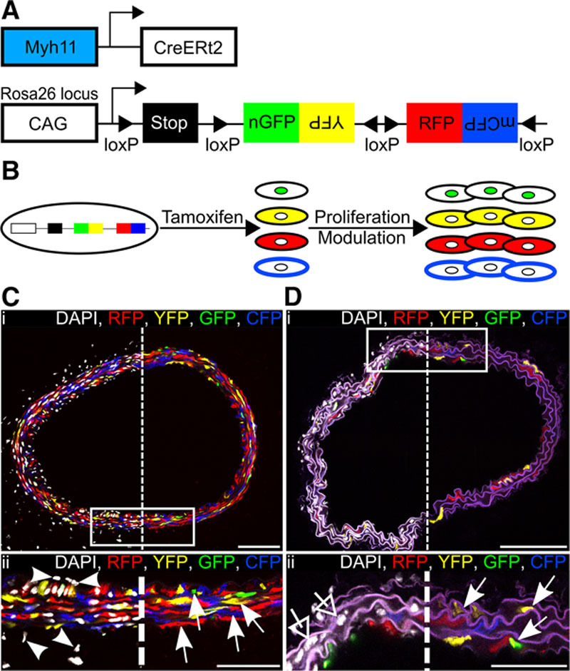
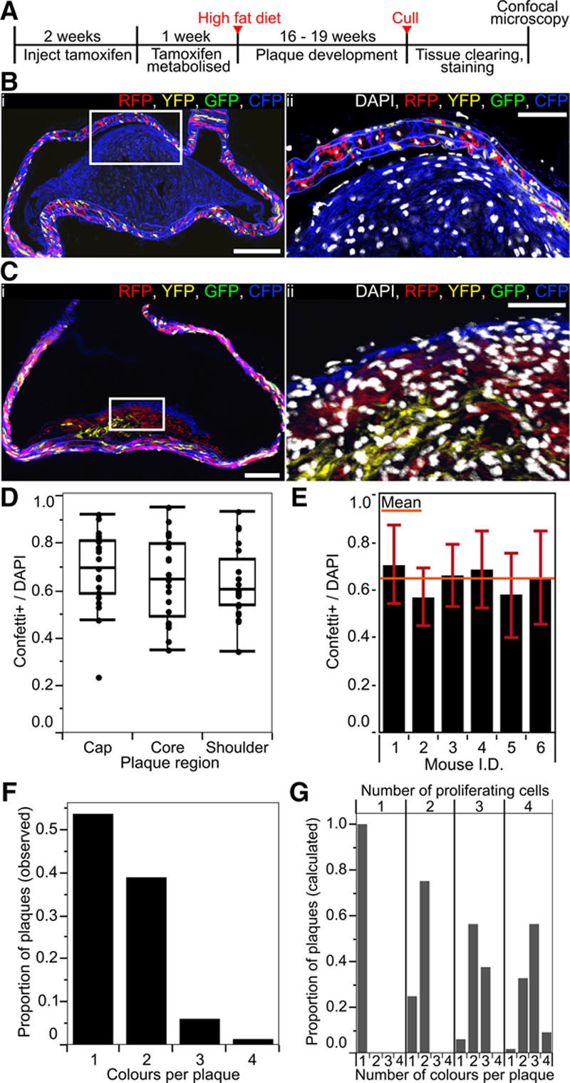
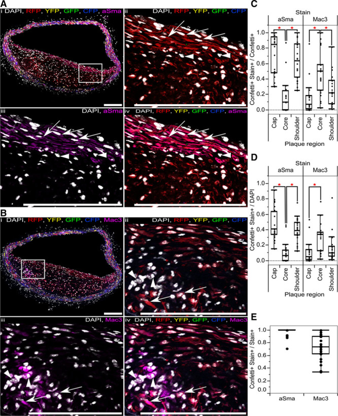
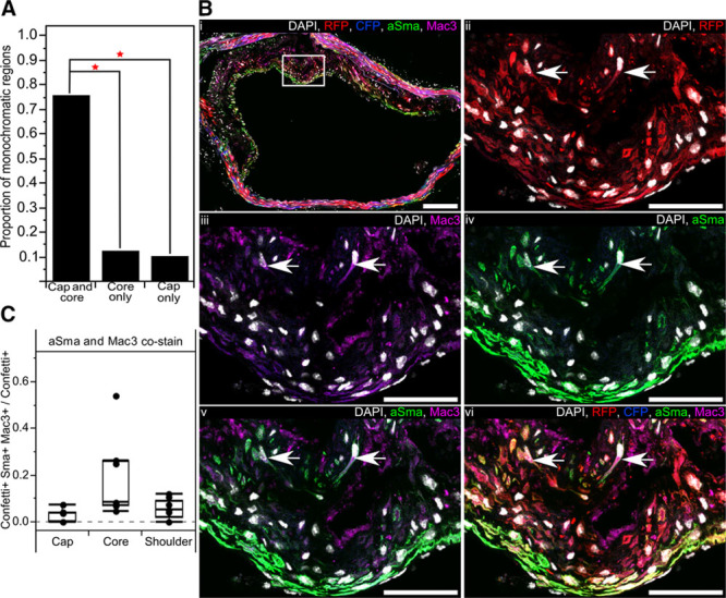
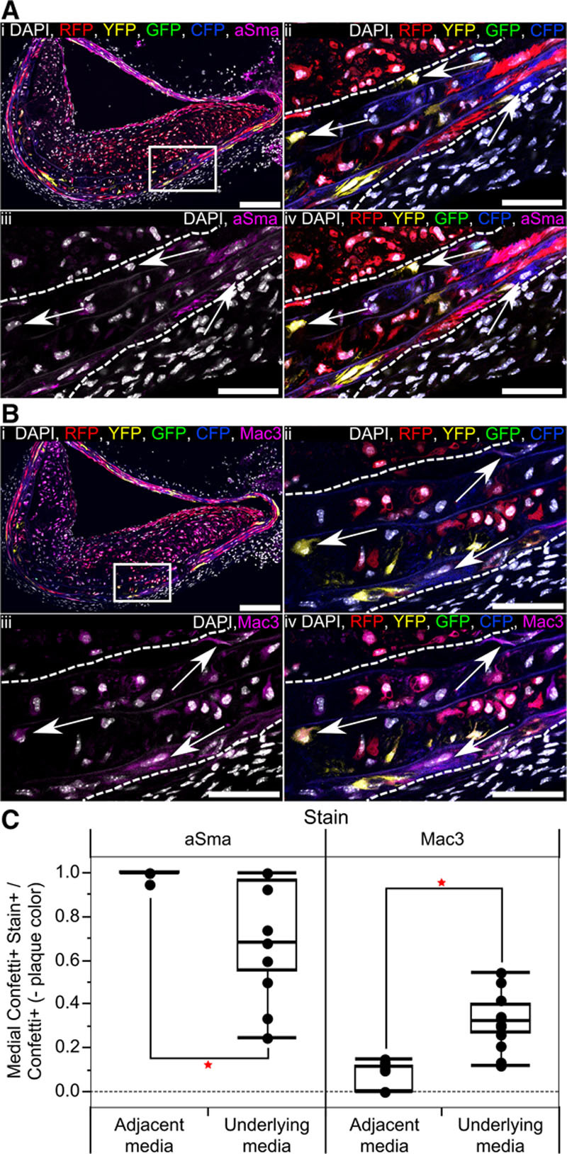
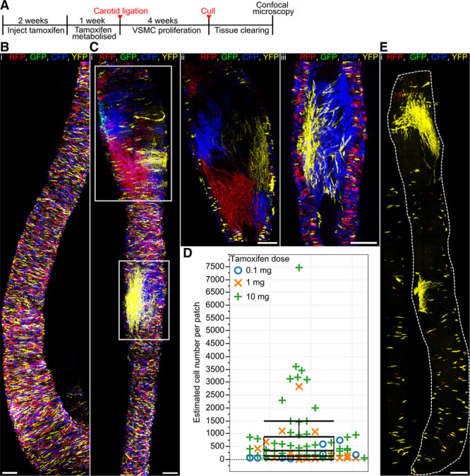
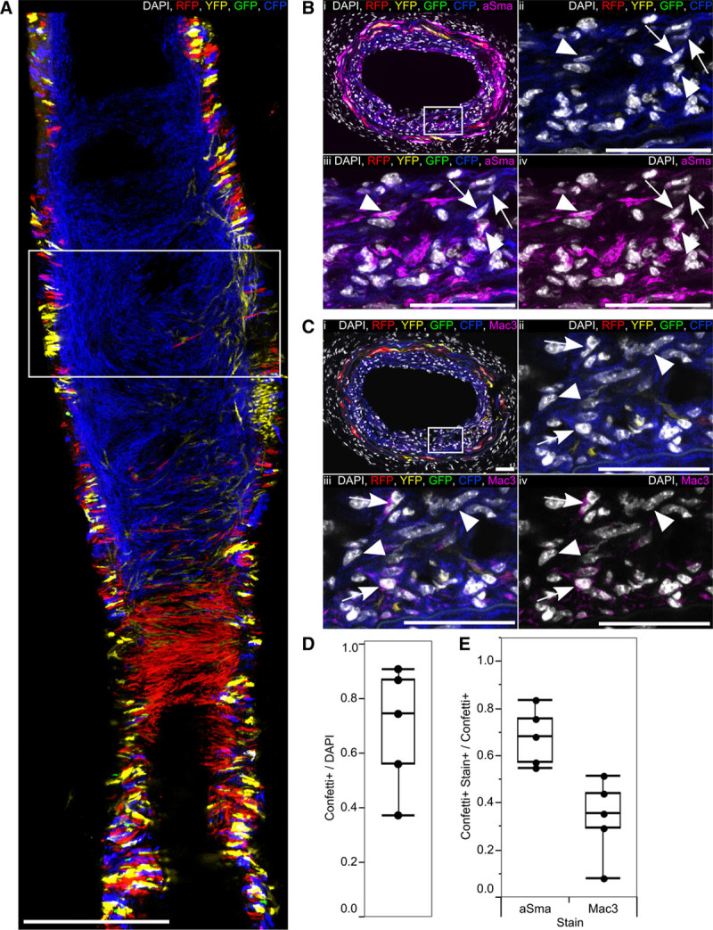
Methods and results: Using multicolor lineage labeling, we demonstrate that VSMCs in injury-induced neointimal lesions and in atherosclerotic plaques are oligoclonal, derived from few expanding cells. Lineage tracing also revealed that the progeny of individual VSMCs contributes to both alpha smooth muscle actin (aSma)-positive fibrous cap and Mac3-expressing macrophage-like plaque core cells. Costaining for phenotypic markers further identified a double-positive aSma+ Mac3+ cell population, which is specific to VSMC-derived plaque cells. In contrast, VSMC-derived cells generating the neointima after vascular injury generally retained the expression of VSMC markers and the upregulation of Mac3 was less pronounced. Monochromatic regions in atherosclerotic plaques and injury-induced neointima did not contain VSMC-derived cells expressing a different fluorescent reporter protein, suggesting that proliferation-independent VSMC migration does not make a major contribution to VSMC accumulation in vascular disease.

Conclusions: We demonstrate that extensive proliferation of a low proportion of highly plastic VSMCs results in the observed VSMC accumulation after injury and in atherosclerotic plaques. Therapeutic targeting of these hyperproliferating VSMCs might effectively reduce vascular disease without affecting vascular integrity.

Keywords: atherosclerosis; lineage-tracing; macrophages; neointima; phenotype; vascular diseases vascular smooth muscle.


Conflict of interest statement

None

Figures

Figure 1.
Figure 1.
Efficient and specific multicolor vascular smooth muscle cell (VSMC) labeling in Myh11-CreERt2/Rosa26-Confetti animals. A, Schematic representation of the Myh11-CreERt2 transgene and the Rosa26-Confetti reporter allele. B, Schematic representation illustrating tamoxifen-induced recombination at the Rosa26-Confetti locus, resulting in expression of 1 of 4 fluorescent proteins, which are stably propagated independent of Myh11 expression within progeny. C and D, Carotid artery cross sections from high density–labeled (C; 10× 1 mg tamoxifen) or low density–labeled (D; 1× 0.1 mg tamoxifen) animals; region outlined in (i) is magnified in (ii). Signals for fluorescent proteins are shown with (left) and without (right) nuclear DAPI (4',6-diamidino-2-phenylindole) staining (white). C, VSMCs, indicated by arrows in (ii), are labeled with red fluorescent protein (RFP), yellow fluorescent protein (YFP), nuclear (n) green fluorescent protein (GFP), or membrane-associated (m) cyan fluorescent protein (CFP), whereas cells within the adventitia and endothelium, indicated by arrow heads, are unlabeled. In (D[ii]), arrows point to the few labeled VSMCs, and open arrows point to unlabeled VSMCs within the media. Scale bars are 100 μm in (i) and 50 μm in (ii).
Figure 2.
Figure 2.
Vascular smooth muscle cell (VSMC)–derived cells generate oligoclonal atherosclerotic plaques. A, Experimental protocol for the atherosclerosis studies. B and C, Arterial cryosections from high density–labeled animals (10× 1 mg tamoxifen) presenting plaques containing VSMC-derived cells of a single color (membrane-associated cyan fluorescent protein [CFP]; B) and >1 color (red fluorescent protein [RFP], membrane-associated CFP, and yellow fluorescent protein [YFP]; C). The region outlined in (i) is magnified in (ii). Scale bars are 150 μm (i) and 50 μm (ii). Signals for fluorescent proteins and nuclear DAPI (4',6-diamidino-2-phenylindole; in ii, white) are shown. D, Box plot showing the proportion of the total number of cells (DAPI), which express the Confetti reporter (Confetti+) within each plaque region (in 23 plaques from 6 animals). E, Bar chart showing the proportion of the total number of cells (DAPI), which express the Confetti reporter (Confetti+) within plaques for 6 individual animals (23 plaques from 6 animals). Mean across all animals is indicated by the orange bar, SD±0.16. F, Bar chart showing the number of colors observed per plaque (82 plaques from 16 animals). G, Bar chart showing the theoretical distribution of colors per plaque resulting from proliferation of 1, 2, 3, or 4 VSMCs labeled at the recombination frequency observed in high density–labeled animals. All data are from animals labeled at high density (10× 1 mg tamoxifen).
Figure 3.
Figure 3.
Progeny of a single vascular smooth muscle cell (VSMC) can adopt multiple phenotypes in disease. A and B, Immunostaining for alpha smooth muscle actin (aSma; A) and Mac3 (B) of serial cryosections from high density–labeled animal (10× 1 mg tamoxifen) containing RFP-expressing VSMC-derived cells. Signals for fluorescent proteins, nuclear DAPI (4',6-diamidino-2-phenylindole; white), aSma (magenta; A), and Mac3 (magenta; B) are shown as indicated on each image. Regions outlined in (i) are magnified in panels (ii through v), scale bars are 150 μm. A, Arrows point to RFP+ aSma+ cells, arrow heads point to RFP− aSma− cells. B, Arrows point to RFP+ Mac3+ cells, and arrow heads point to RFP− Mac3+ cells. C through E, Box plot showing proportion of cells that express the Confetti reporter and stain positive for either aSma or Mac3 (Confetti+ Stain+), relative to all cells expressing the Confetti reporter (Confetti+; C), the total number of cells (DAPI; D), or all cells staining positive for aSma or Mac3 markers (Stain+; E). A red star indicates a significant difference (P<0.05) determined by a 2-way analysis of variance. Data in (C through E) are from 23 plaques from 6 animals. All data are from animals labeled at high density (10× 1 mg tamoxifen). CFP indicates cyan fluorescent protein; GFP, green fluorescent protein; RFP, red fluorescent protein; and YFP, yellow fluorescent protein.
Figure 4.
Figure 4.
Progeny of a single vascular smooth muscle cell (VSMC) generates plaque cells with different phenotypes. A, Bar chart showing the proportion of monochromatic regions, which occupy both the cap and core or only a single region within an atherosclerotic plaque (82 plaques from 16 high density–labeled animals). B, Arterial cryosection from artery containing red fluorescent protein (RFP)–expressing VSMC-derived cells, costained for alpha smooth muscle actin (aSma) and Mac3. The region outlined in (i) is magnified in (ii through iv). Arrows point to RFP+ Mac3+ aSma+ cells. Scale bars are 150 μm (i) and 50 μm (ii through vi). Signals for fluorescent proteins, nuclear DAPI (4',6-diamidino-2-phenylindole; white), aSma (green) and Mac3 (magenta) are shown as indicated on each image. C, Box plot showing the proportion of cells expressing the Confetti reporter (Confetti+), which costain for aSma and Mac3 (Confetti+ aSma+ Mac3+) within different plaque regions (7 plaques from 6 mice). Only red and blue plaques were used for this analysis as the 488 channel was required for aSma imaging. All data are from animals labeled at high density (10× 1 mg tamoxifen). CFP indicates cyan fluorescent protein.
Figure 5.
Figure 5.
Vascular smooth muscle cells (VSMCs) within the media directly underlying atherosclerotic plaques undergo phenotypic switching without contributing to the cell mass of the lesion. A and B, Serial cryosections from artery of high density–labeled (10× 1 mg tamoxifen) animal containing a plaque with RFP+ VSMC–derived cells, stained for alpha smooth muscle actin (aSma) (A) and Mac3 (B). The region outlined in (i) is magnified in (ii through iv), white dotted lines outline the media. Signals for fluorescent proteins, nuclear DAPI (4',6-diamidino-2-phenylindole; white), aSma (magenta; A), and Mac3 (magenta; B) are shown as indicated on each image. Arrows in (A) point to aSma-negative cells within the media, which express the Confetti reporter (excluding RFP-expressing cells). Arrows in (B) point to Mac3-positive cells within the media, which express the Confetti reporter (excluding RFP-expressing cells). Scale bars are 150 μm in (i) and 50 μm in (ii through vi). C, Box plot quantifying the proportion of cells expressing the Confetti reporter, which stain positive for aSma or Mac3 (Confetti+ Stain+), relative to all cells expressing Confetti fluorescent proteins not found in the plaque (Confetti+, −plaque color) within the media underlying or adjacent to a plaque (based on 17 plaques from 6 animals). Red stars indicate a significant difference based on a 2-way analysis of variance, P<0.05. All data are from animals labeled at high density (10× 1 mg tamoxifen). CFP indicates cyan fluorescent protein; GFP, green fluorescent protein; RFP, red fluorescent protein; and YFP, yellow fluorescent protein.
Figure 6.
Figure 6.
A subset of vascular smooth muscle cells (VSMCs) proliferate to form the injury-induced neointima. A, Experimental protocol for carotid artery ligation studies. B and C, Whole-mounted control right carotid artery (B) or ligated left carotid artery (C) from a high density–labeled animal (10× 1 mg tamoxifen) 28 days post surgery. Maximal projection of confocal Z-stack covering the entire artery is shown in (B) and (C[i]), whereas (C[ii] and C[iii]) show magnified longitudinal cross section of the regions outlined in (C[i]). D, Box plot showing the size of individual monochromatic patches. The patch sizes of low (blue circles) and medium density–labeled animals (orange crosses) largely fall within the interquartile range observed in high density–labeled animals (green crosses), strongly suggesting that patches result from clonal expansion of a single cell. Large patches (>1500 cells) might arise from merging of same colored clones. Fifty-two patches from 12 high density–labeled (10× 1 mg), 15 patches from 4 medium density–labeled (1× 1 mg), and 8 patches from 4 low density–labeled animals (1× 0.1 mg) were analyzed. E, Maximal confocal Z-stack projection of whole-mounted ligated left carotid artery from a low density–labeled animal (1× 0.1 mg tamoxifen), analyzed 28 days post ligation. Signals for fluorescent proteins are shown. All scale bars are 150 μm. CFP indicates cyan fluorescent protein; GFP, green fluorescent protein; RFP, red fluorescent protein; and YFP, yellow fluorescent protein.
Figure 7.
Figure 7.
Injury-induced vascular smooth muscle cell (VSMC)–derived neointima contains few phenotypically switched VSMCs. A, Maximal projection of 3 central scans (7 μm apart) of a confocal Z-stack of whole-mounted ligated left carotid artery from high density–labeled (10× 1 mg tamoxifen) animal, 28 days post ligation. Scale bar is 300 μm. B and C, Transverse cryosections from the region outlined in (A), stained for alpha smooth muscle actin (aSma; B), or Mac3 (C); the region outlined in (i) is magnified in (ii through iv). Arrows point to cells expressing the Confetti reporter (Confetti+) that do not stain for aSma (B) and Confetti+ Mac3+ cells (C) within the neointima. Arrow heads point to Confetti+ aSma+ cells (B) or Confetti+ Mac3− cells (C) within the neointima. Scale bars are 50 μm. Signals for fluorescent proteins, nuclear DAPI (4',6-diamidino-2-phenylindole; white), aSma (magenta, B), and Mac3 (magenta, C) are shown as indicated on each image. D, Box plot showing the proportion of all cells (DAPI) within the neointima, which express the Confetti reporter (Confetti+). E, Box plot displaying the proportion of Confetti+ neointimal cells that stain positive for aSma or Mac3 (Stain+; based on 5 regions from 4 animals). All data are from high density–labeled (10× 1 mg tamoxifen) animals. CFP indicates cyan fluorescent protein; GFP, green fluorescent protein; RFP, red fluorescent protein; and YFP, yellow fluorescent protein.

Comment in

References

    1. Alexander MR, Owens GK. Epigenetic control of smooth muscle cell differentiation and phenotypic switching in vascular development and disease. Annu Rev Physiol. 2012;74:13–40. doi: 10.1146/annurev-physiol-012110-142315. - PubMed
    1. Nemenoff RA, Horita H, Ostriker AC, Furgeson SB, Simpson PA, VanPutten V, Crossno J, Offermanns S, Weiser-Evans MC. SDF-1α induction in mature smooth muscle cells by inactivation of PTEN is a critical mediator of exacerbated injury-induced neointima formation. Arterioscler Thromb Vasc Biol. 2011;31:1300–1308. doi: 10.1161/ATVBAHA.111.223701. - PMC - PubMed
    1. Yang P, Hong MS, Fu C, Schmit BM, Su Y, Berceli SA, Jiang Z. Preexisting smooth muscle cells contribute to neointimal cell repopulation at an incidence varying widely among individual lesions. Surgery. 2016;159:602–612. doi: 10.1016/j.surg.2015.08.015. - PMC - PubMed
    1. Herring BP, Hoggatt AM, Burlak C, Offermanns S. Previously differentiated medial vascular smooth muscle cells contribute to neointima formation following vascular injury. Vasc Cell. 2014;6:21. doi: 10.1186/2045-824X-6-21. - PMC - PubMed
    1. Campbell JH, Campbell GR. Smooth muscle phenotypic modulation–a personal experience. Arterioscler Thromb Vasc Biol. 2012;32:1784–1789. doi: 10.1161/ATVBAHA.111.243212. - PubMed