

Optimizing dual energy cone beam CT protocols for preclinical imaging and radiation research
- PMID: 27683003
- PMCID: PMC5605023
- DOI: 10.1259/bjr.20160480
Optimizing dual energy cone beam CT protocols for preclinical imaging and radiation research
Abstract
Objective: The aim of this work was to investigate whether quantitative dual-energy CT (DECT) imaging is feasible for small animal irradiators with an integrated cone-beam CT (CBCT) system.
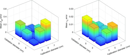
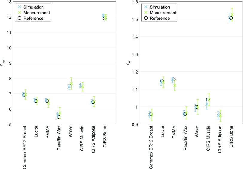
Methods: The optimal imaging protocols were determined by analyzing different energy combinations and dose levels. The influence of beam hardening effects and the performance of a beam hardening correction (BHC) were investigated. In addition, two systems from different manufacturers were compared in terms of errors in the extracted effective atomic numbers (Zeff) and relative electron densities (ρe) for phantom inserts with known elemental compositions and relative electron densities.
Results: The optimal energy combination was determined to be 50 and 90 kVp. For this combination, Zeff and ρe can be extracted with a mean error of 0.11 and 0.010, respectively, at a dose level of 60 cGy.
Conclusion: Quantitative DECT imaging is feasible for small animal irradiators with an integrated CBCT system. To obtain the best results, optimizing the imaging protocols is required. Well-separated X-ray spectra and a sufficient dose level should be used to minimize the error and noise for Zeff and ρe. When no BHC is applied in the image reconstruction, the size of the calibration phantom should match the size of the imaged object to limit the influence of beam hardening effects. No significant differences in Zeff and ρe errors are observed between the two systems from different manufacturers. Advances in knowledge: This is the first study that investigates quantitative DECT imaging for small animal irradiators with an integrated CBCT system.
Figures


References
-
- van Elmpt W, Landry G, Das M, Verhaegen F. Dual energy CT in radiotherapy: current applications and future outlook. Radiother Oncol 2016; 119: 137–44. doi: https://doi.org/10.1016/j.radonc.2016.02.026 - DOI - PubMed
-
- Bazalova M, Carrier JF, Beaulieu L, Verhaegen F. Tissue segmentation in Monte Carlo treatment planning: a simulation study using dual-energy CT images. Radiother Oncol 2008; 86: 93–8. doi: https://doi.org/10.1016/j.radonc.2007.11.008 - DOI - PubMed
-
- Bazalova M, Carrier JF, Beaulieu L, Verhaegen F. Dual-energy CT-based material extraction for tissue segmentation in Monte Carlo dose calculations. Phys Med Biol 2008; 53: 2439–56. doi: https://doi.org/10.1088/0031-9155/53/9/015 - DOI - PubMed
-
- Landry G, Reniers B, Murrer L, Lutgens L, Gurp EB, Pignol JP, et al. . Sensitivity of low energy brachytherapy Monte Carlo dose calculations to uncertainties in human tissue composition. Med Phys 2010; 37: 5188–98. doi: https://doi.org/10.1118/1.3477161 - DOI - PubMed
-
- Bazalova M, Graves EE. The importance of tissue segmentation for dose calculations for kilovoltage radiation therapy. Med Phys 2011; 38: 3039–49. doi: https://doi.org/10.1118/1.3589138 - DOI - PMC - PubMed
Publication types
MeSH terms
LinkOut - more resources
Full Text Sources
Other Literature Sources
