. 2016 Sep 28;36(39):10116-27.
doi: 10.1523/JNEUROSCI.4597-14.2016. Epub 2016 Sep 28.

The Neurotrophic Factor Receptor p75 in the Rat Dorsolateral Striatum Drives Excessive Alcohol Drinking

Affiliations

The Neurotrophic Factor Receptor p75 in the Rat Dorsolateral Striatum Drives Excessive Alcohol Drinking

Emmanuel Darcq et al. J Neurosci. .

Abstract

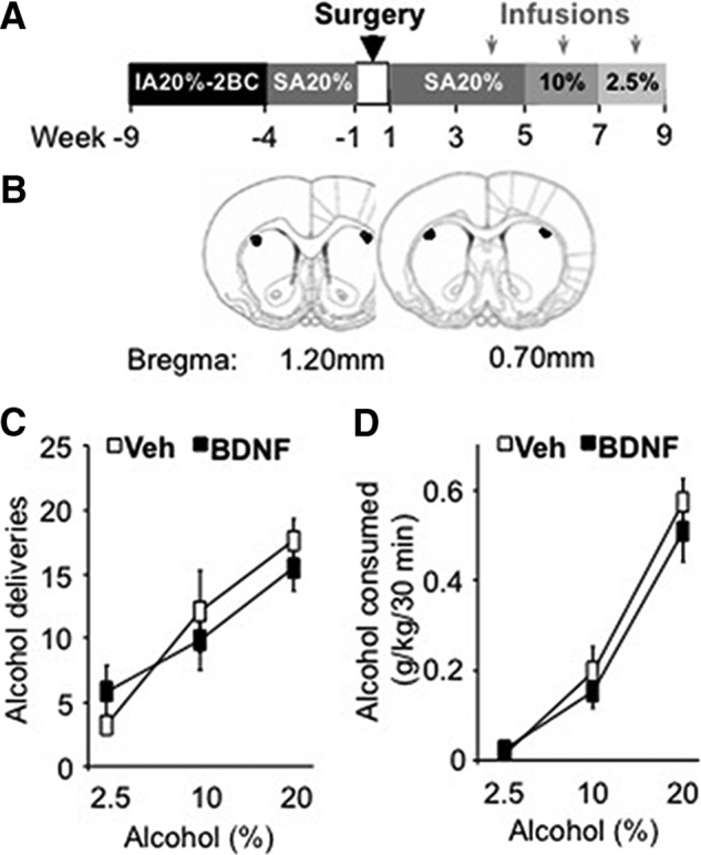
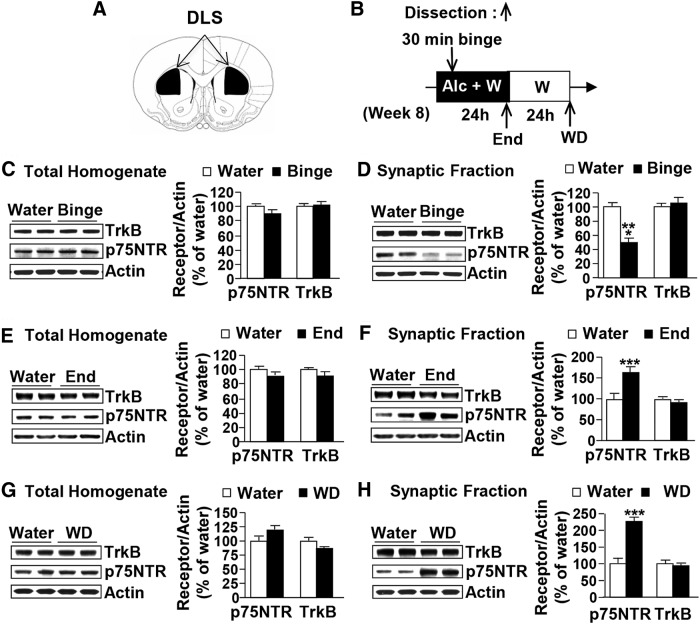
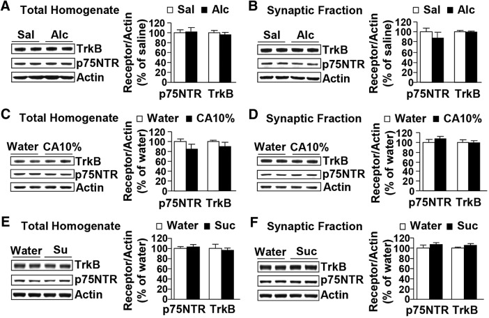
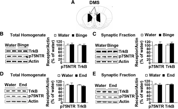
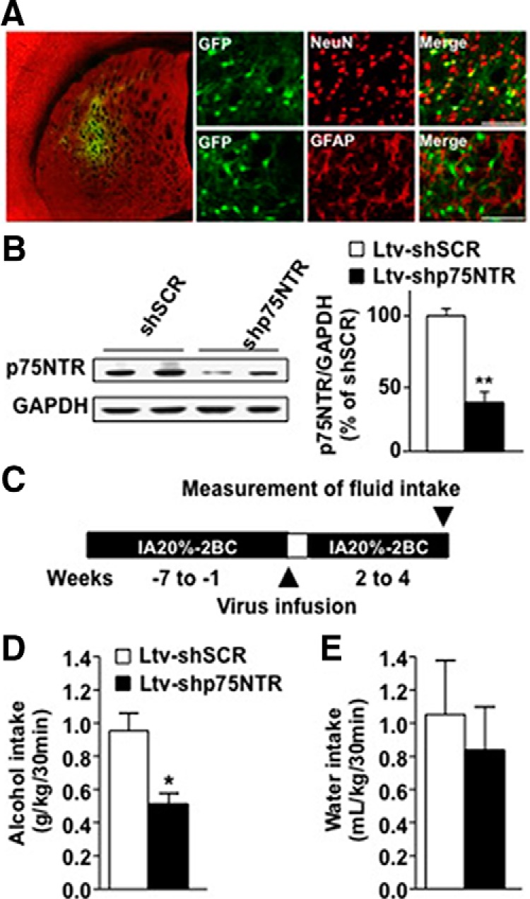
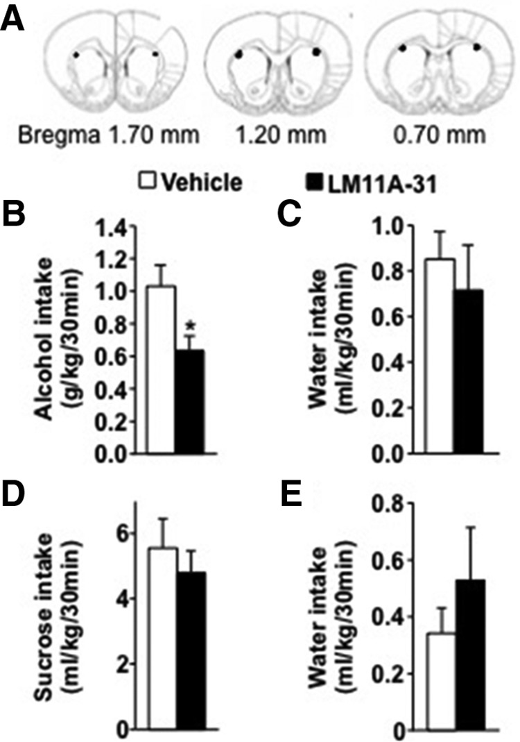
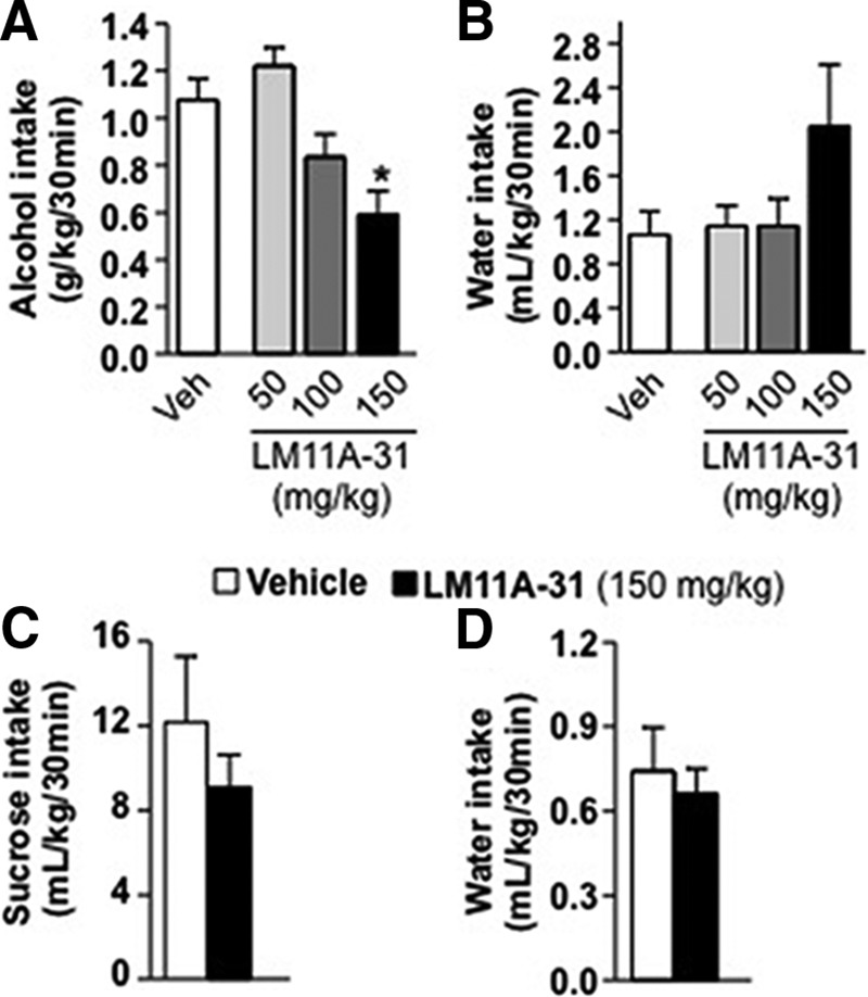
Brain-derived neurotrophic factor (BDNF) signaling in the dorsolateral striatum (DLS) keeps alcohol intake in moderation. For example, activation of the BDNF receptor tropomyosin receptor kinase B (TrkB) in the DLS reduces intake in rats that consume moderate amounts of alcohol. Here, we tested whether long-term excessive consumption of alcohol produces neuroadaptations in BDNF signaling in the rat DLS. We found that BDNF was no longer able to gate alcohol self-administration after a history of repeated cycles of binge alcohol drinking and withdrawal. We then elucidated the possible neuroadaptations that could block the ability of BDNF to keep consumption of alcohol in moderation. We report that intermittent access to 20% alcohol in a two-bottle choice paradigm that models excessive alcohol drinking produces a mobilization of DLS p75 neurotrophin receptor (p75NTR), whose activities oppose those of the Trk receptors, including TrkB. These neuroadaptations were not observed in the DLS of rats exposed to continuous access to 10% alcohol or in rats consuming sucrose. Furthermore, short hairpin RNA (shRNA)-mediated knockdown of the p75NTR gene in the DLS, as well as intra-DLS infusion or systemic administration of the p75NTR modulator, LM11A-31, significantly reduced binge drinking of alcohol. Together, our results suggest that excessive alcohol consumption produces a change in BDNF signaling in the DLS, which is mediated by the recruitment of p75NTR. Our data also imply that modulators of p75NTR signaling could be developed as medications for alcohol abuse disorders.

Significance statement: Neuroadaptations gate or drive excessive, compulsive alcohol drinking. We previously showed that brain-derived neurotrophic factor and its receptor, TrkB, in the dorsolateral striatum (DLS), are part of an endogenous system that keeps alcohol drinking in moderation. Here, we show that a history of excessive alcohol intake produces neuroadaptations in the DLS that preclude BDNF's ability to gate alcohol self-administration in rats by the recruitment of the low-affinity neurotrophin receptor, p75NTR, whose activities opposes those of the Trk receptors. Finally, we show that the administration of the p75NTR modulator, LM11A-31, significantly reduces excessive alcohol intake suggesting that the drug may be developed as a new treatment for alcohol abuse disorders.

Keywords: BDNF; addiction; alcohol; dorsal striatum; neurotrophic factor; p75NTR.


Figures

Figure 1.
Figure 1.
Intra-DLS infusion of BDNF does not reduce alcohol self-administration in rats with a history of excessive alcohol drinking. A, Rats underwent 5 weeks of an IA20%-2BC paradigm and were then trained to lever press for a 20% alcohol solution during 30 min sessions. BDNF (0.75 μg/μl) or vehicle (Veh; PBS) was infused into the DLS 3 h before the beginning of the operant self-administration sessions. The concentration of alcohol was decreased (from 20 to 10% and from 10 to 2.5%) every 2 weeks to allow the rats to reach a new baseline of consumption before BDNF administration. B, Schematic drawings of coronal sections of the rat brain showing the placement of bilateral infusion sites in the DLS (Paxinos and Watson, 2007). C, D, Data are expressed as the mean ± SEM of the number of alcohol deliveries (C) and alcohol consumed in grams per kilogram (D). n = 7 per treatment.
Figure 2.
Figure 2.
Excessive alcohol drinking alters the synaptosomal localization of p75NTR but not TrkB in the DLS. A, Schematic drawings of coronal sections of the rat brain showing the DLS slices in black (Paxinos and Watson, 2007). B, Schematic representation of dissection timeline. Alc, Alcohol; W, water. C–H, Rats received IA20%-2BC (black) or water only (white) for 7 weeks. DLS of high-drinking rats (alcohol intake equal to or >3.5 g/kg/24 h) were dissected immediately after the last 30 min (binge; C, D), at the end of the last 24 h drinking session (end; E, F), or after 24 h of withdrawal (WD; G, H). TrkB and p75NTR levels in total homogenates (C, E, G) and in synaptosomal fractions (D, F, H) were measured by Western blot analysis. Histograms depict the mean ratio of TrkB or p75NTR to actin ± SEM, and values are expressed as percentages of water controls. ***p < 0.0001 versus water (unpaired t test). n = 10–11 (C, D) and 8 per time point of dissection (E–H).
Figure 3.
Figure 3.
Systemic administration of alcohol, moderate alcohol intake, or sucrose consumption do not alter BDNF receptors expression or localization in the DLS. A, B, Rats received a systemic administration of alcohol (Alc; 1.5 g/kg, i.p.; black) or saline (Sal; white), and the DLS was dissected 30 min later. C, D, Rats received continuous access to 10% alcohol (CA10%; black) or water only (white) for 21 d, and the DLS was dissected immediately after the last drinking session. E, F, Rats received 7 weeks of IA of 1% sucrose (Suc) or water only, and the DLS was dissected immediately after the last 30 min drinking session. TrkB and p75NTR levels in total homogenate (A, C, E) or in the synaptosomal fraction (B, D, F) were measured by Western blot analysis. Histograms show mean ratio of TrkB or p75NTR to actin ± SEM, and values are expressed as percentages of water controls. n = 8 per treatment (A, B); n = 8 (C, D) and 4 per drinking regimen (E, F).
Figure 4.
Figure 4.
Excessive alcohol intake does not alter BDNF receptors expression or localization in the DMS. A, Schematic drawings of coronal sections of the rat brain showing the DMS in black (Paxinos and Watson, 2007). B–E, Rats received IA20%-2BC for 7 weeks (black) or water only (white), and the DMS was dissected immediately after the last 30 min drinking session (binge; B, C) or at the end of the 24 h drinking session (end; D, E). TrkB and p75NTR levels in total homogenates (B, D) or in the synaptosomal fraction (C, E) were measured by Western blot analysis. Histograms show the mean ratio of TrkB or p75NTR to actin ± SEM, and values are expressed as percentages of water controls. n = 8 per time point of dissection.
Figure 5.
Figure 5.
Knockdown of p75NTR in the DLS reduces binge drinking of alcohol. A, B, Ltv-shp75NTR or Ltv-shSCR was bilaterally infused at a titer of 2 × 10⁷ pg/ml into the DLS. A, Ltv-shp75NTR infects DLS neurons. Four weeks after virus infusion, slices containing the DLS were costained with anti-GFP (green) and anti-NeuN (red) or costained with anti-GFP and anti-GFAP antibodies. The left image depicts the specificity of the site of infection. The right images depict Ltv-shp75NTR infection of neurons (costaining of GFP with NeuN; top) but not glia (costaining GFP with GFAP; bottom). B, Ltv-shp75NTR-mediated knockdown of p75NTR. The DLS was dissected 4 weeks after virus infusion, and p75NTR and GAPDH protein levels were measured by Western blot analysis. Data are expressed as mean ratio of p75NTR to GAPDH ± SEM and presented as percentages of Ltv-shSCR control. C, Schematic representation of the behavioral experiment. Rats underwent IA20%-2BC alcohol for 7 weeks. High-drinking rats (baseline level of alcohol intake equal or higher than 3.5 g/kg/24 h) received a bilateral infusion of Ltv-shSCR or Ltv-shp75NTR into the DLS, and after 1 week of recovery, the alcohol-drinking procedure resumed. D, E, Alcohol intake (grams per kilogram per 30 min; D) and water intake (milliliters per kilogram per 30 min; E) were recorded 4 weeks after viral infection. Results are expressed as mean ± SEM. *p < 0.01; **p < 0.001 versus Ltv-shSCR (unpaired t test). n = 4 (B) or 7 per treatment (D, E).
Figure 6.
Figure 6.
Intra-DLS infusion of LM11A-31 reduces binge drinking of alcohol but not sucrose. A–E, Rats underwent IA20%-2BC for 7 weeks, and bilateral guide cannula were then surgically implanted in the DLS. After 1 week of recovery, rats were administered with LM11A-31 (30 μg/μl; Tep et al., 2013) or vehicle (PBS) into the DLS 2 h before the beginning of the drinking session and consumption was recorded. A, Schematic drawings of coronal sections of the rat brain showing the placement of bilateral infusion sites in the DLS (Paxinos and Watson, 2007). B, C, Alcohol (grams per kilogram per 30 min; B) and water (milliliters per kilogram per 30 min; C) intake after LM11A-31 or vehicle infusion. D, E, After 1 week of withdrawal from alcohol consumption, rats underwent 2 weeks of IA-2BC of 1% sucrose and then received intra-DLS administration of LM11A-31 (30 μg/μl) or vehicle. Sucrose consumption is presented as milliliters per kilogram per min; D and water (milliliters per kilogram per 30 min; E). Results are expressed as mean ± SEM. *p < 0.05 versus vehicle (paired t test). n = 11 (B, C) and 10 per treatment (D, E).
Figure 7.
Figure 7.
Systemic administration of LM11A-31 reduces binge drinking of alcohol but not sucrose. A–D, Rats underwent an IA-2BC paradigm with 20% alcohol (A, B) or 1% sucrose (C, D) for 7 weeks. LM11A-31 (50–150 mg/kg) or vehicle (0.9% NaCl) was administered intraperitoneally 2 h before the beginning of the drinking session. Alcohol (grams per kilogram per 30 min; A) and water (milliliters per kilogram per 30 min; B) or sucrose (C) and water (both milliliters per kilogram per 30 min; D) intake were recorded. Results are expressed as mean ± SEM. *p < 0.01 versus vehicle (one-way ANOVA with SNK post hoc test). A, B, Vehicle, n = 24 per dose; LM11A-31, n = 12 per dose; C, D, n = 7–8 per treatment.

References

    1. Andero R, Choi DC, Ressler KJ. BDNF-TrkB receptor regulation of distributed adult neural plasticity, memory formation, and psychiatric disorders. Prog Mol Biol Trans Sci. 2014;122:169–192. doi: 10.1016/B978-0-12-420170-5.00006-4. - DOI - PubMed
    1. Autry AE, Monteggia LM. Brain-derived neurotrophic factor and neuropsychiatric disorders. Pharmacol Rev. 2012;64:238–258. doi: 10.1124/pr.111.005108. - DOI - PMC - PubMed
    1. Bronfman FC, Tcherpakov M, Jovin TM, Fainzilber M. Ligand-induced internalization of the p75 neurotrophin receptor: a slow route to the signaling endosome. J Neurosci. 2003;23:3209–3220. - PMC - PubMed
    1. Buckley PF, Mahadik S, Pillai A, Terry A., Jr Neurotrophins and schizophrenia. Schizophr Res. 2007;94:1–11. doi: 10.1016/j.schres.2007.01.025. - DOI - PubMed
    1. Bull C, Freitas KC, Zou S, Poland RS, Syed WA, Urban DJ, Minter SC, Shelton KL, Hauser KF, Negus SS, Knapp PE, Bowers MS. Rat nucleus accumbens core astrocytes modulate reward and the motivation to self-administer ethanol after abstinence. Neuropsychopharmacology. 2014;39:2835–2845. doi: 10.1038/npp.2014.135. - DOI - PMC - PubMed

Substances