

Early risk prognosis of free-flap transplant failure by quantitation of the macrophage colony-stimulating factor in patient plasma using 2-dimensional liquid-chromatography multiple reaction monitoring-mass spectrometry
- PMID: 27684807
- PMCID: PMC5265900
- DOI: 10.1097/MD.0000000000004808
Early risk prognosis of free-flap transplant failure by quantitation of the macrophage colony-stimulating factor in patient plasma using 2-dimensional liquid-chromatography multiple reaction monitoring-mass spectrometry
Abstract
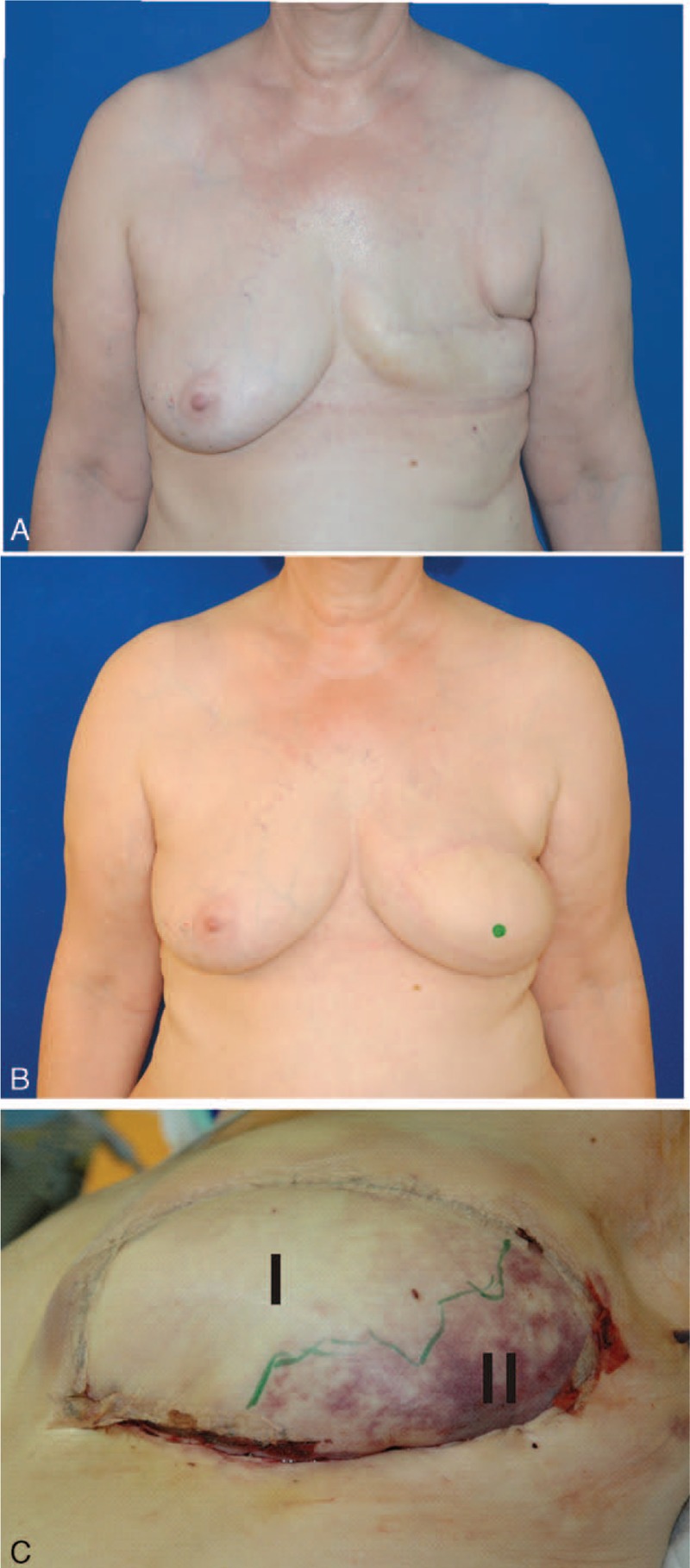
Although great success of microvascular free-flap transplantation surgery has been achieved in recent years, between 1.5% and 15% of flaps are still lost due to vascular occlusion. The clinical challenge remains to salvage a transplant in the case of vascular complications. Since flap loss is devastating for the patient, it is of utmost importance to detect signs of complications or of conspicuities as soon as possible. Rescue success rates highly depend on early revision. In this study, we collected blood samples during transplantation surgery from either the contributory artery or the effluent vein of the flap and applied a targeted mass spectrometry-based approach to quantify 24 acute phase proteins, cytokines, and growth factors in 63 plasma samples from 21 hospitalized patients, generating a dataset with 9450 protein concentration values. Biostatistical analyses of the targeted plasma protein concentrations in all 63 plasma samples showed that venous concentrations of macrophage colony-stimulating factor (M-CSF) provided the highest accuracy for discriminating patients with either clinical conspicuities or complications from control individuals. Using 21.33 ng/mL of M-CSF as the diagnostic threshold when analyzing venous blood plasma samples, the assay obtained a sensitivity of 0.93 and a specificity of 0.85 with an area under the curve value of 0.902 in the receiver operating characteristic analysis. Overall, our results indicate that M-CSF is a potential molecular marker for early risk prognosis of free-flap transplant failure.
Conflict of interest statement
The authors have no conflicts of interest to disclose.
Figures


References
-
- Bizeau A, Guelfucci B, Giovanni A, et al. 15 years experience with microvascular free tissue transfert for repair of head and neck cancer defects. Ann Otolaryngol Chir Cervicofac 2002; 119:31–38. - PubMed
-
- Bui DT, Cordeiro PG, Hu QY, et al. Free flap reexploration: indications, treatment, and outcomes in 1193 free flaps. Plast Reconstr Surg 2007; 119:2092–2100. - PubMed
-
- Vega S, Smartt JM, Jr, Jiang S, et al. 500 Consecutive patients with free TRAM flap breast reconstruction: a single surgeon's experience. Plast Reconstr Surg 2008; 122:329–339. - PubMed
-
- Genden EM, Rinaldo A, Suarez C, et al. Complications of free flap transfers for head and neck reconstruction following cancer resection. Oral Oncol 2004; 40:979–984. - PubMed
-
- Classen DA, Ward H. Complications in a consecutive series of 250 free flap operations. Ann Plast Surg 2006; 56:557–561. - PubMed
Publication types
MeSH terms
Substances
LinkOut - more resources
Full Text Sources
Other Literature Sources
Research Materials
