. 2016 Sep 29;12(9):e1005914.
doi: 10.1371/journal.ppat.1005914. eCollection 2016 Sep.

Inactivation of Prions and Amyloid Seeds with Hypochlorous Acid

Affiliations

Inactivation of Prions and Amyloid Seeds with Hypochlorous Acid

Andrew G Hughson et al. PLoS Pathog. .

Abstract

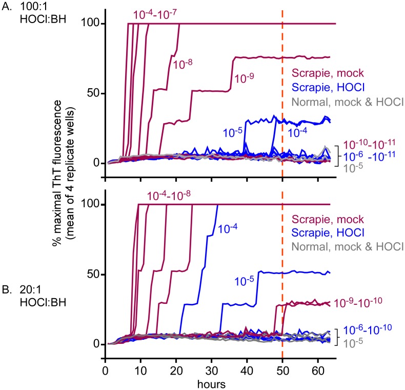
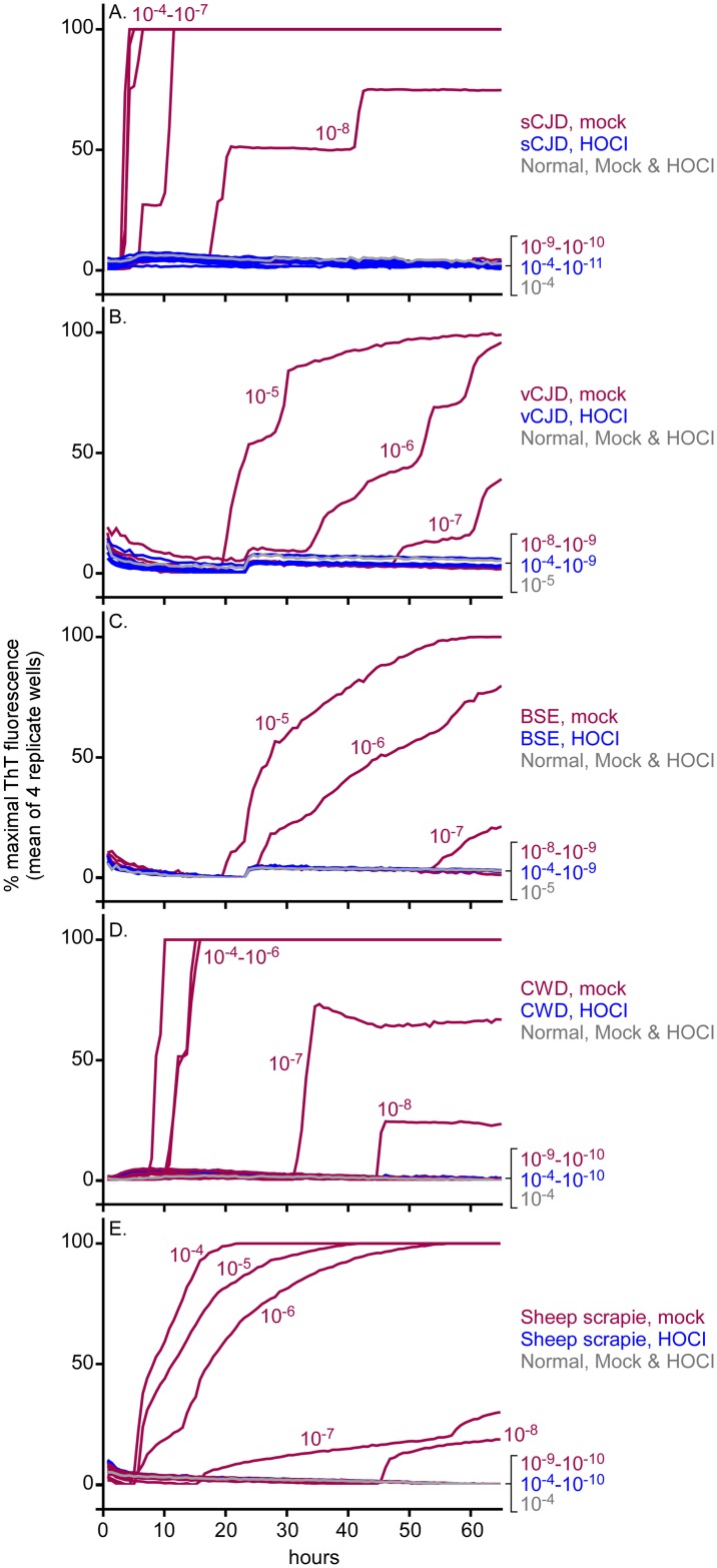
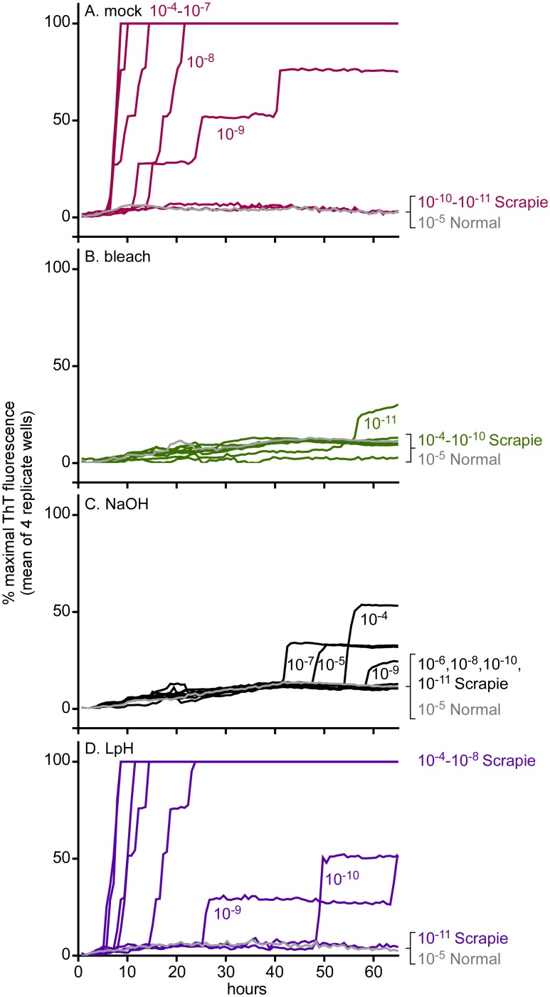
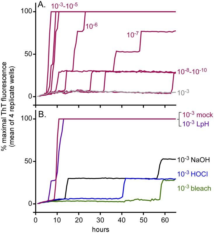
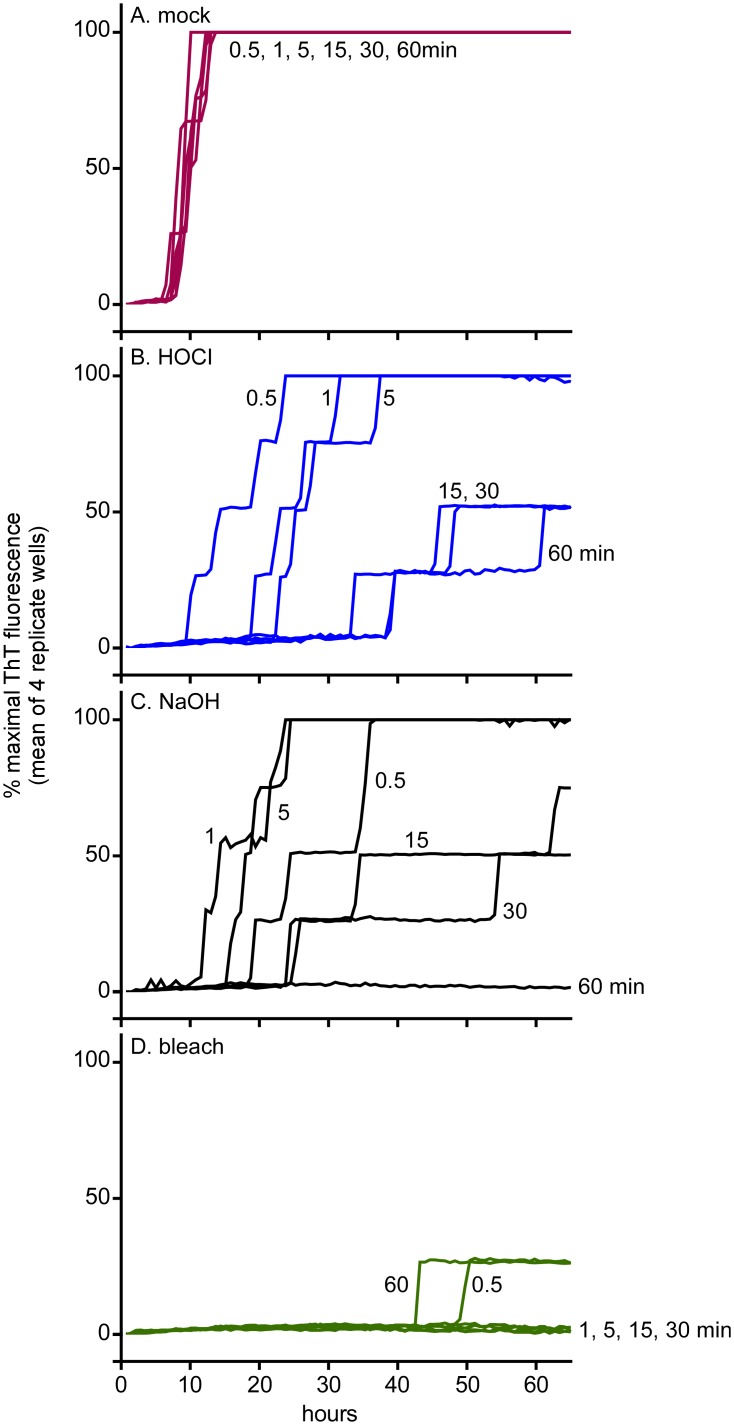
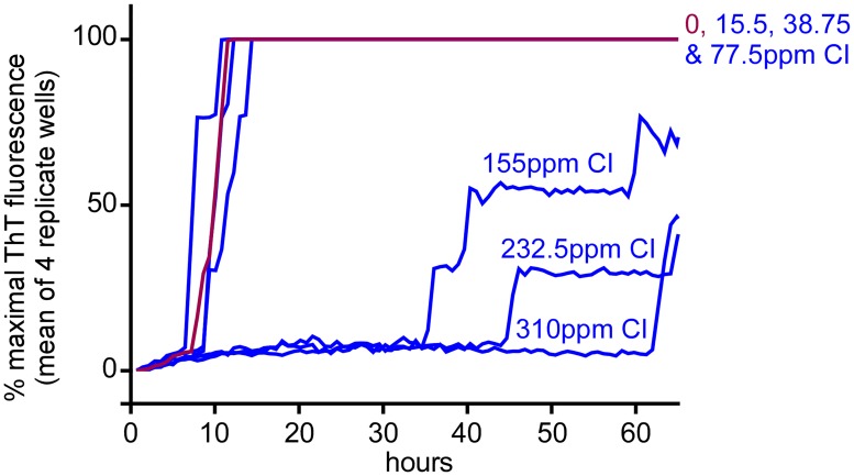
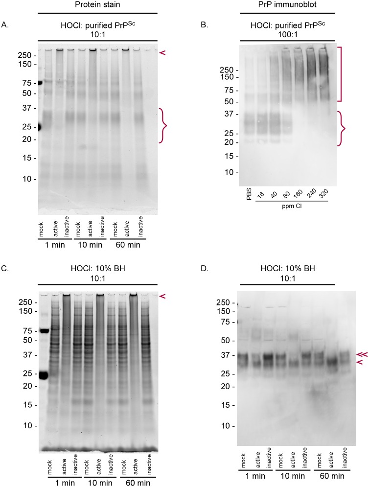
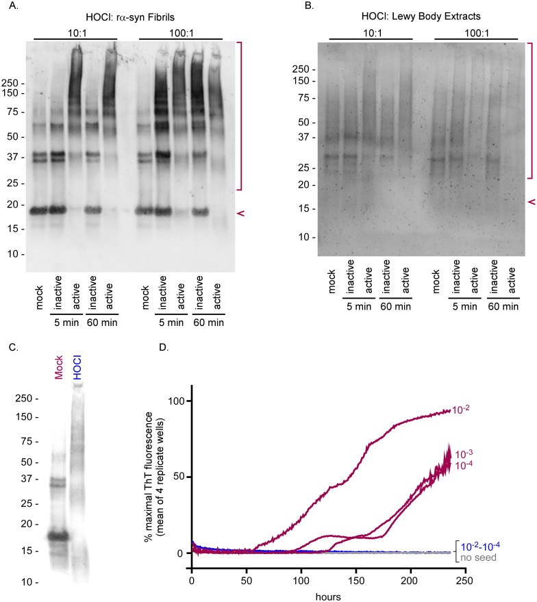
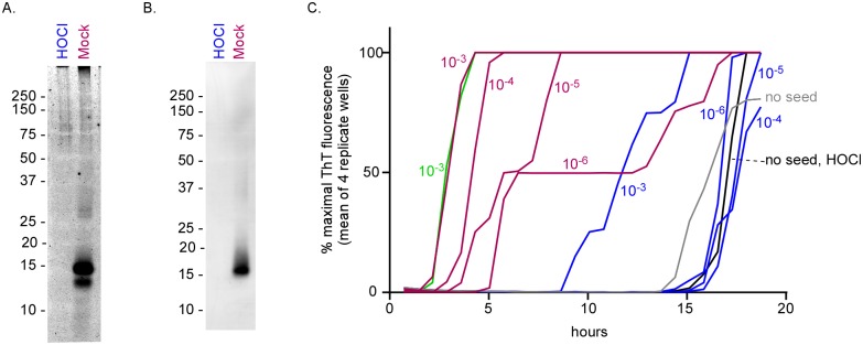
Hypochlorous acid (HOCl) is produced naturally by neutrophils and other cells to kill conventional microbes in vivo. Synthetic preparations containing HOCl can also be effective as microbial disinfectants. Here we have tested whether HOCl can also inactivate prions and other self-propagating protein amyloid seeds. Prions are deadly pathogens that are notoriously difficult to inactivate, and standard microbial disinfection protocols are often inadequate. Recommended treatments for prion decontamination include strongly basic (pH ≥~12) sodium hypochlorite bleach, ≥1 N sodium hydroxide, and/or prolonged autoclaving. These treatments are damaging and/or unsuitable for many clinical, agricultural and environmental applications. We have tested the anti-prion activity of a weakly acidic aqueous formulation of HOCl (BrioHOCl) that poses no apparent hazard to either users or many surfaces. For example, BrioHOCl can be applied directly to skin and mucous membranes and has been aerosolized to treat entire rooms without apparent deleterious effects. Here, we demonstrate that immersion in BrioHOCl can inactivate not only a range of target microbes, including spores of Bacillus subtilis, but also prions in tissue suspensions and on stainless steel. Real-time quaking-induced conversion (RT-QuIC) assays showed that BrioHOCl treatments eliminated all detectable prion seeding activity of human Creutzfeldt-Jakob disease, bovine spongiform encephalopathy, cervine chronic wasting disease, sheep scrapie and hamster scrapie; these findings indicated reductions of ≥103- to 106-fold. Transgenic mouse bioassays showed that all detectable hamster-adapted scrapie infectivity in brain homogenates or on steel wires was eliminated, representing reductions of ≥~105.75-fold and >104-fold, respectively. Inactivation of RT-QuIC seeding activity correlated with free chlorine concentration and higher order aggregation or destruction of proteins generally, including prion protein. BrioHOCl treatments had similar effects on amyloids composed of human α-synuclein and a fragment of human tau. These results indicate that HOCl can block the self-propagating activity of prions and other amyloids.


Conflict of interest statement

I have read the journal's policy and the authors of this manuscript have the following competing interests: DT is founder and CEO of BrioTech Inc, which sells BrioHOClTM. A gift from BrioTech was used to support the work at UW Bothell lead by LR. JFW is Chief Scientific Officer, corporate executive and shareholder of Briotech Inc.

Figures

Fig 1
Fig 1. Inactivation of prion seeding activity in hamster ScBH by BrioHOCl.
ScBH was pretreated for 1 h in BrioHOCl at 100:1 v/v BrioHOCl to 10% BH (panel A, blue), 20:1 BrioHOCl (panel B, blue) or corresponding saline treatments (panels A and B, red). Similar BrioHOCl and saline pretreatments of NBH are indicated in gray. Resulting samples were then subjected to serial 10-fold dilutions and RT-QuIC analysis was performed with hamster (90–231) rPrPC substrate using 2 μl per well of the indicated tissue dilutions as reaction seeds. The dilutions noted refer to the final dilution of original brain mass used to seed the reaction. The orange dashed line indicates the 50-h cutoff time at which seeding activity was quantified by end-point dilution under these assay conditions, after which unseeded control reactions occasionally gave false-positive reactions (see Material and Methods). Each trace represents the average ThT fluorescence of 4 technical replicate wells normalized between baseline and maximal signal and graphed as a function of time.
Fig 2
Fig 2. Inactivation of sCJD (A), vCJD (B), BSE (C), CWD (D) & sheep scrapie (E) seeding activity in brain homogenates by BrioHOCl.
The indicated BH samples (10%) were pretreated for 1 h with 100 volumes of BrioHOCl (blue), or saline (red) as a mock treatment control. Similar HOCl and saline pretreatments of NBH are indicated in gray. Resulting samples were then subjected to serial 10-fold dilutions and RT-QuIC analysis was performed using 10−4 through 10−11 tissue dilutions as indicated. Hamster (90–231) rPrPC was used as substrate for the sCJD and CWD reactions while chimeric hamster-sheep rPrPC was used as substrate for the vCJD, BSE and sheep scrapie reactions. Each trace represents the average normalized ThT fluorescence of 4 replicate wells.
Fig 3
Fig 3. Effects of bleach, NaOH and Environ LpH on hamster scrapie seeding activity.
ScBH was pretreated for 1 h in (A) saline (mock disinfectant) (red), (B) 40% bleach (2.4% hypochlorite) (green), (C) 1 N NaOH (black) or (D) 2% Environ LpH (purple) at a ratio (v/v) of 100:1 disinfectant to 10% ScBH. Similar disinfectant pretreatments of NBH are indicated in gray. Resulting samples were then subjected to serial 10-fold dilutions and RT-QuIC analysis was performed with hamster (90–231) rPrPC substrate using the designated tissue dilutions as seeds. Each trace represents the average normalized ThT fluorescence of 4 replicate wells.
Fig 4
Fig 4. Inactivation of steel-bound prion seeding activity.
A. RT-QuIC reaction wells were seeded with a 3–4 mm segment of stainless steel wire pre-coated with hamster scrapie (red) or normal BH (gray) at tissue dilutions of 10−3–10−10 as indicated. B. Wire segments pre-coated with ScBH at a 10−3 tissue dilution were submersed for 1 h in saline (mock disinfectant; red), BrioHOCl (blue), 40% bleach (2.4% hypochlorite; green), 1 N NaOH (black) or 2% Environ LpH (purple) as indicated prior to RT-QuIC analysis using hamster (90–231) rPrPC substrate. Each trace represents the average normalized ThT fluorescence of 4 replicate wells.
Fig 5
Fig 5. Effect of disinfectant exposure time on inactivation of hamster scrapie seeding activity on wires coated with ScBH.
Stainless steel wire segments (3–4mm) pre-coated with hamster ScBH at a 10−3 tissue dilution were submersed in (A) saline (mock disinfectant), (B) BrioHOCl, (C) 1 N NaOH or (D) 40% bleach (2.4% hypochlorite) for 0.5–60 min prior to a quick rinse and RT-QuIC analysis using hamster (90–231) rPrPC substrate. Each trace represents the average normalized ThT fluorescence of 4 replicate wells.
Fig 6
Fig 6. Effect of BrioHOCl free Cl concentration on scrapie seeding activity.
Hamster ScBH was pretreated for 5 min in saline (mock disinfectant) (red) or BrioHOCl formulations (blue) containing the designated ppm of free Cl at a ratio (v/v) of 100:1 disinfectant to 10% ScBH. Resulting samples were then subjected to serial 10-fold dilutions and RT-QuIC analysis was performed with hamster (90–231) rPrPC substrate using 10−4 through 10−7 tissue dilutions as seeds. For simplicity, only the 10−6 dilutions are shown here. See Table 3 for summary of all results. Each trace represents the average normalized ThT fluorescence of 4 replicate wells.
Fig 7
Fig 7. Chemical stability of BrioHOCl.
A. Concentration of active chlorine as measured by iodometric titration (days 14–151). The data were fit to an exponential decay (y = 159.78e(-0.00157x)). B. Active chlorine concentrations over initial 13 days. C. UV-VIS measurements of samples after 10 (dark blue), 14 (pink), and 88 (light blue) days.
Fig 8
Fig 8. SDS-PAGE analysis of BrioHOCl treatments on purified PrPSc and ScBH.
(A) Purified PrPSc was treated with 10 volume equivalents of saline (mock), active (260 ppm Cl) or inactive (30 ppm Cl) BrioHOCl solutions for 1, 10 or 60 min as indicated. BrioHOCl activity or inactivity was determined by its ability or inability to reduce prion-seeding activity in the RT-QuIC in other experiments. Samples were run on denaturing protein gels and visualized using Deep Purple total protein stain. PrP monomer (curly bracket) and multimeric aggregates (arrowhead) are marked. (B) Purified PrPSc (at 3 mg/mL) was treated for 5 minutes in saline (mock disinfectant) or BrioHOCl formulations containing the designated ppm free Cl at a ratio (v/v) of 100:1 disinfectant to PrPSc prior to immunoblotting using R30 antiserum against PrP residues 90–104. PrP monomer (curly bracket) and PrP aggregates (square bracket) are marked. Gels shown are representative of three independent experiments. (C) ScBH was treated with saline (mock disinfectant), or active or inactive BrioHOCl solutions at a ratio (v/v) of 10:1 disinfectant to 10% ScBH for the designated time and analyzed by SDS-PAGE with Deep Purple total protein stain. Insoluble aggregates are indicated with an arrowhead. (D) The samples from panel C were analyzed by immunoblot using R30 PrP antiserum. Full length, diglycosylated PrP monomer (double arrowhead); Truncated, and/or less glycosylated PrP monomer (single arrowhead) are identified.
Fig 9
Fig 9. Effects of BrioHOCl on recombinant α-synuclein seeds and Lewy bodies.
Recombinant α-syn (rα-syn) fibrils generated in vitro (A) or Lewy Bodies isolated from a patient with Lewy Body Dementia (B) were treated with an active (190 ppm Cl) or inactive (30 ppm Cl) BrioHOCl solution at a 10:1 or 100:1 disinfectant to α-syn ratio for 5 or 60 min, as indicated, and probed for α-syn by immunoblot. Samples treated 10:1 were diluted an additional 10 fold in 1x sample buffer prior to loading the gel to match the protein concentrations of the 100:1 treated samples on the immunoblot. The arrow indicates monomeric α-syn protein and the bracket denotes aggregates and degradation products (A & B). Recombinant α-syn fibrils generated in vitro were treated with either a mock solution or an active BrioHOCl solution at 100:1 disinfectant to α-syn ratio for 60 min and probed for α-syn by immunoblot (C). These mock (red) and HOCl (blue) treated samples were subjected to 10 fold serial dilutions and analyzed for recombinant α-syn seeding activity (D). 20 μl per well of 10−2 through 10−4 sample dilutions were used as reaction seeds as indicated. Negative control reactions were run with no seed (gray). Other controls indicated that direct addition of 10−3 and 10−4 dilutions of BrioHOCl to the seeded polymerization reactions without preincubation with the α-syn seed had no effect on the reaction kinetics, whereas a 10−2 dilution partially interfered with the reaction (S3 Fig). Each trace represents the average ThT fluorescence of 4 replicate wells. Similar results were obtained in two additional independent experiments.
Fig 10
Fig 10. Inactivation of tau peptide amyloid seeds by BrioHOCl.
Synthetic tau seeds were generated with recombinant K19 Cys-free tau fragment and treated with or without a 100-fold excess of BrioHOCl. The seed preparations were subjected to SDS-PAGE with non-specific staining for protein (Deep Purple) (A), immunoblotting probed with anti-tau antibody (B) or a seeded polymerization assay (C). In the seeded polymerization assay, the designated dilutions of the untreated (red) or BrioHOCl-treated (blue) seed samples were tested. To control for potential effects of BrioHOCl on the assay itself without allowing time for prior interactions the amyloid seed on its own, an amount of BrioHOCl comparable to that in a 10−3 dilution of treated seed was added directly to reactions solutions that were either left unseeded (black) or seeded with a 10−3 dilution of untreated seed (green). Reactions mock-seeded with 10−5 dilutions of NBH samples that were treated or not with 20 or 100 volumes of BrioHOCl are all shown in gray. Similar effects on K19 Cys-free seeding activity were obtained in at least five independent experiments.

References

    1. Parchi P, de BL, Saverioni D, Cohen ML, Ferrer I, Gambetti P, et al. Consensus classification of human prion disease histotypes allows reliable identification of molecular subtypes: an inter-rater study among surveillance centres in Europe and USA. Acta Neuropathol. 2012;124(4):517–29. 10.1007/s00401-012-1002-8 - DOI - PMC - PubMed
    1. Brown P, Brandel JP, Sato T, Nakamura Y, MacKenzie J, Will RG, et al. Iatrogenic Creutzfeldt-Jakob disease, final assessment. Emerging Infect Dis. 2012;18(6):901–7. 10.3201/eid1806.120116 - DOI - PMC - PubMed
    1. Zanusso G, Monaco S, Pocchiari M, Caughey B. Advanced tests for early and accurate diagnosis of Creutzfeldt-Jakob disease. Nat Rev Neurol. 2016;12(6):325–33. 10.1038/nrneurol.2016.65 . - DOI - PubMed
    1. Zanusso G, Monaco S. Bovine Spongiform Encephalopathy In: Zou W, Gambetti P, editors. Prions and Diseases. 2 New York: Springer; 2013. p. 1–14.
    1. Fast C, Groschup M. Classical and Atypical Scrapie in Sheep and Goats In: Zou W, Gambetti P, editors. Prions and Diseases. 2 New York: Springer; 2013. p. 15–44.