. 2016 Sep 29;7(9):e2388.
doi: 10.1038/cddis.2016.260.

Hsa-miR-623 suppresses tumor progression in human lung adenocarcinoma

Affiliations

Hsa-miR-623 suppresses tumor progression in human lung adenocarcinoma

Shuang Wei et al. Cell Death Dis. .

Erratum in

Retraction in

Abstract

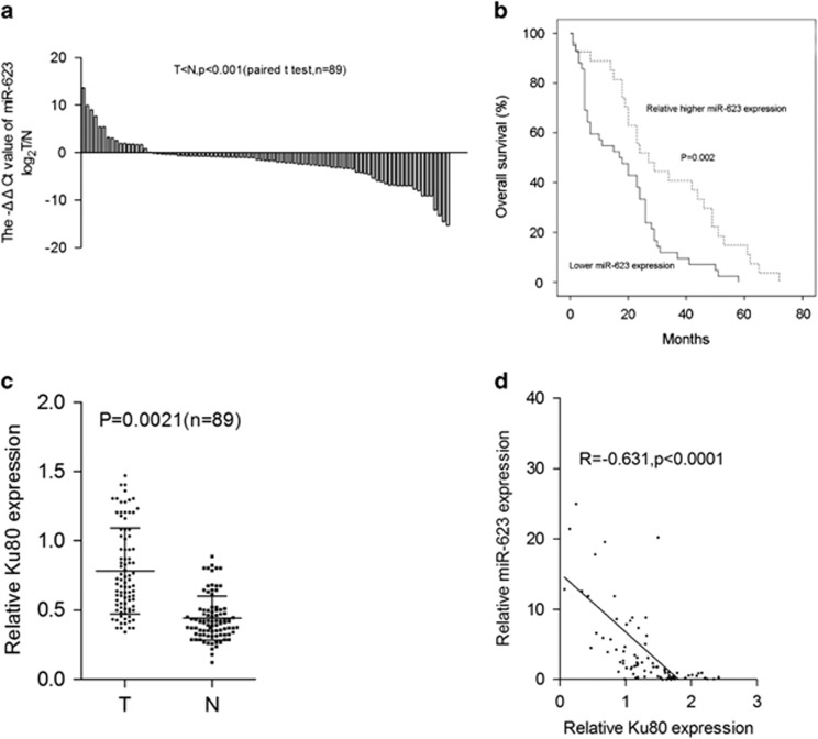
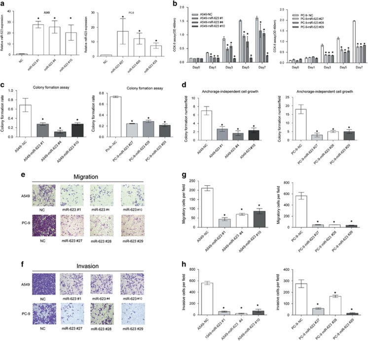
Our previous study revealed that Ku80 was overexpressed in lung cancer tissues and hsa-miR-623 regulated the Ku80 expression; however, the detailed function of hsa-miR-623 in lung cancer was unclear. We identified that hsa-miR-623 bound to the 3'-UTR of Ku80 mRNA, thus significantly decreasing Ku80 expression in lung adenocarcinoma cells. Hsa-miR-623 was downregulated in lung adenocarcinoma tissues compared with corresponding non-tumorous tissues, and its expression was inversely correlated with Ku80 upregulation. Downregulation of hsa-miR-623 was associated with poor clinical outcomes of lung adenocarcinoma patients. Hsa-miR-623 suppressed lung adenocarcinoma cell proliferation, clonogenicity, migration and invasion in vitro. Hsa-miR-623 inhibited xenografts growth and metastasis of lung adenocarcinoma in vivo. Ku80 knockdown in lung adenocarcinoma cells suppressed tumor properties in vitro and in vivo similar to hsa-miR-623 overexpression. Further, hsa-miR-623 overexpression decreased matrix metalloproteinase-2 (MMP-2) and MMP-9 expression levels, with decreased ERK/JNK phosphorylation. Inhibition of hsa-miR-623 or overexpression of Ku80 promoted lung adenocarcinoma cell invasion, activated ERK/JNK phosphorylation and increased MMP-2/9 expressions, which could be reversed by ERK kinase inhibitor or JNK kinase inhibitor. In summary, our results showed that hsa-miR-623 was downregulated in lung adenocarcinoma and suppressed the invasion and metastasis targeting Ku80 through ERK/JNK inactivation mediated downregulation of MMP-2/9. These findings reveal that hsa-miR-623 may serve as an important therapeutic target in lung cancer therapy.


Figures

Figure 1
Figure 1
Hsa-miR-623 directly targets Ku80 in lung adenocarcinoma cells. (a) Predicted consequential pairing of wild type or mutant 3′-UTR of Ku80 mRNA and hsa-miR-623 by Targetscan. (b) Luciferase assays in A549 and PC-9 cells. PLUC, pLUC-wtKu80 and pLUC-mutKu80 vectors were co-transfected with pre-miR-623 or pre-miR-NC. Relative suppression of luciferase expression was standardized to β-gal signal. Luciferase activity in the pLUC-wtKu80 group displayed a significant decrease following ectopic expression of hsa-miR-623. (c and d) Ku80 protein was measured by western blot analysis 48 h after transfection. (c) Ku80 protein was downregulated in A549 and PC-9 cells transfected with hsa-miR-623 mimics. (d) Ku80 protein was upregulated in A549 and PC-9 cells with hsa-miR-623 inhibitor. *P<0.05
Figure 2
Figure 2
Hsa-miR-623 is downregulated in lung adenocarcinoma tissues and its downregulation is inversely correlated with Ku80 overexpression. (a) Downregulation of hsa-miR-623 in lung adenocarcinoma tissues compared with adjacent lung tissues (measured by TaqMan qRT-PCR, P<0.001 compared with controls). (b) Kaplan–Meier analysis of overall survival in all lung adenocarcinoma patients according to hsa-miR-623 expression level. (c) Quantitative analysis of Ku80 expression in 89 cases of paired lung adenocarcinoma tissues and their corresponding adjacent lung tissues by western blot analysis. The P-value (P=0.0021) corresponds to the comparison of Ku80 expression between the lung adenocarcinoma tissues and corresponding adjacent lung tissues. (d) Downregulation of hsa-miR-623 is inversely correlated with Ku80 upregulation (R=−0.631, P<0.0001, Pearson's correlation)
Figure 3
Figure 3
Hsa-miR-623 suppresses growth, migration and invasion of lung adenocarcinoma cells in vitro. (a) Relative expression of hsa-miR-623 detected by TaqMan qRT-PCR in two lung adenocarcinoma cell lines stably transfected with hsa-miR-623 or hsa-miR-NC. (b) Hsa-miR-623 overexpression significantly suppressed the proliferation of A549 and PC-9 cells by using CCK8 assay. (c) Histograms indicated that hsa-miR-623 can markedly inhibit the plate colony formation in A549 and PC-9 cells. (d) Histograms showed that hsa-miR-623 significantly suppressed anchorage-independent cell growth in A549 and PC-9 cells. (e) and (f) represent the results of cell migration and invasion in transwell assays with or without Matrigel. (g) and (h) Histograms indicate the relative number of migratory or invasive cells across a membrane with 8 mm pores with or without Matrigel, respectively. The results are representative of three independent experiments. Data are given as means±S.D., *P<0.05 compared with controls
Figure 4
Figure 4
The suppressive role of hsa-miR-623 on xenografts growth in vivo and metastasis in orthotopic lung cancer nude mice model. (a and b) The subcutaneous tumors derived from the hsa-miR-623-overexpressing clones were smaller than those from the NC-transfected clones in A549 and PC-9 cells. (c and d) Tumor growth curves showed that tumors derived from the hsa-miR-623-overexpressing A549 or PC-9 cells grew significantly slower than those from the control cells at all time points past 12 days after injection. (e) Representative photographs of mouse two lungs, mediastinum and liver, as well as images of the histological inspection of mouse two lungs, mediastinum and liver for the presence of microscopic lesions at 4 weeks after thoracic injection with A549 cells stably expressing miR-623 or the negative control lentiviral vector. (fh) Quantification of microscopic nodules and incidence of metastasis in the mediastinum, contralateral lung and liver of each group, respectively. The results shown represent the mean±S.D. of triplicate experiments. *P<0.05
Figure 5
Figure 5
Hsa-miR-623 suppresses lung adenocarcinoma cell growth, migration and invasion by downregulating Ku80 expression. (a) Cell proliferation detected in different cell groups at 0, 1, 3 and 5 days after transfection. (b) Histogram illustrated the relative colony numbers per field of indicated cell groups. (c and d) Histograms of migratory (c) and invasive (d) cell numbers per field of the indicated cell groups. (e) Subcutaneous tumors derived from indicated cell groups (n=4 each). (f) Tumor growth curves demonstrating that transfection of GV320-Ku80 in A549-miR-623 #1 cells significantly promoted subcutaneous tumor growth, while intratumoral injection of cholesterol-conjugated Ku80 siRNA significantly suppressed subcutaneous tumor growth compared with control groups. The mean and S.D. for tumor volumes were determined for each group. NS, P>0.05; *P<0.05
Figure 6
Figure 6
Hsa-miR-623 decreases MMP-2/9 expression and inhibits activation of the ERK/JNK pathway. (a) Western blot analysis showing the expressions of E-cadherin, N-cadherin, Vimentin, Snail, MMP-2 and MMP-9 in A549 and PC-9 cells stably transfected with the miR-623-expressing vector or empty vector. β-Actin was used as a loading control. (b) Conditioned medium from each group were collected and analyzed for MMP-2 and MMP-9 expression by gelatin zymography. (c) Western blot analysis showing the expression levels of Ku80, MMP-2, MMP-9, as well as activities of ERK1/2, p38 and JNK for total and phosphorylated forms. (d) Immunohistochemistry showing the expression status of Ku80, p-JNK, p-ERK, MMP-2 and MMP-9 in the xenograft tumor tissues. Data are given as means±S.D. *P<0.05
Figure 7
Figure 7
Hsa-miR-623 may inhibit cell invasion by MMP-2/9 downregulation through ERK/JNK pathway. (a) A549 cells were transfected with negative control RNA, hsa-miR-623 mimics or Ku80 siRNA, followed by JNK inhibitor (SP600125) and the ERK inhibitor (PD98059) treatment. Whole-cell lysates were analyzed for the protein levels of Ku80, MMP-2, MMP-9, p-JNK, p-ERK, t-JNK and t-ERK. (b) Cell invasion was evaluated using Matrigel-coated filters for 24 h after miRNA mimics treatment or 2 h after inhibitors treatment. (c) HBE cells were transfected with negative control RNA, hsa-miR-623 inhibitor or Ku80-overexpressed vector, followed by JNK inhibitor (SP600125) and the ERK inhibitor (PD98059) treatment. Expression levels of Ku80, MMP-2, MMP-9, p-JNK, p-ERK, t-JNK and t-ERK were determined by western blot analysis. (d) Matrigel cell invasion was evaluated. Data are given as means±S.D. invaded cells per field. *P<0.05

References

    1. Ferlay J, Shin HR, Bray F, Forman D, Mathers C, Parkin DM. Estimates of worldwide burden of cancer in 2008: GLOBOCAN 2008. Int J Cancer 2010; 127: 2893–2917. - PubMed
    1. Siegel R, Naishadham D, Jemal A. Cancer statistics, 2012. CA Cancer J Clin 2012; 62: 10–29. - PubMed
    1. Gupta GP, Massague J. Cancer metastasis: building a framework. Cell 2006; 127: 679–695. - PubMed
    1. Mehlen P, Puisieux A. Metastasis: a question of life or death. Nat Rev Cancer 2006; 6: 449–458. - PubMed
    1. Ross GM, Eady JJ, Mithal NP, Bush C, Steel GG, Jeggo PA et al. DNA strand break rejoining defect in xrs-6 is complemented by transfection with the human Ku80 gene. Cancer Res 1995; 55: 1235–1238. - PubMed

Publication types

MeSH terms