

Hemodynamics in Coronary Arterial Tree of Serial Stenoses
- PMID: 27685989
- PMCID: PMC5042402
- DOI: 10.1371/journal.pone.0163715
Hemodynamics in Coronary Arterial Tree of Serial Stenoses
Abstract
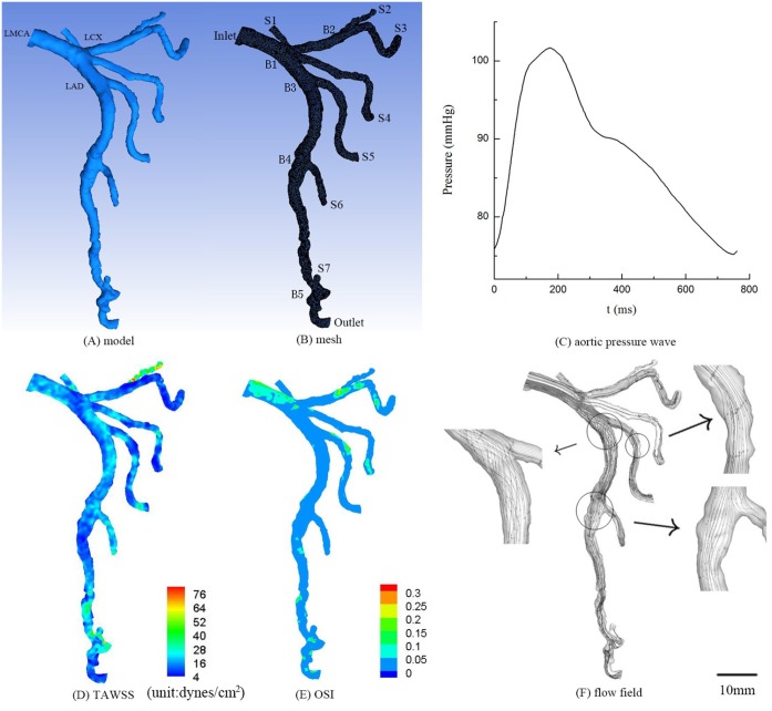
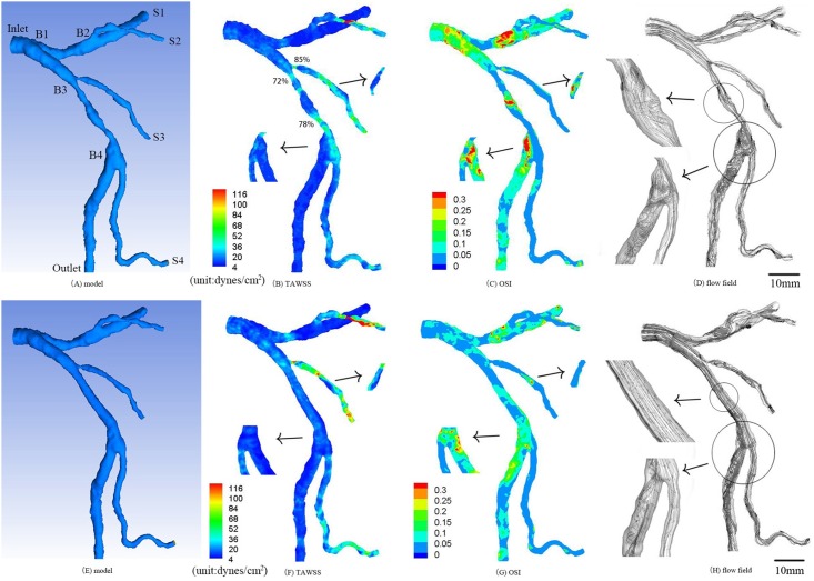
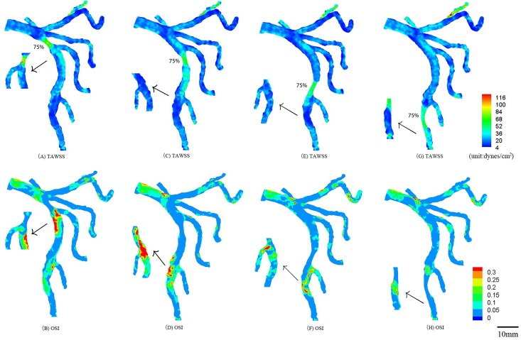
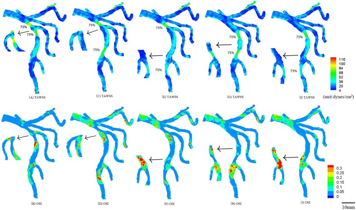
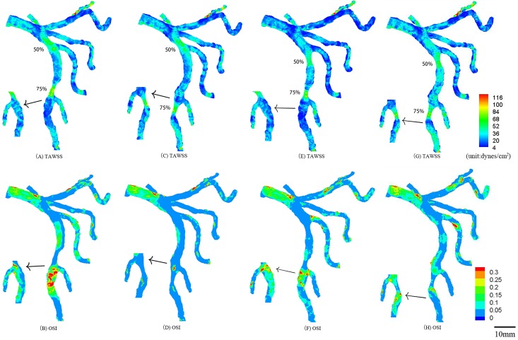
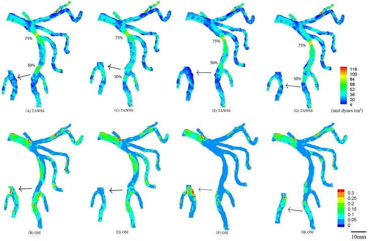
Serial segmental narrowing frequently occurs in humans, which alters coronary hemodynamics and further affects atherosclerotic progression and plaque formation. The objective of this study was to understand the distribution of hemodynamic parameters in the epicardial left main coronary arterial (LMCA) tree with serial stenoses reconstructed from patient computer tomography angiography (CTA) images. A finite volume method was used in conjunction with the inlet pressure wave and outlet flow resistance. The time-averaged wall shear stress (TAWSS) and oscillatory shear index (OSI) were determined from the flow field. A stenosis at a mother vessel mainly deteriorated the hemodynamics near the bifurcation while a stenosis at a daughter vessel affected the remote downstream bifurcation. In comparison with a single stenosis, serial stenoses increased the peak pressure gradient along the main trunk of the epicardial left anterior descending arterial tree by > 50%. An increased distance between serial stenoses further increased the peak pressure gradient. These findings have important implications on the diagnosis and treatment of serial coronary stenoses.
Conflict of interest statement
The California Medical Innovations Institute does not alter our adherence to PLOS ONE policies on sharing data and materials.
Figures
Similar articles
-
The improvement of the shear stress and oscillatory shear index of coronary arteries during Enhanced External Counterpulsation in patients with coronary heart disease.PLoS One. 2020 Mar 19;15(3):e0230144. doi: 10.1371/journal.pone.0230144. eCollection 2020. PLoS One. 2020. PMID: 32191730 Free PMC article.
-
Hemodynamics of left internal mammary artery bypass graft: Effect of anastomotic geometry, coronary artery stenosis, and postoperative time.J Biomech. 2016 Mar 21;49(5):645-652. doi: 10.1016/j.jbiomech.2016.01.031. Epub 2016 Feb 8. J Biomech. 2016. PMID: 26900034
-
The effects of plaque morphological characteristics on the post-stenotic flow in left main coronary artery bifurcation.Biomed Phys Eng Express. 2021 Sep 2;7(6). doi: 10.1088/2057-1976/ac202c. Biomed Phys Eng Express. 2021. PMID: 34425569
-
Hemodynamics and atherosclerosis. Insights and perspectives gained from studies of human arteries.Arch Pathol Lab Med. 1988 Oct;112(10):1018-31. Arch Pathol Lab Med. 1988. PMID: 3052352 Review.
-
Regulation of coronary blood flow during exercise.Physiol Rev. 2008 Jul;88(3):1009-86. doi: 10.1152/physrev.00045.2006. Physiol Rev. 2008. PMID: 18626066 Review.
Cited by
-
Vertebral Artery Stenoses Contribute to the Development of Diffuse Plaques in the Basilar Artery.Front Bioeng Biotechnol. 2020 Mar 6;8:168. doi: 10.3389/fbioe.2020.00168. eCollection 2020. Front Bioeng Biotechnol. 2020. PMID: 32211395 Free PMC article.
-
Influence of microcirculation load on FFR in coronary artery stenosis model.BMC Cardiovasc Disord. 2020 Mar 21;20(1):144. doi: 10.1186/s12872-020-01437-w. BMC Cardiovasc Disord. 2020. PMID: 32199456 Free PMC article.
-
Myocardial infarction impaired wall mechanics and hemodynamics in peripheral arteries.Front Physiol. 2023 Aug 29;14:1266568. doi: 10.3389/fphys.2023.1266568. eCollection 2023. Front Physiol. 2023. PMID: 37705604 Free PMC article.
-
Hepatic Hemangiomas Alter Morphometry and Impair Hemodynamics of the Abdominal Aorta and Primary Branches From Computer Simulations.Front Physiol. 2018 Apr 5;9:334. doi: 10.3389/fphys.2018.00334. eCollection 2018. Front Physiol. 2018. PMID: 29674973 Free PMC article.
-
Flow Characteristics by Blood Speckle Imaging in Non-Stenotic Congenital Aortic Root Disease Surrounding Valve-Preserving Operations.Bioengineering (Basel). 2025 Jul 17;12(7):776. doi: 10.3390/bioengineering12070776. Bioengineering (Basel). 2025. PMID: 40722468 Free PMC article.
References
-
- Kim HL, Koo BK, Nam CW, Doh JH, Kim JH, Yang HM, et al. Clinical and physiological outcomes of fractional flow reserve-guided percutaneous coronary intervention in patients with serial stenoses within one coronary artery. JACC Cardiovasc Interv. 2012;5(10):1013–8. 10.1016/j.jcin.2012.06.017 . - DOI - PubMed
LinkOut - more resources
Full Text Sources
Other Literature Sources
