. 2016 Sep 30:7:12865.
doi: 10.1038/ncomms12865.

Lateral opening in the intact β-barrel assembly machinery captured by cryo-EM

Affiliations

Lateral opening in the intact β-barrel assembly machinery captured by cryo-EM

Matthew G Iadanza et al. Nat Commun. .

Abstract

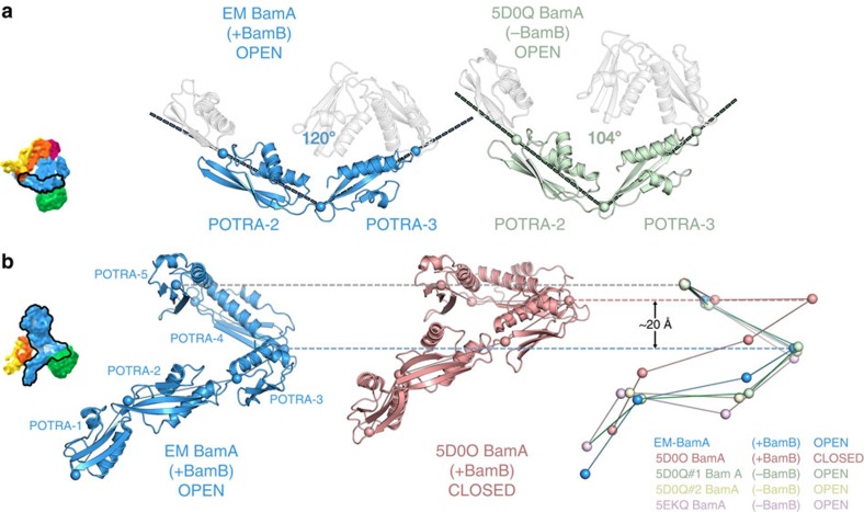
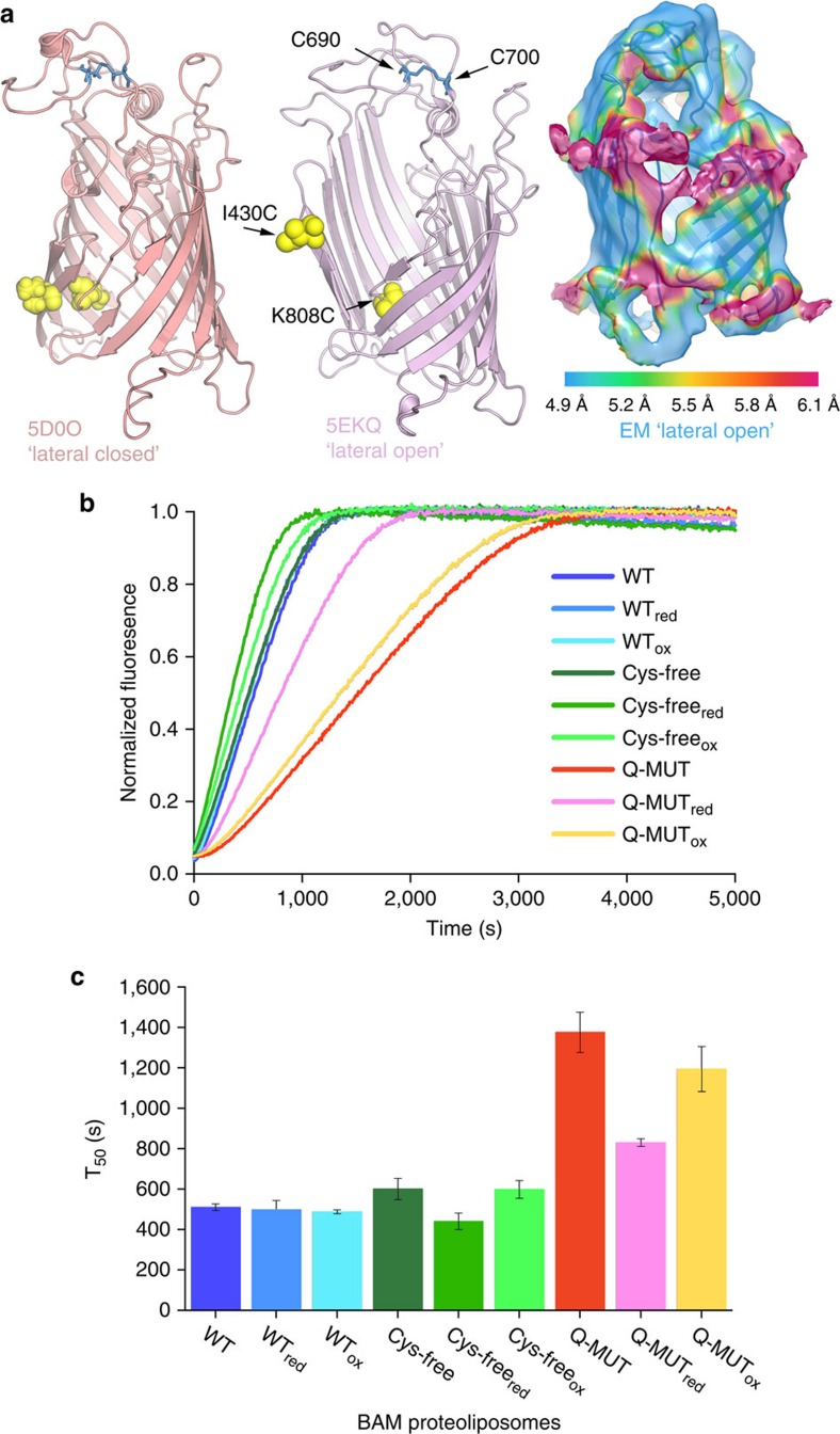
The β-barrel assembly machinery (BAM) is a ∼203 kDa complex of five proteins (BamA-E), which is essential for viability in E. coli. BAM promotes the folding and insertion of β-barrel proteins into the outer membrane via a poorly understood mechanism. Several current models suggest that BAM functions through a 'lateral gating' motion of the β-barrel of BamA. Here we present a cryo-EM structure of the BamABCDE complex, at 4.9 Å resolution. The structure is in a laterally open conformation showing that gating is independent of BamB binding. We describe conformational changes throughout the complex and interactions between BamA, B, D and E, and the detergent micelle that suggest communication between BAM and the lipid bilayer. Finally, using an enhanced reconstitution protocol and functional assays, we show that for the outer membrane protein OmpT, efficient folding in vitro requires lateral gating in BAM.


Figures

Figure 1
Figure 1. Characterization of the intact BAM complex and optimization of its activity in E. coli polar lipids.
(a) SDS–polyacrylamide gel showing that all five subunits are present in the purified BAM complex reconstituted into DDM micelles (analysed after boiling (+)) or into liposomes formed from E. coli polar lipids by extensive dialysis. The unboiled and boiled samples show a differential electrophoretic mobility (band-shift) for BamA, consistent with this subunit being folded in the proteoliposomes. The full gels are shown in Supplementary Fig. 10. (b) Intact electrospray ionization–mass spectrum of the BAM complex. Charge states from the intact complex, as well as subcomplexes formed by gas phase ionization (inset) are indicated. The intact complex elutes as a single peak on size exclusion chromatography, indicating that it is intact in solution (Supplementary Fig. 1). (c) Optimization of BAM complex-containing proteoliposomes for activity. Denatured OmpT in solution in the presence of a seven-fold molar excess of SurA was added to liposomes that are empty or contain BAM. Successful folding results in an increase in fluorescence by cleavage of the fluorogenic substrate. Controls were performed in the absence of OmpT (green), SurA (blue), fluorogenic peptide (cyan) and using empty liposomes (pink). BAM proteoliposomes generated by dilution or dialysis are compared (see Methods).
Figure 2
Figure 2. Cryo-EM structure of the BAM complex.
(a) Views of the front and back face of the cryo-EM structure of the intact BAM complex at 4.9 Å resolution. BamA is coloured blue, BamB in green, BamC in yellow, BamD in orange and BamE in magenta. Density corresponding to the micelle of n-dodecyl-β-D-maltopyranoside (DDM) in the structure is shown as a pale grey mesh. (b) Flexible fitting of a hybrid X-ray structure into the EM density. The views and colouring are identical, but density for the micelle has been masked and the EM density made transparent, showing the fitted pseudo-atomic model within. This colour scheme is maintained for all further figures showing the cryo-EM structure. The figure was made using UCSF Chimera.
Figure 3
Figure 3. Resolution of secondary structure in individual subunits of the BAM complex.
The EM map has a mean resolution of 4.9 Å, allowing resolution of secondary structure throughout the BAM structure, including (a) the β-barrel of BamA, which is in a ‘lateral open' conformation, (b) the β-propeller of BamB, in which β-sheets are well resolved, (c) the ‘lasso' of BamC, (d) the α-helical region of BamD and (e) α-helices and β-sheets in BamE. All panels were made using PyMOL v1.7 (ref. 65).
Figure 4
Figure 4. The effect of BamB binding and β-barrel conformation on the BamA POTRA domains.
(a) The presence of BamB correlates with a more obtuse angle between POTRA domains 2 and 3. On the left, is the EM structure (‘lateral open', +BamB; blue), and on the right 5D0Q (a ‘lateral open', -BamB, X-ray structure; pale green). The view is from outside the bacterial cell, looking approximately down the axis of the β-barrel (see thumbnail image). Both structures have the BamA β-barrel in a ‘lateral open' conformation; thus, the barrel opening and a wide POTRA 2–3 angle do not correlate, but BamB binding and a wide POTRA angle do correlate. The remaining POTRA domains are shown in pale grey. The angle is measured between identical points in the hinge regions between each POTRA domain, indicated by spheres. (b) Vertical extension of the POTRA domains correlates with β-barrel state. The open barrel of the EM structure (‘lateral open', +BamB; blue) and all ‘lateral open' X-ray structures, correlates with a vertically extended conformation of the POTRA chain, whereas the ‘lateral closed' barrel of 5D0O (+BamB; pink) has a much more compact POTRA chain. Both structures contain BamB; hence, BamB binding does not appear to correlate with the extension of the POTRA chain. Thumbnail images are in the appropriate view and coloured with the same colour scheme as Fig. 2. For overall comparisons of BamA, see Supplementary Fig. 9.
Figure 5
Figure 5. Interactions between BAM components and the detergent micelle.
(a) A hydrophobic loop within the body of BamA POTRA 3 (blue, white arrow) is buried within the micelle (grey mesh), with details of the hydrophobic residues inset. The N terminus of BamB (green, black arrow), which is unmodelled in the X-ray structures of BAM and its subcomplexes, also dips into the micelle. (b) A hydrophobic 310 helix in BamD (orange) inserts into the micelle, with hydrophobic residues buried in the hydrocarbon tail groups of the detergent (see inset) and polar residues flanking the helix placed to interact with the polar head groups of the detergent. (c) The N terminus of BamE (magenta), which is the site of the lipid anchor, also inserts into the micelle, well away from the body of the BamA β-barrel.
Figure 6
Figure 6. Interactions with the micelle may alter BamD conformation.
(a) Residues 32–85 in the BamC lasso region are similar in all structures (EM, yellow) except for the ‘lateral closed' structure of intact BAM (5D0O (pink)), where an extra helical segment is seen. (b) This extra helical segment would clash with the 310 helix of BamD (residues 123–132), which is consequently disordered in 5D0O (red/pink), which is compared with 5D0Q (pale green and green). BamC is pale (pink/green) and BamD is strongly coloured (red/green). (c) Conformational change in BamD. BamE of the EM structure is aligned to the equivalent region from the ‘lateral open' BamACDE (5D0Q33) structure. The C-terminal half of BamD (left hand side) overlaps almost exactly in the two structures, showing that the interface between BamD and E is preserved. The structures deviate around residue 157 (indicated by black arrow). A helix runs directly from this position to the micelle/membrane. The rest of BamD is displaced as a rigid body by ∼6° at the N terminus.
Figure 7
Figure 7. β-Barrel gating of BamA is required for full BAM activity in vitro.
(a) Cys residues introduced into the BamA β-barrel at positions 430 (I430C) and 808 (K808C) (yellow spheres) are hypothesized to be able to form a disulfide in the ‘lateral closed' structure (5D0O33) (left image), but not in a ‘lateral open' barrel (for example, 5EKQ32) (centre image). The two natural Cys residues (C690 and C700), which were removed, are shown in blue ball and stick. On the right hand side, the EM density is in a similar orientation and coloured according to local resolution, showing that the density around β1–β16 is at a lower resolution and thus more mobile than the body of the barrel. (b) Example kinetic traces of OmpT folding measured by its proteolytic activity in the presence of BAM complexes containing wild-type BamA, BamAC690S/C700S (Cys-free) or BamAC690S/C700S/I430C/K808C (Q-MUT; see Supplementary Fig. 1). All experiments were performed with final concentrations of 0.25 μM BAM proteoliposomes, 5 μM OmpT, 1 mM fluorogenic peptide, 35 μM SurA, in oxidizing (1 mM CuSO4) or reducing (50 mM dithiothreitol (DTT)) conditions. All experiments were performed in 50 mM glycine-NaOH pH 9.5, 25 °C. (c) Bar chart of the average half-time for each folding reaction. These show the mean and s.e.m. from four repeats, across two proteoliposome preparations (see also Supplementary Table 3).

References

    1. Bos M. P., Robert V. & Tommassen J. Biogenesis of the gram-negative bacterial outer membrane. Annu. Rev. Microbiol. 61, 191–214 (2007). - PubMed
    1. Fairman J. W., Noinaj N. & Buchanan S. K. The structural biology of beta-barrel membrane proteins: a summary of recent reports. Curr. Opin. Struct. Biol. 21, 523–531 (2011). - PMC - PubMed
    1. Hagan C. L., Silhavy T. J. & Kahne D. beta-Barrel membrane protein assembly by the Bam complex. Annu. Rev. Biochem. 80, 189–210 (2011). - PubMed
    1. Kim K. H., Aulakh S. & Paetzel M. The bacterial outer membrane beta-barrel assembly machinery. Protein Sci. 21, 751–768 (2012). - PMC - PubMed
    1. Rollauer S. E., Sooreshjani M. A., Noinaj N. & Buchanan S. K. Outer membrane protein biogenesis in Gram-negative bacteria. Philos. Trans. R Soc. Lond. B Biol. Sci. 370, 1–10 (2015). - PMC - PubMed

LinkOut - more resources