. 2017 Jan;38(1):119-126.
doi: 10.3174/ajnr.A4951. Epub 2016 Sep 29.

Flow Conditions in the Intracranial Aneurysm Lumen Are Associated with Inflammation and Degenerative Changes of the Aneurysm Wall

Affiliations

Flow Conditions in the Intracranial Aneurysm Lumen Are Associated with Inflammation and Degenerative Changes of the Aneurysm Wall

J Cebral et al. AJNR Am J Neuroradiol. 2017 Jan.

Abstract

Background and purpose: Saccular intracranial aneurysm is a common disease that may cause devastating intracranial hemorrhage. Hemodynamics, wall remodeling, and wall inflammation have been associated with saccular intracranial aneurysm rupture. We investigated how saccular intracranial aneurysm hemodynamics is associated with wall remodeling and inflammation of the saccular intracranial aneurysm wall.

Materials and methods: Tissue samples resected during a saccular intracranial aneurysm operation (11 unruptured, 9 ruptured) were studied with histology and immunohistochemistry. Patient-specific computational models of hemodynamics were created from preoperative CT angiographies.

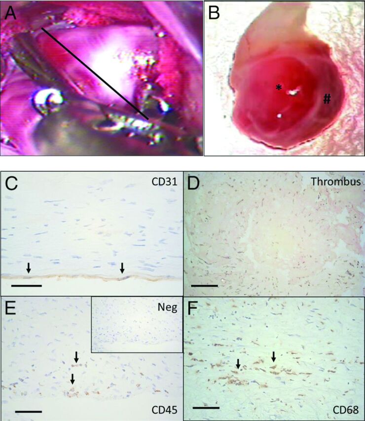
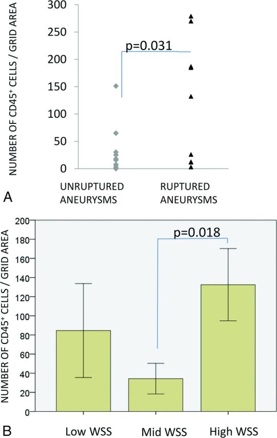
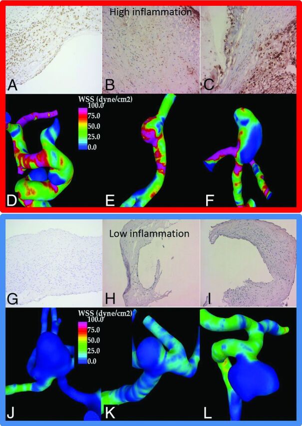
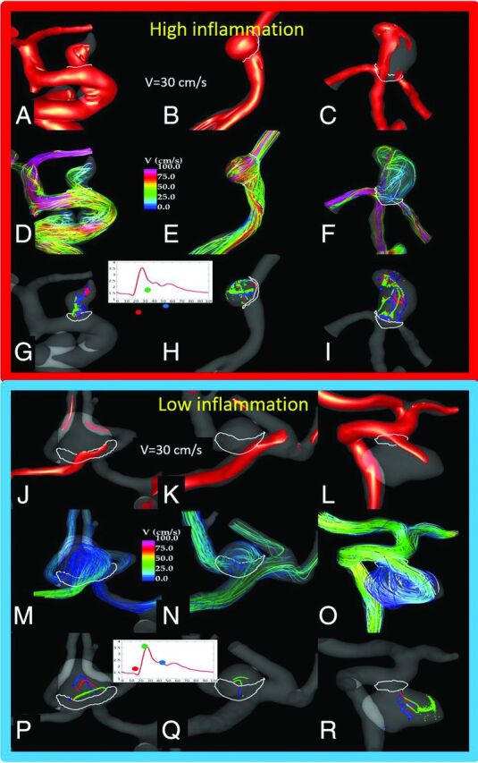
Results: More stable and less complex flows were associated with thick, hyperplastic saccular intracranial aneurysm walls, while slower flows with more diffuse inflow were associated with degenerated and decellularized saccular intracranial aneurysm walls. Wall degeneration (P = .041) and rupture were associated with increased inflammation (CD45+, P = .031). High wall shear stress (P = .018), higher vorticity (P = .046), higher viscous dissipation (P = .046), and high shear rate (P = .046) were associated with increased inflammation. Inflammation was also associated with lack of an intact endothelium (P = .034) and the presence of organized luminal thrombosis (P = .018), though overall organized thrombosis was associated with low minimum wall shear stress (P = .034) and not with the flow conditions associated with inflammation.

Conclusions: Flow conditions in the saccular intracranial aneurysm are associated with wall remodeling. Inflammation, which is associated with degenerative wall remodeling and rupture, is related to high flow activity, including elevated wall shear stress. Endothelial injury may be a mechanism by which flow induces inflammation in the saccular intracranial aneurysm wall. Hemodynamic simulations might prove useful in identifying saccular intracranial aneurysms at risk of developing inflammation, a potential biomarker for rupture.


Figures

Fig 1.
Fig 1.
Tissue samples were harvested after aneurysm clipping by cutting through the aneurysm dome distal to the clip, as demonstrated in A (black line represents the estimated site of cut). B, The tissue samples obtained varied significantly in size (from small, approximately 2 × 1 mm tissue samples to half domes with a >10-mm radius). In addition to variation in size, many of the aneurysm samples had local variation in the appearance of the wall (B) with translucent areas (asterisk) and thicker wall regions (hash tag). C, In histology, endothelium (CD31+ luminal cells with endothelial cells morphology) was present in only 3/10 of the studied aneurysm walls. D, In 8/20 aneurysms, the luminal surface was instead covered by thrombus, and in 7/20 aneurysms, some degree of thrombus organization was observed (hematoxylin-eosin staining). E, Loss of endothelium and organizing luminal thrombosis were associated with inflammatory cell infiltration (CD45+) in the aneurysm wall. F, Many of the inflammatory cells were macrophages (CD68+). Immunostaining protocol for CD68 is described by Ollikainen et al. Scale bars represent a 50-μm scale. Negative control for immunostaining is given for CD45 staining (Neg). The black arrows indicate positive cells.
Fig 2.
Fig 2.
A, Infiltration of inflammatory cells (CD45+) associated with rupture and high mean wall shear stress. B, WSS grouping is as follows: mean WSS of < 4.5 dyne/cm2 = low; 4.5 < mean WSS < 18 dyne/cm2 = mid; mean WSS > 18 dyne/cm2 = high. Bars display means, and error bars, standard error of mean.
Fig 3.
Fig 3.
A–F, Three aneurysms from the high WSS group with substantial wall inflammation. A–C, CD45 immunostaining and inflammatory cells in brown along with 3 aneurysms from the low WSS group (G–L) with little wall inflammation (J–L, negative CD45 immunostaining).
Fig 4.
Fig 4.
Flow conditions in the 6 sample aneurysms (Fig 3) with high or no inflammation in the wall. The visualizations show the following: the inflow jet at peak systole (A–C, J–L), the flow pattern at peak systole (D–F, M–O), and vortex core lines at 3 instances during the cardiac cycle (G–I, P–R), to illustrate both the complexity of the flow pattern and its change during the cycle (instability). An aneurysm with inflammation (upper panel) had a strong inflow jet impacting the aneurysm dome and producing complex (but somewhat stable) flow structures and associated WSS distributions with regions of high WSS and regions of low WSS near the outflow. In contrast, aneurysms with no inflammation had more diffuse inflows entering the aneurysms at lower velocity and diffusing into a simpler but more variable (unstable) flow pattern, with associated WSS distribution characterized by a fairly uniformly low WSS.

References

    1. Vlak MH, Algra A, Brandenburg R, et al. . Prevalence of unruptured intracranial aneurysms, with emphasis on sex, age, comorbidity, country, and time period: a systematic review and meta-analysis. Lancet Neurol 2011;10:626–36 10.1016/S1474-4422(11)70109-0 - DOI - PubMed
    1. Karamanakos PN, von Und Zu Fraunberg M, Bendel S, et al. . Risk factors for three phases of 12-month mortality in 1657 patients from a defined population after acute aneurysmal subarachnoid hemorrhage. World Neurosurg 2012;78:631–39 10.1016/j.wneu.2011.08.033 - DOI - PubMed
    1. Korja M, Lehto H, Juvela S. Lifelong rupture risk of intracranial aneurysms depends on risk factors: a prospective Finnish cohort study. Stroke 2014;45:1958–63 10.1161/STROKEAHA.114.005318 - DOI - PubMed
    1. Kotowski M, Naggara O, Darsaut TE, et al. . Safety and occlusion rates of surgical treatment of unruptured intracranial aneurysms: a systematic review and meta-analysis of the literature from 1990 to 2011. J Neurol Neurosurg Psychiatry 2013;84:42–48 10.1136/jnnp-2011-302068 - DOI - PubMed
    1. Kataoka K, Taneda M, Asai T, et al. . Structural fragility and inflammatory response of ruptured cerebral aneurysms: a comparative study between ruptured and unruptured cerebral aneurysms. Stroke 1999;30:1396–401 10.1161/01.STR.30.7.1396 - DOI - PubMed

MeSH terms