. 2016 Nov;80(5):776-790.
doi: 10.1002/ana.24791. Epub 2016 Oct 25.

Neuroinflammatory component of gray matter pathology in multiple sclerosis

Affiliations

Neuroinflammatory component of gray matter pathology in multiple sclerosis

Elena Herranz et al. Ann Neurol. 2016 Nov.

Abstract

Objective: In multiple sclerosis (MS), using simultaneous magnetic resonance-positron emission tomography (MR-PET) imaging with 11 C-PBR28, we quantified expression of the 18kDa translocator protein (TSPO), a marker of activated microglia/macrophages, in cortex, cortical lesions, deep gray matter (GM), white matter (WM) lesions, and normal-appearing WM (NAWM) to investigate the in vivo pathological and clinical relevance of neuroinflammation.

Methods: Fifteen secondary-progressive MS (SPMS) patients, 12 relapsing-remitting MS (RRMS) patients, and 14 matched healthy controls underwent 11 C-PBR28 MR-PET. MS subjects underwent 7T T2*-weighted imaging for cortical lesion segmentation, and neurological and cognitive evaluation. 11 C-PBR28 binding was measured using normalized 60- to 90-minute standardized uptake values and volume of distribution ratios.

Results: Relative to controls, MS subjects exhibited abnormally high 11 C-PBR28 binding across the brain, the greatest increases being in cortex and cortical lesions, thalamus, hippocampus, and NAWM. MS WM lesions showed relatively modest TSPO increases. With the exception of cortical lesions, where TSPO expression was similar, 11 C-PBR28 uptake across the brain was greater in SPMS than in RRMS. In MS, increased 11 C-PBR28 binding in cortex, deep GM, and NAWM correlated with neurological disability and impaired cognitive performance; cortical thinning correlated with increased thalamic TSPO levels.

Interpretation: In MS, neuroinflammation is present in the cortex, cortical lesions, deep GM, and NAWM, is closely linked to poor clinical outcome, and is at least partly linked to neurodegeneration. Distinct inflammatory-mediated factors may underlie accumulation of cortical and WM lesions. Quantification of TSPO levels in MS could prove to be a sensitive tool for evaluating in vivo the inflammatory component of GM pathology, particularly in cortical lesions. Ann Neurol 2016;80:776-790.


Figures

Figure 1
Figure 1
A) Mean normalized 11C-PBR28 standardized uptake values (SUVR) in healthy controls (n=14) and patients (n=27) with either relapsing-remitting or secondary-progressive multiple sclerosis (RRMS, n=12; SPMS, n=15) across different tissue compartments in gray and white matter. Horizontal bars indicate group averages for HAB= high-affinity binder, MAB= mixed-affinity binder. Connector-lines connect brain tissue compartments that were compared in the whole MS cohort relative to controls using linear regression and covarying for age and affinity binding. In MS, mean SUVR in intracortical and leukocortical lesions were compared with mean cortical SUVR from controls. The asterisks denote when the specific comparison, connector-line, between healthy controls and MS patients was statistically significant. B) Histogram showing mean 11C-PBR28 normalized volume of distributions (DVR) and standard deviations in different brain regions for both multiple sclerosis (MS) subjects (n=15) and controls (n=11). 11C-PBR28 DVR were significantly increased in MS subjects compared to controls in all regions assessed. DVR cortical lesions analysis was performed in 9 MS patients. *p<0.05, **p<0.005, by linear regression, adjusting for binding genotype and age, and corrected for multiple comparisons. HC= healthy controls; HAB= high affinity binder; MAB= mixed affinity binder; ICL= intracortical lesions; LCL= leukocortical lesions; Hipp= hippocampus; BG= basal ganglia; NAWM= normal appearing white matter; WML= white matter lesions; CL= cortical lesions.
Figure 2
Figure 2
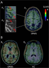
A) Fused magnetic resonance and positron emission tomography (MR-PET) images in a subject with secondary-progressive multiple sclerosis (SPMS) and a high affinity binding (HAB) genotype (A1) showing cortical areas of increased 11C-PBR28 standardized uptake values normalized by a pseudo-reference region (SUVR). (A2) Detail of cortical lesions visible on the 3 Tesla (T) anatomical scan fused with SUVR images and (A3) on the co-registered 7T T2* images. B) Fused 11C-PBR28 SUVR MR-PET images in a HAB MS subject (B1) and in a HAB age-matched healthy control (B2) showing increased 11C-PBR28 SUVR uptake in the thalamus of the patient.
Figure 3
Figure 3
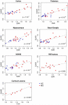
Plots illustrating, in the whole study cohort, the correlation between 11C-PBR28 normalized volume of distributions (DVR) and standardized uptake values (SUVR) across different brain tissue compartments by linear regression, adjusting for binding genotype and correcting for multiple comparisons. HC= healthy controls; MS= multiple sclerosis; NAWM= normal-appearing white matter; WM= white matter.
Figure 4
Figure 4
Plots illustrating, in the whole multiple sclerosis (MS) cohort, correlations between 11C-PBR28 normalized standardized uptake values (SUVR) and neurological disability and cognitive test scores (linear regression analysis, adjusting for binding genotype, age. All p values are corrected for multiple comparisons). EDSS= Expanded Disability Status Scale; NAWM= normal-appearing white matter; WM= white matter; SDMT= Symbol Digit Modalities Test, RRMS= relapsing-remitting MS; SPMS= secondary-progressive MS.
Figure 5
Figure 5
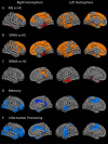
Overlay of the general linear model significance maps (p<0.05, corrected for multiple comparisons, adjusting for age, binding genotype and cortical thickness) on the average pial surface showing, relative to healthy controls (HC, n=14), regions of increased 11C-PBR28 normalized standardized uptake values (SUVR) A) in the whole multiple sclerosis (MS) cohort (n=27), as well as B) in the secondary-progressive MS (SPMS, n=15) and C) in the relapsing-remitting MS (RRMS, n=12) subgroups. Regions in all MS cases where 11C-PBR28 uptake correlated negatively (p<0.05, corrected for multiple comparisons) with D) memory and E) information processing speed function.

Comment in

References

    1. Zivadinov R, Pirko I. Advances in understanding gray matter pathology in multiple sclerosis: are we ready to redefine disease pathogenesis? BMC Neurol. 2012;12:9. - PMC - PubMed
    1. Peterson JW, Bo L, Mork S, Chang A, Trapp BD. Transected neurites, apoptotic neurons, and reduced inflammation in cortical multiple sclerosis lesions. Ann Neurol. 2001 Sep;50(3):389–400. - PubMed
    1. Bo L, Vedeler CA, Nyland H, Trapp BD, Mork SJ. Intracortical multiple sclerosis lesions are not associated with increased lymphocyte infiltration. Mult Scler. 2003 Aug;9(4):323–31. - PubMed
    1. Magliozzi R, Howell O, Vora A, et al. Meningeal B-cell follicles in secondary progressive multiple sclerosis associate with early onset of disease and severe cortical pathology. Brain. 2007 Apr;130(Pt 4):1089–104. - PubMed
    1. Howell OW, Reeves CA, Nicholas R, et al. Meningeal inflammation is widespread and linked to cortical pathology in multiple sclerosis. Brain. 2011 Sep;134(Pt 9):2755–71. - PubMed

MeSH terms

Substances