. 2016 Sep 30:6:33234.
doi: 10.1038/srep33234.

PLGA nanoparticle encapsulation reduces toxicity while retaining the therapeutic efficacy of EtNBS-PDT in vitro

Affiliations

PLGA nanoparticle encapsulation reduces toxicity while retaining the therapeutic efficacy of EtNBS-PDT in vitro

Hsin-I Hung et al. Sci Rep. .

Abstract

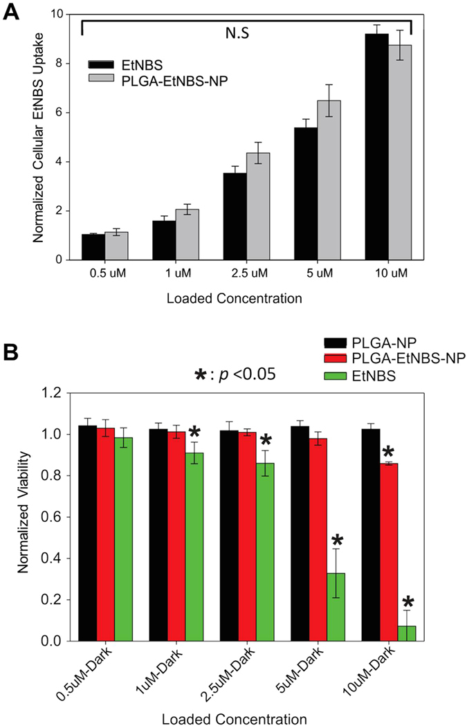
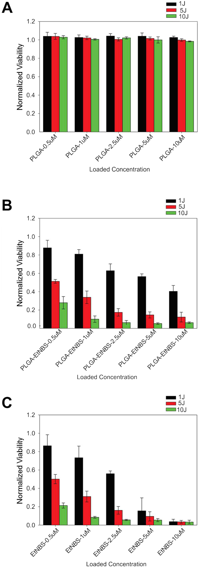
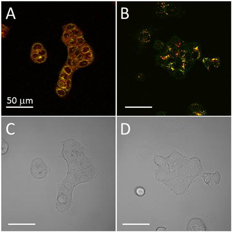
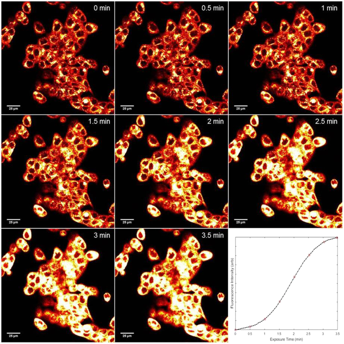
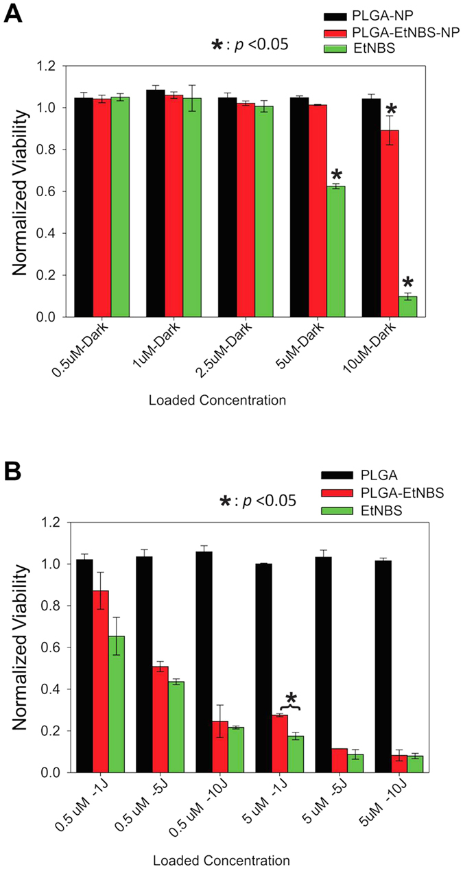
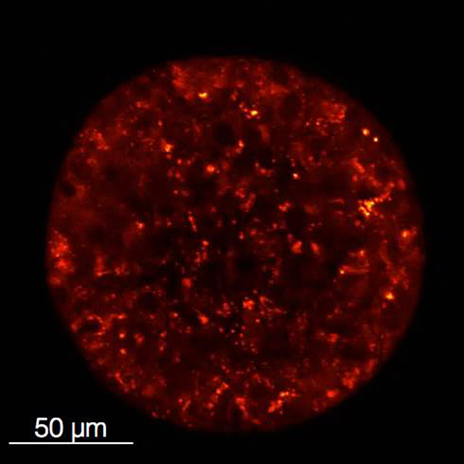
Photodynamic therapy regimens, which use light-activated molecules known as photosensitizers, are highly selective against many malignancies and can bypass certain challenging therapeutic resistance mechanisms. Photosensitizers such as the small cationic molecule EtNBS (5-ethylamino-9-diethyl-aminobenzo[a]phenothiazinium chloride) have proven potent against cancer cells that reside within acidic and hypoxic tumour microenvironments. At higher doses, however, these photosensitizers induce "dark toxicity" through light-independent mechanisms. In this study, we evaluated the use of nanoparticle encapsulation to overcome this limitation. Interestingly, encapsulation of the compound within poly(lactic-co-glycolic acid) (PLGA) nanoparticles (PLGA-EtNBS) was found to significantly reduce EtNBS dark toxicity while completely retaining the molecule's cytotoxicity in both normoxic and hypoxic conditions. This dual effect can be attributed to the mechanism of release: EtNBS remains encapsulated until external light irradiation, which stimulates an oxygen-independent, radical-mediated process that degrades the PLGA nanoparticles and releases the molecule. As these PLGA-encapsulated EtNBS nanoparticles are capable of penetrating deeply into the hypoxic and acidic cores of 3D spheroid cultures, they may enable the safe and efficacious treatment of otherwise unresponsive tumour regions.


Figures

Figure 1
Figure 1. Comparison of uptake and dark toxicity of free EtNBS and PLGA-EtNBS nanoparticles.
(A) Cellular uptake of free EtNBS and PLGA-EtNBS at equivalent cellular incubation concentrations. (B) Dark toxicity caused by PLGA vehicle controls (PA), free EtNBS, and PLGA-EtNBS nanoparticles (PE) at equivalent incubation concentrations/equivalent volumes. Viability was determined via the MTT cell viability assay.
Figure 2
Figure 2. PDT-induced death of OVCAR5 cells in monolayer culture.
(A) Viability of cells to PLGA vehicle only controls, showing no observed toxicity at any light dose. (B) Viability of cells following PLGA-EtNBS PDT across a range of concentrations and light doses. (C) Viability of cells following EtNBS-PDT across a range of concentrations and light doses. Viability was determined via the MTT cell viability assay.
Figure 3
Figure 3. Physically encapsulated (PE) and chemical conjugated (CC) PLGA-EtNBS nanoparticles are both endocytosed into lysosomes.
(A) Uptake of PE nanoparticles into OVCAR5 cells. EtNBS fluorescence is in red, while Lysotracker Green is displayed in green. (B) Uptake of CC nanoparticles, with EtNBS in red and Lysotracker in green. Note that in the CC nanoparticle case, the EtNBS signal is solely localized in lysosomes, while the PE nanoparticles demonstrate more diffuse EtNBS signal. This is thought to arise from minor leakage during the incubation period of EtNBS from the PE nanoparticles into the endoplasmic reticulum. (C,D) Trans-illumination images of (A,B), respectively.
Figure 4
Figure 4. Photobrightening of PLGA-EtNBS nanoparticles over time.
Cells were continuously observed over 3.5 min on a confocal microscope using 1.4 mW of focused 635 nm excitation raster-scanned across the field of view to visualize EtNBS via its 670 nm-centred fluorescence (via a 60X, 1.20NA objective, corresponding to a field of view size of 212 × 212 μm2, 512 × 512 pixels, a pixel dwell time of 4 μs/pixel, and 3x line averaging, resulting in an acquisition time of 3.3 seconds per frame). Initially, EtNBS fluorescence is weak and localized to lysosomes. As the 635 nm light is continuously scanned, the fluorescence intensity increases and eventually plateaus, as shown in the inset depicting the increase in fluorescence intensity across the field of view.
Figure 5
Figure 5. PDT of EtNBS and PLGA-EtNBS nanoparticles at equivalent loading concentrations under hypoxic conditions.
(A) Viability of cells following incubation of each agent under hypoxia without the application of light. (B) Viability of cells under hypoxia following PDT at 0.5 μM and 5 μM equivalent EtNBS doses at different light doses. Note that PLGA-EtNBS nanoparticles have similar efficacy to free EtNBS even under hypoxic conditions. Viability was determined via the MTT cell viability assay.
Figure 6
Figure 6. Uptake of PLGA-EtNBS nanoparticles into ovarian cancer in vitro spheroids (OVCAR5).
PLGA-EtNBS nanoparticles are distributed throughout all cells within in vitro tumour spheroids, indicating that the nanoparticles diffuse and are transported through multiple cell layers. The dimmer appearance of EtNBS fluorescence in the centre of the spheroid relative to the periphery is not reflective of the actual cellular uptake and is the result of light scattering within the solid cellular mass. The distribution of PLGA-EtNBS under confocal microscopy matches that observed under identical conditions for free EtNBS, where the photosensitizer was found to be taken up throughout the spheroid.

References

    1. Foster R., Buckanovich R. J. & Rueda B. R. Ovarian cancer stem cells: working towards the root of stemness. Cancer letters 338, 147–157, doi: 10.1016/j.canlet.2012.10.023 (2013). - DOI - PubMed
    1. Dong Y. et al.. Paclitaxel resistance and multicellular spheroid formation are induced by kallikrein-related peptidase 4 in serous ovarian cancer cells in an ascites mimicking microenvironment. PloS one 8, e57056, doi: 10.1371/journal.pone.0057056 (2013). - DOI - PMC - PubMed
    1. Agarwal R. & Kaye S. B. Ovarian cancer: strategies for overcoming resistance to chemotherapy. Nature reviews. Cancer 3, 502–516, doi: 10.1038/nrc1123 (2003). - DOI - PubMed
    1. Murakami A. et al.. Hypoxia increases gefitinib-resistant lung cancer stem cells through the activation of insulin-like growth factor 1 receptor. PloS one 9, e86459, doi: 10.1371/journal.pone.0086459 (2014). - DOI - PMC - PubMed
    1. De Milito A. & Fais S. Tumor acidity, chemoresistance and proton pump inhibitors. Future oncology 1, 779–786, doi: 10.2217/14796694.1.6.779 (2005). - DOI - PubMed