

L-DOPA in the hu man ovarian follicular fluid acts as an antioxidant factor on granulosa cells
- PMID: 27686972
- PMCID: PMC5043631
- DOI: 10.1186/s13048-016-0269-0
L-DOPA in the hu man ovarian follicular fluid acts as an antioxidant factor on granulosa cells
Abstract
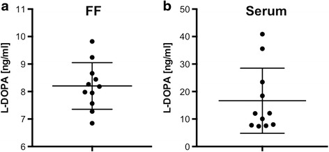
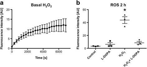
Background: A previous study showed that dopamine (DA), which is contained in follicular fluid (FF) from IVF patients, strongly increased the production of reactive oxygen species (ROS) by cultured human granulosa cells (GCs). ROS, including H2O2, are assumed to play roles in ovarian physiology and pathology. Ovarian DA could be derived from the circulation, ovarian innervation and/or unknown ovarian sources. L-DOPA is the direct precursor of DA in its synthetic pathway. It was not yet described in FF. We examined L-DOPA levels in FF from IVF patients. As it may exert anti-oxidative and ROS-scavenging functions, we studied whether it exerts such actions in human GCs and whether DOPA-decarboxylase (DDC), the enzyme converting L-DOPA to DA, is expressed in the human ovary.
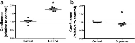
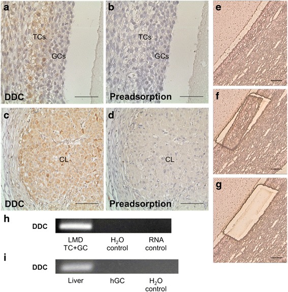
Results: ELISA measurements revealed that human IVF-derived FF contains L-DOPA. In cultured human GCs automated confluence analyses showed that L-DOPA enhanced their survival. This is in contrast to the actions of DA, which reduced cell survival. A dose-dependent mode of action of L-DOPA was identified using a fluorescent ROS indicator. The results showed that it antagonized intracellular ROS accumulation induced by exogenous H2O2. DDC was absent in follicular GCs, but immunohistochemistry identified it in theca cells (TCs) of large follicles in the human ovary. Laser micro-dissection followed by RT-PCR corroborated the expression. DDC was also identified in the steroidogenic cells of the corpus luteum.
Conclusions: L-DOPA in FF is an antioxidant factor and exerts positive influences on GCs. Ovarian DA is derived from L-DOPA and has opposite actions. Exogenous L-DOPA is a standard therapy for Parkinson's disease, and the results raise the possibility that it may be able to exert positive actions as an antioxidant in ovarian conditions, as well.
Keywords: Granulosa cells; L-DOPA; Reactive oxygen species.
Figures
References
-
- Saller S, Kunz L, Berg D, Berg U, Lara H, Urra J, et al. Dopamine in human follicular fluid is associated with cellular uptake and metabolism-dependent generation of reactive oxygen species in granulosa cells: implications for physiology and pathology. Hum Reprod. 2014;29:555–67. doi: 10.1093/humrep/det422. - DOI - PubMed
-
- Saller S, Merz-Lange J, Raffael S, Hecht S, Pavlik R, Thaler C, et al. Norepinephrine, active norepinephrine transporter, and norepinephrine-metabolism are involved in the generation of reactive oxygen species in human ovarian granulosa cells. Endocrinology. 2012;153:1472–83. doi: 10.1210/en.2011-1769. - DOI - PubMed
LinkOut - more resources
Full Text Sources
Other Literature Sources
Research Materials
