. 2016 Sep 29;4(1):106.
doi: 10.1186/s40478-016-0378-4.

Evaluation of the brain-penetrant microtubule-stabilizing agent, dictyostatin, in the PS19 tau transgenic mouse model of tauopathy

Affiliations

Evaluation of the brain-penetrant microtubule-stabilizing agent, dictyostatin, in the PS19 tau transgenic mouse model of tauopathy

Vishruti Makani et al. Acta Neuropathol Commun. .

Abstract

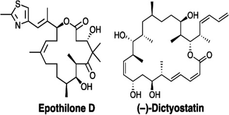
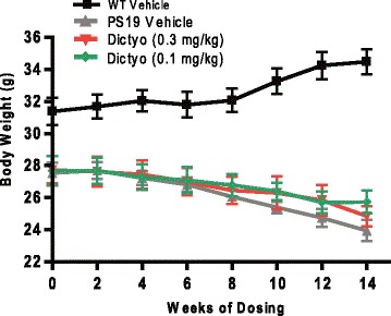
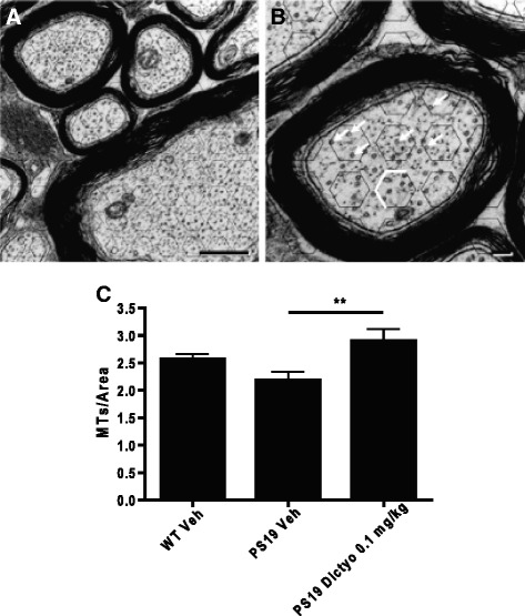
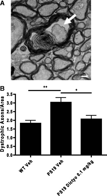
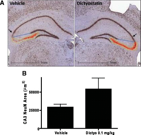
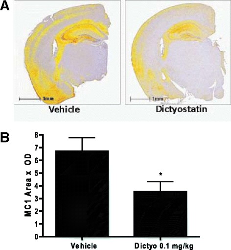
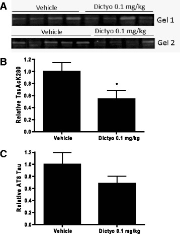
Neurodegenerative disorders referred to as tauopathies, which includes Alzheimer's disease (AD), are characterized by insoluble deposits of the tau protein within neuron cell bodies and dendritic processes in the brain. Tau is normally associated with microtubules (MTs) in axons, where it provides MT stabilization and may modulate axonal transport. However, tau becomes hyperphosphorylated and dissociates from MTs in tauopathies, with evidence of reduced MT stability and defective axonal transport. This has led to the hypothesis that MT-stabilizing drugs may have potential for the treatment of tauopathies. Prior studies demonstrated that the brain-penetrant MT-stabilizing drug, epothilone D, had salutary effects in transgenic (Tg) mouse models of tauopathy, improving MT density and axonal transport, while reducing axonal dystrophy. Moreover, epothilone D enhanced cognitive performance and decreased hippocampal neuron loss, with evidence of reduced tau pathology. To date, epothilone D has been the only non-peptide small molecule MT-stabilizing agent to be evaluated in Tg tau mice. Herein, we demonstrate the efficacy of another small molecule brain-penetrant MT-stabilizing agent, dictyostatin, in the PS19 tau Tg mouse model. Although dictyostatin was poorly tolerated at once-weekly doses of 1 mg/kg or 0.3 mg/kg, likely due to gastrointestinal (GI) complications, a dictyostatin dose of 0.1 mg/kg was better tolerated, such that the majority of 6-month old PS19 mice, which harbor a moderate level of brain tau pathology, completed a 3-month dosing study without evidence of significant body weight loss. Importantly, as previously observed with epothilone D, the dictyostatin-treated PS19 mice displayed improved MT density and reduced axonal dystrophy, with a reduction of tau pathology and a trend toward increased hippocampal neuron survival relative to vehicle-treated PS19 mice. Thus, despite evidence of dose-limiting peripheral side effects, the observed positive brain outcomes in dictyostatin-treated aged PS19 mice reinforces the concept that MT-stabilizing compounds have significant potential for the treatment of tauopathies.

Keywords: Alzheimer’s; Drug; Microtubule; Mouse; Pathology; Tauopathy; Transgenic.


Figures

Fig. 1
Fig. 1
Structures of epothilone D and dictyostatin
Fig. 2
Fig. 2
Representative hemisphere brain sections from 6- and 9-month old male B6 PS19 months stained for MC1-positive tau pathology. Bregma-matched sections from male PS19 mice were immunostained with the MC1 antibody and whole hemispheres were imaged. Scale bar = 1 mm
Fig. 3
Fig. 3
Dictyostatin doses of 0.3 mg/kg biweekly or 0.1 mg/kg weekly did not exacerbate body weight loss in 6-month old PS19 mice. Male 6-month old PS19 mice received either biweekly ip injections of 0.3 mg/kg dictyostatin, weekly injections of 0.1 mg/kg dictyostatin, or weekly injections of vehicle (n = 11/treatment). In addition, wild-type male 6-month old B6 mice received weekly injections of vehicle. Body weights were recorded weekly. A total of 6 mice in the 0.3 mg/kg dictyostatin group and 3 mice in the 0.1 mg/kg dictyostatin group died before study completion. Error bars represent SEM
Fig. 4
Fig. 4
Dictyostatin treatment increased MT density in optic nerves of aged PS19 mice. MTs were counted in electron micrographs (50,000X) of cross-sections of optic nerves (a bar = 0.5 μm) from dictyostatin- or vehicle-treated aged PS19, or vehicle-treated WT mice, as previously described [46]. b An expanded imaged showing the grid of hexagons applied to optic nerve EM images (bar = 0.1 μm). To avoid repeat counting, only MTs that resided within the hexagon or were on one of the three indicated borders of each hexagon were counted. MTs are identified by arrows. c Quantification of MTs/area in the optic nerves of mice from each treatment group. Error bars denote SEM, with group sizes of 8–11. **, p < 0.01 as determined by one-way ANOVA with a post-hoc Tukey’s multiple comparison test
Fig. 5
Fig. 5
Dictyostatin treatment decreased axonal dystrophy in optic nerves of aged PS19 mice. a A representative electron micrograph showing an example of a dystrophic axon within the optic nerve from a study mouse (bar = 0.5 μm). b The number of dystrophic axons were counted in electron micrographs of cross-sections of optic nerves from dictyostatin- or vehicle-treated aged PS19 or WT mice, as previously described [46]. Error bars denote SEM, with group sizes of 8–11. *, p < 0.05; **, p < 0.01 as determined by one-way ANOVA with a post-hoc Tukey’s multiple comparison test
Fig. 6
Fig. 6
Dictyostatin treatment resulted in a trend towards reduced hippocampal CA3 neuron loss in aged PS19 mice. a Bregma-matched hemisphere brain sections from dictyostatin- or vehicle-treated aged PS19 mice (two sections/mouse) were immunostained with NeuN antibody and the area occupied by NeuN staining was determined within the demarcated CA3 region. The end boundary of the CA3 region was defined by drawing a line from the dentate to the start of the CA2 region (blue line), also using the demarcation of the end of the CA2 region (black arrow) as a landmark. Representative images with the quantified area are depicted. Scale bar = 1 mm. b A comparison of the results of NeuN staining in vehicle- and dictyostatin-treated PS19 mice (n = 7 for the dictyostatin group and n = 9 for the vehicle group). Error bars denote SEM
Fig. 7
Fig. 7
Dictyostatin treatment reduced MC1-positive tau pathology. a Bregma-matched hemisphere brain sections from dictyostatin- or vehicle-treated aged PS19 mice (two sections/mouse) were immunostained with MC1 antibody, and the total integrated MC1 signal over the section was obtained. Representative images with quantified areas and relative staining intensity are depicted, where the intensity of staining is shown as a yellow (low OD) to red (high OD) spectra. Scale bar = 1 mm. b A comparison of the results of MC1 staining in vehicle- and dictyostatin-treated PS19 mice (n = 7 for the dictyostatin group and n = 10 for the vehicle group). Error bars denote SEM. *, p < 0.05 as determined by a two-tailed t-test
Fig. 8
Fig. 8
Dictyostatin treatment caused a significant reduction of insoluble TauAcK280. a The high salt buffer-insoluble fraction from combined cortical and hippocampal homogenates of vehicle- and dictyostatin-treated aged PS19 mice were analyzed for the amount of TauAcK280 by immunoblotting. Samples from PS19 mice receiving vehicle (n = 9) or 0.1 mg/kg of dicytostatin (n = 8) were analyzed on two separate gels, with the densitometric values for TauAcK280 bands from the vehicle and dictyostatin samples (a) compared and normalized for each gel after immunoblotting. b A plot of the relative TauAcK280 values in the vehicle- and dictyostatin-treated PS19 mice. c A similar analysis was performed in which immunoblots were probed with the AT8 antibody that recognizes tau that is phosphorylated at Ser202/Thr205. Error bars denote SEM. *, p < 0.05 as determined by a two-tailed t-test

Similar articles

Cited by

References

    1. Alonso AD, GrundkeIqbal I, Iqbal K. Abnormally phosphorylated-tau from Alzheimer-disease brain depolymerizes microtubules. Neurobiol Aging. 1994;15:S37.
    1. Alonso AD, GrundkeIqbal I, Iqbal K. Alzheimer's disease hyperphosphorylated tau sequesters normal tau into tangles of filaments and disassembles microtubules. Nat Med. 1996;2:783–7. doi: 10.1038/nm0796-783. - DOI - PubMed
    1. Arriagada PV, Growdon JH, Hedleywhyte ET, Hyman BT. Neurofibrillary tangles but not senile plaques parallel duration and severity of Alzheimers disease. Neurology. 1992;42:631–9. doi: 10.1212/WNL.42.3.631. - DOI - PubMed
    1. Ballatore C, Lee VMY, Trojanowski JQ. Tau-mediated neurodegeneration in Alzheimer's disease and related disorders. Nat Rev Neurosci. 2007;8:663–72. doi: 10.1038/nrn2194. - DOI - PubMed
    1. Barten DM, Fanara P, Andorfer C, Hoque N, Wong PYA, Husted KH, Cadelina GW, Decarr LB, Yang L, Liu V, et al. Hyperdynamic microtubules, cognitive deficits, and pathology Are improved in Tau transgenic mice with Low doses of the microtubule-stabilizing agent BMS-241027. J Neurosci. 2012;32:7137–45. doi: 10.1523/JNEUROSCI.0188-12.2012. - DOI - PMC - PubMed