. 2016 Sep 30:6:34501.
doi: 10.1038/srep34501.

Ectopic expression of Cripto-1 in transgenic mouse embryos causes hemorrhages, fatal cardiac defects and embryonic lethality

Affiliations

Ectopic expression of Cripto-1 in transgenic mouse embryos causes hemorrhages, fatal cardiac defects and embryonic lethality

Xiaolin Lin et al. Sci Rep. .

Abstract

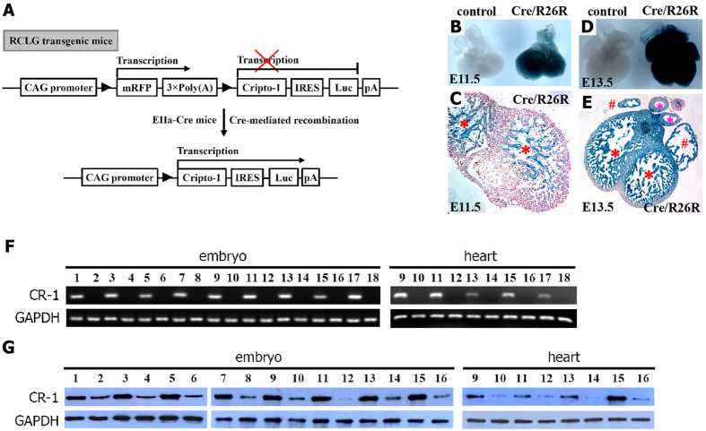
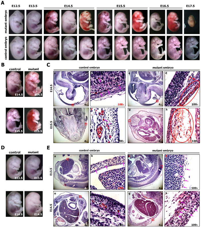
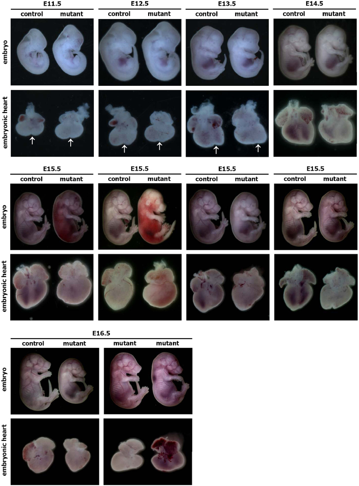
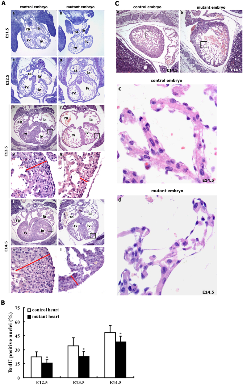
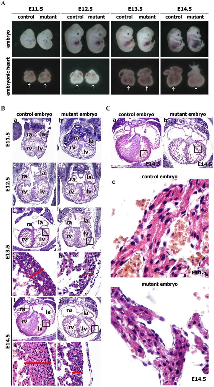
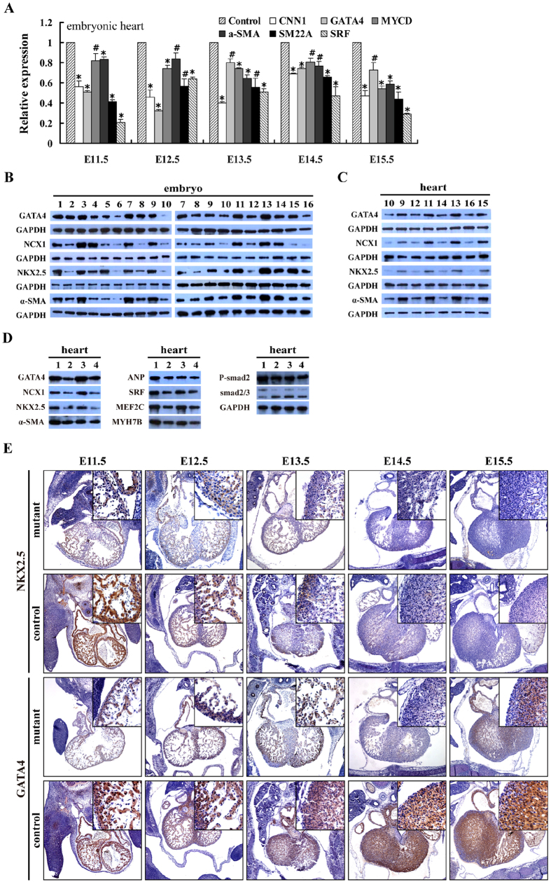
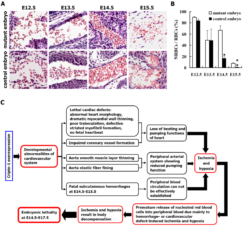
Targeted disruption of Cripto-1 in mice caused embryonic lethality at E7.5, whereas we unexpectedly found that ectopic Cripto-1 expression in mouse embryos also led to embryonic lethality, which prompted us to characterize the causes and mechanisms underlying embryonic death due to ectopic Cripto-1 expression. RCLG/EIIa-Cre embryos displayed complex phenotypes between embryonic day 14.5 (E14.5) and E17.5, including fatal hemorrhages (E14.5-E15.5), embryo resorption (E14.5-E17.5), pale body surface (E14.5-E16.5) and no abnormal appearance (E14.5-E16.5). Macroscopic and histological examination revealed that ectopic expression of Cripto-1 transgene in RCLG/EIIa-Cre embryos resulted in lethal cardiac defects, as evidenced by cardiac malformations, myocardial thinning, failed assembly of striated myofibrils and lack of heartbeat. In addition, Cripto-1 transgene activation beginning after E8.5 also caused the aforementioned lethal cardiac defects in mouse embryos. Furthermore, ectopic Cripto-1 expression in embryonic hearts reduced the expression of cardiac transcription factors, which is at least partially responsible for the aforementioned lethal cardiac defects. Our results suggest that hemorrhages and cardiac abnormalities are two important lethal factors in Cripto-1 transgenic mice. Taken together, these findings are the first to demonstrate that sustained Cripto-1 transgene expression after E11.5 causes fatal hemorrhages and lethal cardiac defects, leading to embryonic death at E14.5-17.5.


Figures

Figure 1
Figure 1. Cripto-1 (CR-1) transgene activation in embryos upon Cre-mediated excision by EIIa-Cre mice.
(A) The strategy for the conditional expression of Cripto-1 and Luc transgenes. Please see the Materials and Methods for additional details on the strategy for the conditional expression of the transgenes. (B–E) Cre-mediated excision efficiency in embryonic hearts. (B,D) Whole-mount blue-stained embryonic hearts. (C,E) Histological sections of E11.5 (C) and E13.5 (E) embryonic hearts. The ventricular surface is indicated with a “*”, the atrial appendage is indicated with a “#”, and arteries are indicated with a “*”. (F) RT-PCR analysis using human-specific primer for human Cripto-1 transgene expression in RCLG/EIIa-Cre transgenic embryos and embryonic hearts. The cropped gels are used in Fig. 1F, and the full-length gel images are available in Supplementary Figure S28. The gels have been run under the same experimental conditions. (G) Western blot for the Cripto-1 protein in RCLG/EIIa-Cre transgenic embryos and their hearts. For (F,G), lane 1: E7.5+RCLG/Cre; lane 2: E7.5+Cre; lane 3: E8.5+RCLG/Cre; lane 4: E8.5+Cre; lane 5: E9.5+RCLG/Cre; lane 6: E9.5+Cre; lane 7: E10.5+RCLG/Cre; lane 8: E10.5+Cre; lane 9: E11.5+RCLG/Cre; lane 10: E11.5+Cre; lane 11: E12.5+RCLG/Cre; lane 12: E12.5+Cre; lane 13: E13.5+RCLG/Cre; lane 14: E13.5+Cre; lane 15: E14.5+RCLG/Cre; lane 16: E14.5+Cre; lane 17: E15.5+RCLG/Cre; lane 18: E15.5+Cre. The cropped blots are used in Fig. 1G, and the quantification of Cripto-1 expression and the full-length gel images are available in Supplementary Figures S6 and S28, respectively. The blots have been run under the same experimental conditions.
Figure 2
Figure 2. The EIIa-Cre-mediated activation of Cripto-1 leads to hemorrhaging and embryonic lethality.
(A) Whole-mount views of representative control (EIIa-Cre) and mutant (RCLG/EIIa-Cre) embryos at E12.5 to E17.5. (B,C) Histological analysis of control (a,b,e,f) and mutant (c,d,g,h) embryos at E14.5 and E15.5. E14.5 and E15.5 mutant embryos (B) demonstrated hemorrhages. Sagittal sections of E14.5 control and mutant embryos (indicated in B) were stained with H&E (a–d), while transverse sections of E15.5 control and mutant embryos (indicated in B) were stained with H&E (e–h). In Fig. 2C(b,d,f,h) are higher magnifications of the red rectangular regions indicated in (a,c,e,g), respectively. The pink arrows indicate normal capillaries (b,f) with erythrocytes in the control embryos. The red ovals in (d,h) outline hemorrhaging of the body surface capillaries of mutant embryos, while the red arrow shows a normal great vessel (d) in a mutant embryo. (D,E) Histological analysis of control (a,b,e,f) and mutant (c,d,g,h) embryos at E13.5 and E14.5. No significant difference was found in the body surfaces of E13.5 control and mutant embryos (D, upper image), whereas the E14.5 mutant embryo appears pale compared to the E14.5 control embryo (D, lower image). Sagittal section of E13.5 and E14.5 control and mutant embryos (indicated in D) were stained with H&E. In Fig. 2E,(b,d,f,h) are higher magnifications of the red boxes indicated in (a,c,e,g), respectively. The pink arrows indicate normal capillaries (b,d,f,h) with or without erythrocytes, which are nucleated in both control and mutant embryos at this stage of development.
Figure 3
Figure 3. Whole-mount views of representative hearts obtained from control (EIIa-Cre) and mutant (RCLG/EIIa-Cre) embryos at E11.5 to E16.5.
The lower images show the hearts of the E11.5 to E16.5 control and mutant embryos shown in the upper images. At E14.5, E15.5 and E16.5, all of the mutant embryos exhibiting hemorrhages, a pale body surface or no abnormal appearance displayed abnormal heart morphology. The arrows indicate the interventricular sulcus.
Figure 4
Figure 4. Cardiac defects in mutant embryos (RCLG/EIIa-Cre).
(A) H&E staining of transverse sections of control and mutant embryos. The upper images show the entire hearts of E13.5 and E14.5 embryo, and the lower images (g,h,k,l) are higher magnifications of the rectangular regions indicated in images (e,f,i,j), respectively. Higher magnification of E13.5 and E14.5 hearts revealed thinning due to a decrease in the myocardial cell layers in mutant embryos. The red lines show the thickness of the myocardial layer of the left ventricle (lv). Abbreviations: ra, right atrium; la, left atrium; rv, right ventricle; lv, left ventricle. (B) The cardiomyocyte proliferation index was calculated by dividing the number of BrdU positive nuclei by the total number of nuclei in the E12.5, E13.5 and E14.5 mutant hearts. Representative BrdU staining of embryonic heart sections at E12.5-E14.5 is indicated in Supplementary Figure S16. (C) The cardiomyocytes in the mutant embryos failed to assemble striated myofibrils. (c,d) are higher magnifications of the regions indicated in (a,b), respectively.
Figure 5
Figure 5. Temporally regulated Cripto-1 activation causes the cardiac defects in mutant embryos (RCLG/hUb-CreERT2).
(A) Whole-mount views of representative hearts obtained from control (RCLG) and mutant (RCLG/hUb-CreERT2) embryos at E11.5 to E14.5. The lower images show the hearts of the E11.5 to E14.5 control and mutant embryos shown in the upper images. The arrows indicate the interventricular sulcus. (B) H&E staining of transverse sections of control and mutant embryos. The upper images show the entire hearts of E13.5 and E14.5 embryo, and the lower images (g,h,k,l) are higher magnifications of the rectangular regions indicated in images (e,f,i,j), respectively. Other details as in Fig. 4. (C) The cardiomyocytes in the mutant embryos failed to assemble striated myofibrils. (c,d) are higher magnifications of the regions indicated in (a,b), respectively.
Figure 6
Figure 6. Mutant embryonic hearts exhibit altered cardiac gene expression.
(A) qRT-PCR for the indicated gene expression in mutant embryonic hearts (RCLG/EIIa-Cre). The cropped gels are used in Fig. 6A, and the full-length gel images are available in Supplementary Figure S28. The gels have been run under the same experimental conditions. (B,C) Western blot for the indicated proteins in E7.5-E14.5 mutant embryos and E11.5-E14.5 hearts (RCLG/EIIa-Cre). GAPDH was used as a control for the Western blot. Lane 1: Cre(E7.5); lane 2: RCLG/Cre(E7.5); lane 3: Cre(E8.5); lane 4: RCLG/Cre(E8.5); lane 5: Cre(E9.5); lane 6: RCLG/Cre(E9.5); lane 7: Cre(E10.5); lane 8: RCLG/Cre(E10.5); lane 9: Cre(E11.5); lane 10: RCLG/Cre(E11.5); lane 11: Cre(E12.5); lane 12: RCLG/Cre(E12.5); lane 13: Cre(E13.5); lane 14: RCLG/Cre (E13.5); lane 15: Cre(E14.5); lane 16: RCLG/Cre(E14.5). The cropped blots are used in Fig. 6B,C, and the full-length gel images are available in Supplementary Figure S28. The blots have been run under the same experimental conditions. (D) Western blot analysis of the indicated proteins in E11.5 and E14.5 mutant embryonic hearts (RCLG/hUb-CreERT2). Lane 1: RCLG (E11.5); lane 2: RCLG/Cre(E11.5); lane 3: RCLG (E14.5); lane 4: RCLG/Cre(E14.5). The cropped blots are used in Fig. 6D, and the full-length gel images are available in Supplementary Figure S28. The blots have been run under the same experimental conditions. (E) Immunohistochemical (IHC) analyses of Nkx2.5 and GATA4 expression in RCLG/EIIa-Cre embryonic hearts. ANF, atrial natriuretic peptide; α-SMA, α-smooth muscle actin; CNN1, calponin 1; MEF2C, Myocyte-specific enhancer factor 2C; MYCD, myocardin; MYH7B, myosin, heavy chain 7B; NCX1, cardiac sodium–calcium exchanger; SM22A, smooth muscle protein 22 alpha; SRF: serum response factor.
Figure 7
Figure 7
(A,B) Mutant embryos (RCLG/EIIa-Cre) exhibit aberrantly elevated levels of primitive erythrocytes at E14.5 and E15.5. (A) H&E staining of paraffin sections of representative E12.5 to E15.5 control and mutant embryos were stained with H&E. These representative sections focused on showing the nucleated red blood cells (NRBCs) in mutant and control embryos between E12.5 and E15.5. (B) The percentage of nucleated red blood cells (NRBCs) in E12.5 to E15.5 embryos. RBCs: red blood cells. (C) The mechanisms underlying embryonic lethality due to constitutive Cripto-1 expression in transgenic mice.

Similar articles

Cited by

References

    1. Beachy P. A., Karhadkar S. S. & Berman D. M. Tissue repair and stem cell renewal in carcinogenesis. Nature 432, 324–331, 10.1038/nature03100 (2004). - DOI - PubMed
    1. Gidekel S., Pizov G., Bergman Y. & Pikarsky E. Oct-3/4 is a dose-dependent oncogenic fate determinant. Cancer Cell 4, 361–370 (2003). - PubMed
    1. Hochedlinger K., Yamada Y., Beard C. & Jaenisch R. Ectopic expression of Oct-4 blocks progenitor-cell differentiation and causes dysplasia in epithelial tissues. Cell 121, 465–477, 10.1016/j.cell.2005.02.018 (2005). - DOI - PubMed
    1. Bianco C. et al. Role of Cripto-1 in stem cell maintenance and malignant progression. Am J Pathol 177, 532–540, 10.2353/ajpath.2010.100102 (2010). - DOI - PMC - PubMed
    1. de Castro N. P., Rangel M. C., Nagaoka T., Salomon D. S. & Bianco C. Cripto-1: an embryonic gene that promotes tumorigenesis. Future Oncol 6, 1127–1142, 10.2217/fon.10.68 (2010). - DOI - PubMed

LinkOut - more resources