. 2016 Sep 30:6:34508.
doi: 10.1038/srep34508.

SMC4, which is essentially involved in lung development, is associated with lung adenocarcinoma progression

Affiliations

SMC4, which is essentially involved in lung development, is associated with lung adenocarcinoma progression

Chengli Zhang et al. Sci Rep. .

Abstract

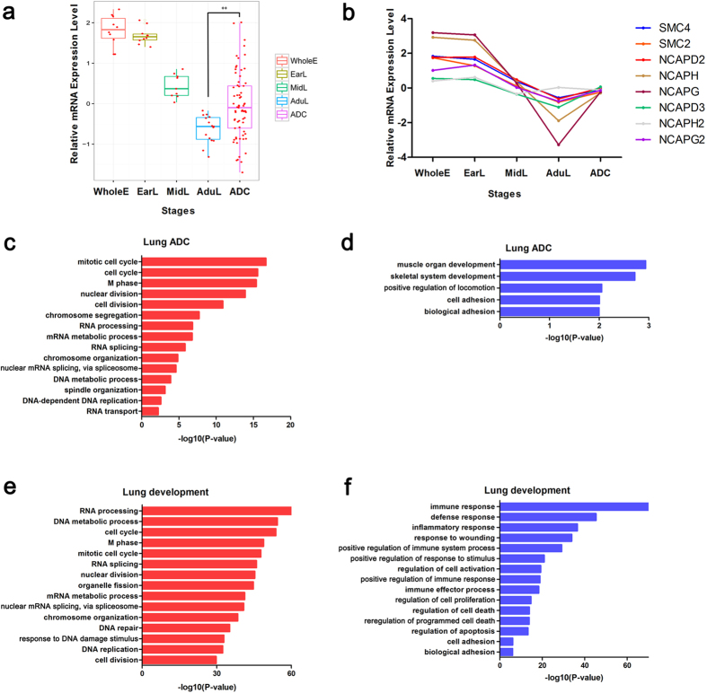
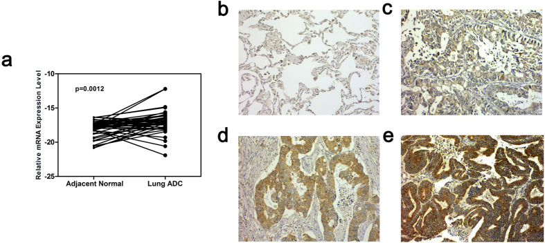
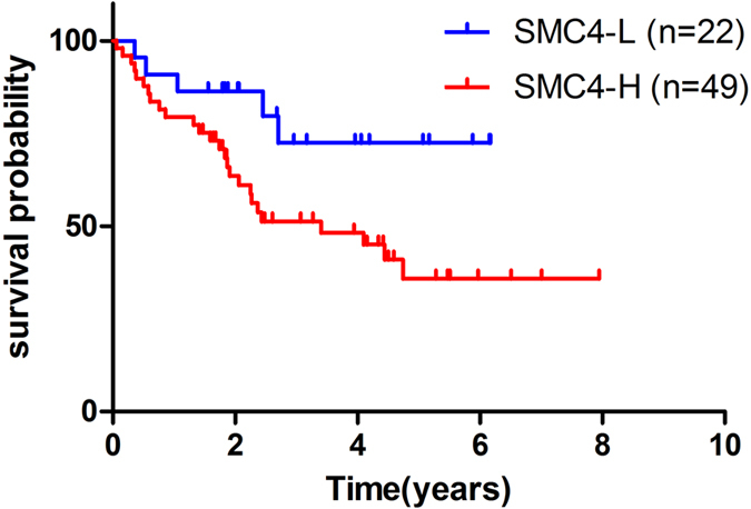
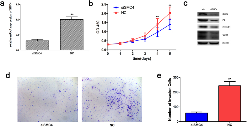
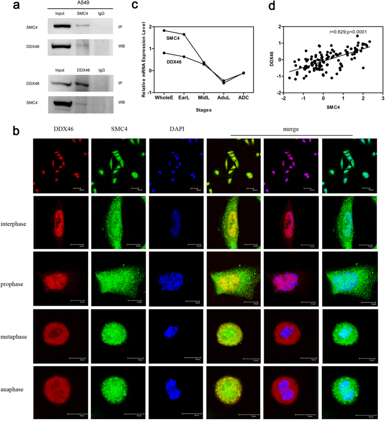
Structural maintenance of chromosome 4 (SMC4) is a core subunit of condensin complexes that mainly contributes to chromosome condensation and segregation. Our previous study demonstrated that the gene expression profile during lung development is of great values for the study of lung cancer. In this study, we identified SMC4 through co-expression network analysis and clique percolation clustering using genes that constant changes during four stages of lung development. Gene ontology and KEGG pathway enrichment analysis demonstrated that SMC4 is closely related to cell cycle, cell adhesion, and RNA processing in lung development and carcinogenesis. Moreover, SMC4 is overexpressed in lung adenocarcinoma tissues and acts as an independent prognostic factor. SMC4 knockdown significantly inhibits the proliferation and invasion of A549 cells. Furthermore, we found that SMC4 interacts with DDX46 (DEAD-box helicase 46). In conclusion, the pivotal role of SMC4 in lung development and carcinogenesis suggests that genes with a similar expression pattern to SMC4 in lung development may also contribute to lung cancer progression. The identification of genes that are essentially involved in development through a comparative study between development and cancer may be a practical strategy for discovering potential biomarkers and illuminating the mechanisms of carcinogenesis.


Figures

Figure 1
Figure 1. Identification of SMC4 through construction of a co-expression network using lung development data.
(a) The co-expression network contained 1288 nodes and 8860 edges. (b) Scattergram of node degrees in the co-expression network. The co-expression network was scale-free. (c) The subnetwork contained 63 hub nodes with the top 5% of degrees. The nodes represent the genes, and the edges indicate the interactions between genes. The genes upregulated during lung development are labeled in red, whereas the downregulated genes are labeled in blue. All of the nodes are marked with node degrees. (d) K-clique community with 18 nodes (k = 16). These genes are closely involved in the cell cycle and chromosome organization.
Figure 2
Figure 2. SMC4 participates in multiple aspects of lung development and tumorigenesis.
(a) Expression levels of SMC4 during four stages of lung development and in lung ADC samples. The bars represent the minimum and maximum normalized signals. Two-tailed unpaired t-test, **p < 0.01. (b) Median expression level of all eight condensin subunits at four stages of lung development and in lung ADC samples. (c,d) Representative GO biological process (BP) terms of the genes positively correlated with SMC4. (e,f) Representative GO BP terms of the genes negatively correlated with SMC4. The −log10 (enrichment P-value) is displayed on the x-axis.
Figure 3
Figure 3. SMC4 is overexpressed in lung ADC tissues compared with adjacent normal lung tissues at both the mRNA and protein levels.
(a) Expression levels of SMC4 mRNA in 43 lung ADC tissues and paired normal lung tissues. The expression levels were determined by real-time PCR, and the y-axis shows the −ΔCt values. 18S ribosomal RNA was used as reference gene. Two-tailed paired t-test, **p < 0.01. (b–e) Representative immunohistochemical staining results for SMC4 protein in (b) adjacent normal lung tissue with negative staining (×200), (c) lung ADC tissue with weak positive staining (×200), (d) lung ADC tissue with moderate positive staining (×200), and (e) lung ADC tissue with strong positive staining (×200).
Figure 4
Figure 4. Association between SMC4 expression and lung ADC prognosis.
71 ADC patients was divided into two groups based on the immunostaining score: 0–1 points represents the SMC4-L group (n = 22, blue line), and 2–3 points represents the SMC4-H group (n = 49, red line). Higher SMC4 protein expression level is a poor prognostic factor for overall survival, as indicated by Kaplan-Meier analysis.
Figure 5
Figure 5. Downregulation of SMC4 inhibits the proliferation and invasion of A549 cells.
The mRNA level (a) and protein level (c) of SMC4 in A549 cells were determined by real-time PCR and Western blot analysis, respectively, 48 h after the cells were transfected with siSMC4 or NC. (b) The CCK-8 assay was used to evaluate the proliferation of A549 cells after transfection. (c) Western blots showed the effect of SMC4 knockdown on mitosis-related genes in tumor cells after 48 h. β-actin was used as a control to ensure equal loading. (d,e) Transwell invasion assay of A549 cells 48 h after transfection with siSMC4 or NC. **P < 0.01. The data are presented as the means ± SEM from three independent experiments.
Figure 6
Figure 6. SMC4-DDX46 protein interactions in vivo.
(a) Nuclear extracts of A459 cells were immunoprecipitated with anti-SMC4 or anti-DDX46 antibodies or control Ig. Co-IP samples were analyzed by Western blotting with the indicated antibodies. (b) Double immunofluorescence staining for endogenous SMC4 (green) and DDX46 (red) in A549 cells. Merged images are also shown. (c) mRNA expression levels of SMC4 and DDX46 during four stages of lung development and in lung ADC samples. The bars represent the median normalized signals. (d) Linear relationship of SMC4 and DDX46 in lung development samples and lung ADC samples at the mRNA expression levels, as determined by Spearman’s correlation coefficient test.

References

    1. Sardenberg R. A., Mello E. S. & Younes R. N. The lung adenocarcinoma guidelines: what to be considered by surgeons. Journal of thoracic disease 6, S561–567, doi: 10.3978/j.issn.2072-1439.2014.08.25 (2014). - DOI - PMC - PubMed
    1. Torre L. A. et al.. Global cancer statistics, 2012. CA: a cancer journal for clinicians 65, 87–108, doi: 10.3322/caac.21262 (2015). - DOI - PubMed
    1. Galvan A. et al.. Gene expression signature of non-involved lung tissue associated with survival in lung adenocarcinoma patients. Carcinogenesis 34, 2767–2773, doi: 10.1093/carcin/bgt294 (2013). - DOI - PubMed
    1. Goldstraw P. et al.. Non-small-cell lung cancer. Lancet (London, England) 378, 1727–1740, doi: 10.1016/s0140-6736(10)62101-0 (2011). - DOI - PubMed
    1. Marin J. J., Briz O., Monte M. J., Blazquez A. G. & Macias R. I. Genetic variants in genes involved in mechanisms of chemoresistance to anticancer drugs. Current cancer drug targets 12, 402–438 (2012). - PubMed

LinkOut - more resources