. 2016 Oct:12:161-169.
doi: 10.1016/j.ebiom.2016.09.020. Epub 2016 Sep 21.

Zika Virus Strains Potentially Display Different Infectious Profiles in Human Neural Cells

Affiliations

Zika Virus Strains Potentially Display Different Infectious Profiles in Human Neural Cells

Yannick Simonin et al. EBioMedicine. 2016 Oct.

Abstract

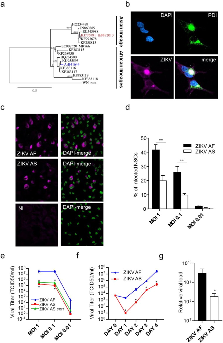
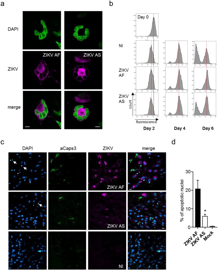
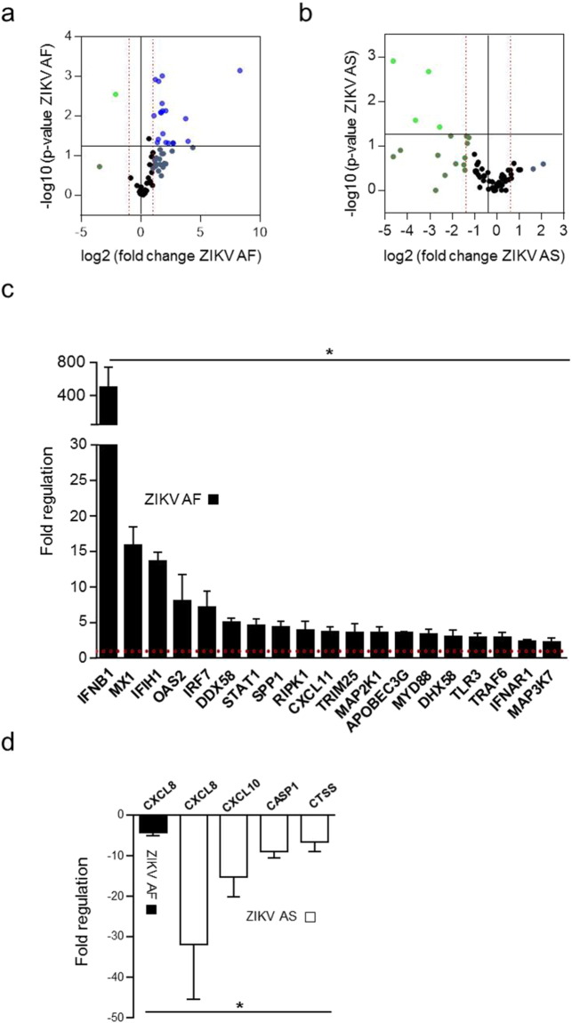
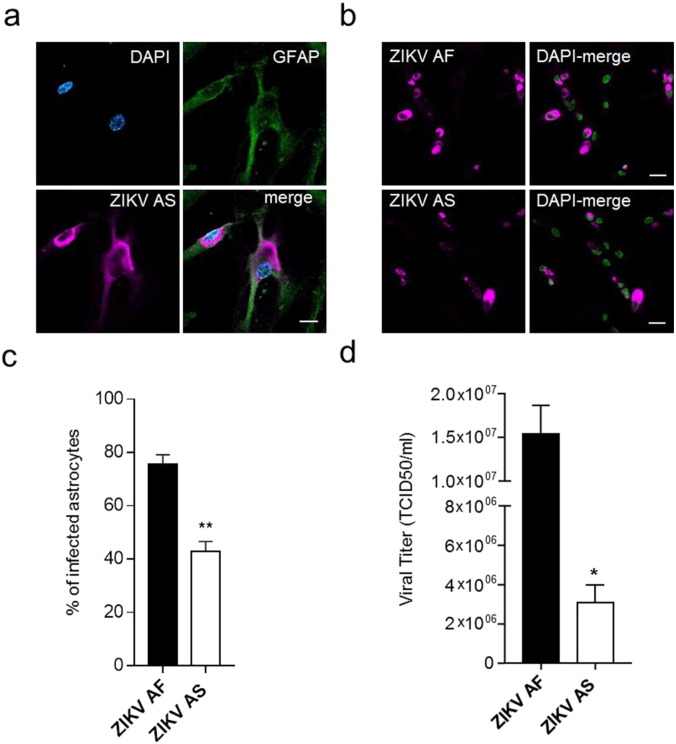
The recent Zika virus (ZIKV) epidemic has highlighted the poor knowledge on its physiopathology. Recent studies showed that ZIKV of the Asian lineage, responsible for this international outbreak, causes neuropathology in vitro and in vivo. However, two African lineages exist and the virus is currently found circulating in Africa. The original African strain was also suggested to be neurovirulent but its laboratory usage has been criticized due to its multiple passages. In this study, we compared the French Polynesian (Asian) ZIKV strain to an African strain isolated in Central African Republic and show a difference in infectivity and cellular response between both strains in human neural stem cells and astrocytes. Consistently, this African strain led to a higher infection rate and viral production, as well as stronger cell death and anti-viral response. Our results highlight the need to better characterize the physiopathology and predict neurological impairment associated with African ZIKV.

Keywords: Astrocytes; Lineages; Neural stem cells; Zika virus.


Figures

Fig. 1
Fig. 1
Differential infectivity of ZIKV AF and AS in human IPSc-derived NSCs. (a) Phylogenetic analysis of ZIKV strains used in this study was performed through a consensus flavivirus RT-PCR fragment that targeted a conserved NS5 gene region. Bootstrap values are indicated at main nodes. (b) Indirect immunofluorescence (IF) of ZIKV AS-infected NSCs (MOI 1, day 4 post-infection) where the nucleus is stained using DAPI (cyan), the endoplasmic reticulum is stained using an anti-PDI (green) and the virus is labeled using a pan-flavivirus antibody (magenta). Scale bar = 10 μm. (c) IF of non-infected (NI), ZIKV AF- and ZIKV AS-infected NSCs (MOI 1, day 4 post-infection). The virus is labeled using a pan-flavivirus antibody (magenta) and nuclei by DAPI (false colored in green). Scale bar = 10 μm. (d) Quantitative analyses show differences in rate of infection between ZIKV AF and ZIKV AS that is consistently seen using different MOIs. 4 independent experiments were quantified with 200–600 cells per conditions. Results are expressed as means ± SEM and analyzed using an unpaired t-test **p < 0.01. (e) Viral titers were determined by TCID50 on Vero cells. Supernatants from NSCs infected at different MOIs and collected day 4 post-infection show differential titers between ZIKV AF and AS, even when % of infected cells were taken in account (green curve, ZIKV AS corrected, titer normalized assuming same infection rate than ZIKV AF (i.e. 41%). Results are expressed as means ± SEM, for three independent experiments and analyzed using Mann-Whitney test *p < 0.05. (f) Kinetics of viral production show early dichotomy in viral replication between ZIKV AF and ZIKV AS. Day 0 corresponds to the input inoculum. Results are expressed as means ± SEM, for three independent experiments and analyzed using Mann-Whitney test *p < 0.05. (g) Quantitative RT-PCR done on extracts of supernatants from NSCs infected at MOI 1 by the different ZIKV strains and collected at day 4 post-infection shows also a difference in the relative physical viral load between ZIKV AF and AS. CTs from amplifying curves were arbitrarily converted using internal control (see Material and Methods section). Results of three independent experiments are expressed as means of relative charges ± SEM and analyzed using Mann-Whitney test. *p < 0.05.
Fig. 2
Fig. 2
Effect of ZIKV AF and AS on NSC cell cycle and survival. (a) Nuclear fragmentation or endoreplication in ZIKV-infected cells. IF of ZIKV-infected NSCs (ZIKV AF or AS MOI 1 day 4 post-infection). The nucleus is labeled with DAPI (false colored in green) and viruses with a pan-flavivirus antibody (magenta). Scale bar = 5 μm. (b) Flow cytometry analyses of NSCs incubated with CFSE at day 0 and infected with ZIKV AF and AS for 2 to 6 days. The shift in fluorescence is consistent with cell division. (c) IF of non-infected (NI), ZIKV AF and ZIKV AS-infected NSCs (MOI 1, day 4 post-infection) show the presence of apoptotic cells in infected cells (white arrows show condensed nuclei and anti-activated caspase 3 is in green). Scale bar = 10 μm. (d) Apoptotic nuclei were quantified in ZIKV AF and AS-infected NSCs at day 4 post-infection. Three independent experiments were quantified with 200–400 nuclei per condition. Results are expressed as means ± SEM and analyzed using an unpaired t-test *p < 0.05.
Fig. 3
Fig. 3
Anti-viral response in NSCs infected by ZIKV AF or AS. (a) and (b) Volcano plots of genes modulated upon ZIKV infection in NSCs. mRNA from supernatants of NSCs infected with ZIKV AF (a) or ZIKV AS (b) at MOI 1 for 4 days were subjected to qRT-PCR analyses. The experiment was performed in triplicates and each point represents the mean. Statistically significant changes in fold regulation appear in the top-right cadran (genes upregulated) and top-left cadran (genes downregulated). (c) and (d) Fold regulation of statistically significant genes modulated (upregulated (c) or downregulated (d)) upon ZIKV AF and AS-infection shown in (a) and (b). Results are expressed as means ± SD and analyzed using an unpaired t-test *p < 0.05.
Fig. 4
Fig. 4
Human astrocytes are permissive to ZIKV infection. (a) IF of ZIKV AS-infected astrocytes (MOI 1, day 4 post-infection). The astrocytic marker GFAP is detected using a specific antibody (green) and the virus is labeled with a pan-flavivirus antibody (magenta) and nuclei by DAPI (cyan). Scale bar = 10 μm. (b) IF of ZIKV AF and ZIKV AS-infected astrocytes (MOI 0.1, day 4 post-infection). The virus is labeled using a pan-flavivirus antibody (magenta) and nuclei by DAPI (false colored in green). Scale bar = 20 μm. (c) Quantitative analyses show differences in rate of infection between ZIKV AF and ZIKV AS (MOI 0.1, day 4 post-infection). Three independent experiments were quantified with 150–200 cells per condition. Results are expressed as means ± SEM and analyzed using an unpaired t-test **p < 0.01. (d) Viral titers from supernatant of ZIKV AF and AS-infected astrocytes (MOI 0.1, day 4 post-infection) were determined by TCID50 on Vero cells. Three independent experiments were quantified. Results are expressed as means ± SEM and analyzed using an unpaired t-test *p < 0.05.

References

    1. Aliota M.T. Characterization of lethal Zika virus infection in AG129 mice. PLoS Negl. Trop. Dis. 2016;10(4) - PMC - PubMed
    1. Baraka V., Kweka E.J. The threat of Zika virus in Sub-Saharan Africa — the need to remain vigilant. Front. Public Health. 2016;4(May):1–3. - PMC - PubMed
    1. Bayer A. Type III interferons produced by human placental trophoblasts confer protection against Zika virus infection. Cell Host Microbe. 2016:1–8. - PMC - PubMed
    1. Beasley D.W.C. Mouse neuroinvasive phenotype of West Nile virus strains varies depending upon virus genotype. Virology. 2002;296(1):17–23. - PubMed
    1. Beasley D.W.C. Envelope protein glycosylation status influences mouse neuroinvasion phenotype of genetic lineage 1 West Nile virus strains. J. Virol. 2005;79(13):8339–8347. - PMC - PubMed