. 2016 Oct:12:55-67.
doi: 10.1016/j.ebiom.2016.07.013. Epub 2016 Jul 14.

The miR-367-3p Increases Sorafenib Chemotherapy Efficacy to Suppress Hepatocellular Carcinoma Metastasis through Altering the Androgen Receptor Signals

Affiliations

The miR-367-3p Increases Sorafenib Chemotherapy Efficacy to Suppress Hepatocellular Carcinoma Metastasis through Altering the Androgen Receptor Signals

Junjie Xu et al. EBioMedicine. 2016 Oct.

Abstract

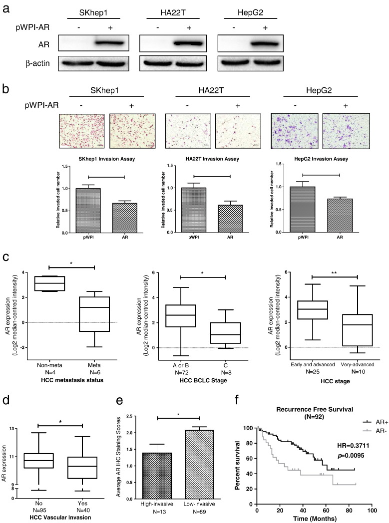
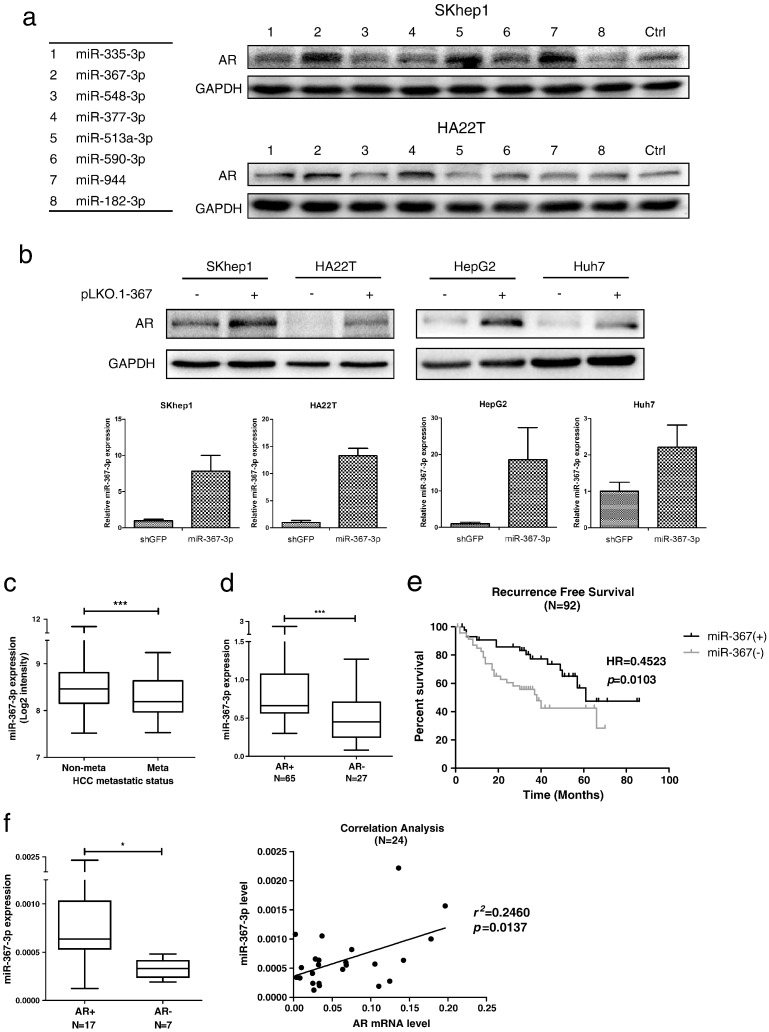
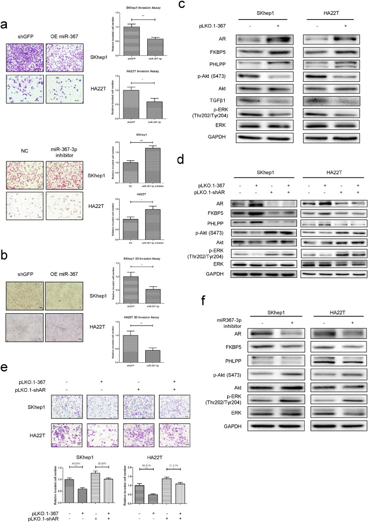
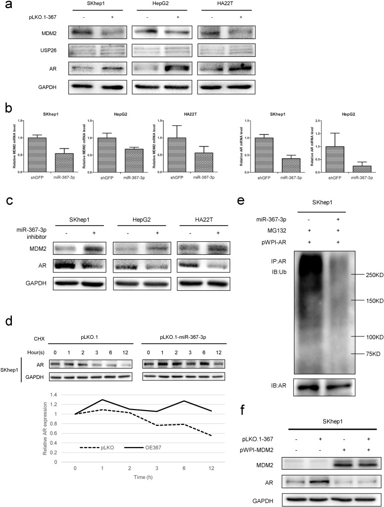
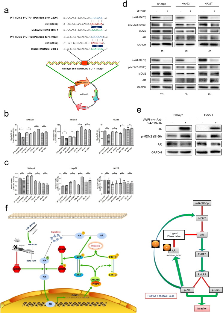
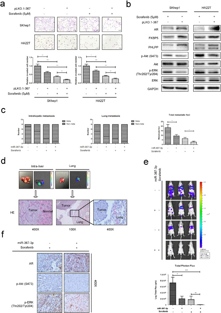
The androgen receptor (AR) was found to suppress hepatocellular carcinoma (HCC) metastasis at late stages. Due to this discovery, we searched for some AR enhancers to increase the efficacy of Sorafenib chemotherapy, and identified the microRNA (miR)-367-3p, whose expression is positively correlated with AR expression in advanced HCC, as an HCC metastasis suppressor. Combining miR-367-3p with Sorafenib showed better efficacy to suppress HCC cell invasion in vitro and in vivo. Mechanism dissection revealed that miR-367-3p could increase AR expression via directly targeting the 3'UTR of MDM2 to decrease MDM2 protein expression. The resultant increase of AR expression might then promote the expression of FKBP5 and PHLPP, thus dephosphorylating and inactivating AKT and ERK, to suppress the HCC cell invasion. Interestingly, the suppression of pAKT by miR-367-3p could subsequently attenuate the phosphorylation of AR and MDM2, giving rise to additional enhancement of AR protein expression, effectively forming a positive feedback loop. Together, these results suggest that miR-367-3p may function as an AR enhancer to increase Sorafenib chemotherapy efficacy via altering the MDM2/AR/FKBP5/PHLPP/(pAKT and pERK) signals to better suppress HCC metastasis. Successful development of this newly combined chemotherapy in the future may help us to better suppress the HCC metastasis at late stages.

Keywords: Androgen receptor; Hepatocellular carcinoma; Metastasis; miR-367-3p.


Figures

Fig. 1
Fig. 1
AR suppresses HCC cells invasion. (a), verification of AR over-expression by western blot assays; (b), chamber-transwell invasion assays showed that over-expression of AR decreased the cell invasion in all 3 HCC cell lines (SKhep1, HA22T, and HepG2). Upper panel, representative images of the chamber-transwell invasion assays; Lower panel, quantification of the invaded cells. The invaded cells were counted in 10 randomly chosen microscopic fields (100 ×) of each experiment and pooled. Each sample was run in triplicate and multiple experiments were performed. (c-d), 4 individual research databases clearly showed that AR levels were lower in advanced stages compared with early stages, implicating that AR played a vital role in late-stage HCC and was negative correlated with HCC metastasis. Data were extracted from Oncomine® Platform (c) and Gene Expression Omnibus Datasets (d). (e), the average AR IHC staining scores of patients were evaluated by experienced pathologists both in high-invasive group (N = 13) and low-invasive group (N = 89). Lower AR IHC staining scores were found in high-invasive group as compared to those found in low-invasive group. (f), recurrence-free survival curve of HCC patients who received surgery (N = 92) indicated that patients with HCC (AR+) (defined by IHC staining) had significant higher recurrence-free survival (HR = 0.3711) than patients with HCC (AR-). p < 0.05 was considered statistically significant. * p < 0.05 and ** p < 0.01.
Fig. 2
Fig. 2
The miR-367-3p increases AR protein levels and is expressed at lower levels in late-stage HCC. (a), western blot assays were applied to check the AR protein levels after over-expressing the 8 miRNA candidates according to the screening strategy in SKhep1 and HA22T cell lines. (b), upper panel: AR levels in 4 different HCC cell lines (SKhep1, HA22T, HepG2, and Huh7) with overexpressed miR-367-3p were checked by western blot assays and were found consistently and significantly increased; lower panel: verification of miR-367-3p over-expression by qRT-PCR. (c), miR-367-3p was expressed at lower levels in metastatic patients than non-metastatic patients according to the non-coding RNA microarray data submitted on public Gene Expression Omnibus Datasets. (d), paraffin-fixed clinical sample surveys disclosed a significantly higher miR-367-3p expression in patients with HCC (AR+, N = 65) than in those with HCC (AR-, N = 27). HCC (AR+) and HCC (AR-) were determined by IHC staining. (e), recurrence-free survival curve of HCC patients who received surgery (N = 92) indicated that patients with higher miR-367-3p expression had significantly higher recurrence-free survival (HR = 0.4523) than patients with lower miR-367-3p expression. (f), left panel: freshly frozen clinical samples showed a significantly higher miR-367-3p expression in patients with HCC (AR+, N = 17) than in those with HCC (AR-, N = 7). HCC (AR+) and HCC (AR-) were determined by IHC staining; right panel: correlation analysis of miR-367-3p and AR mRNA levels from 24 HCC samples by qRT-PCR indicated a significant positive correlation between miR-367-3p and AR mRNA levels. p < 0.05 was considered statistically significant. * p < 0.05 and *** p < 0.001.
Fig. 3
Fig. 3
The miR-367-3p suppresses HCC cell invasion via altering the AR/FKBP5/PHLPP/(pAKT and pERK) signals pathway. (a), chamber-transwell invasion assays on SKhep1 and HA22T cell lines showed that over-expression of miR-367-3p could significantly decrease, while miR-367-3p inhibitor could significantly increase the invasion ability. The invaded cells were counted in 10 randomly chosen microscopic fields (100 ×) of each experiment and pooled for quantification. (b), 3D-invasion assays on SKhep1 and HA22T cell lines also showed that over-expression of miR-367-3p could significantly decrease the invasion ability. The cells with protrusions were regarded as invaded cells and 10 random different fields at 200 × magnification were counted for quantification; (c), we used western blot assays to test downstream altered molecules upon over-expressing miR-367-3p in SKhep1 and HA22T cells. (d, e), rescue assays by knocking down AR showed partially reversed FKBP5-PHLPP-(pAKT/pERK) levels (by western blot assays) and cell invasion ability (by chamber-transwell invasion assays). (f), we used western blot assays to test downstream altered molecules upon transfecting miR-367-3p inhibitor (50 nM) in SKhep1 and HA22T cells.
Fig. 4
Fig. 4
The miR-367-3p mediates AR protein stability and ubiquitination via targeting MDM2. (a), we used western blot assays to test MDM2, USP26 and AR protein levels upon over-expressing miR-367-3p and found MDM2 was consistently decreased in these 3 HCC cell lines (SKhep1, HepG2 and HA22T), whereas USP26 increased slightly after over-expression of miR-367-3p. (b), left panels, transcript levels of MDM2 were consistently decreased after 7 days exogenous expression of miR-367-3p. Right panels, AR mRNA levels decreased after over-expression of miR-367-3p. MDM2 and AR mRNA levels were determined by qRT-PCR. (c), miR-367-3p (50 nM) inhibitor could significantly increase MDM2 and decrease AR at protein levels as shown by western blot assays. (d), the expression of miR-367-3p could increase AR protein stability as determined by western blot assays after cycloheximide (CHX) treatment for 0, 1, 2, 3, 6, and 12 h. (e), in AR over-expressed SKhep1 cells, the expression of miR-367-3p could decrease AR ubiquitination as determined by immunoprecipitation (AR) and western blot (ubiquitin) after 3 h 20 μM MG132 treatment. (f), interruption approach via over-expressing MDM2 revealed that increased MDM2 could significantly decrease AR expression and could partially abolish miR-367-3p-increased AR protein expression.
Fig. 5
Fig. 5
The miR-367-3p directly targets the 3′UTR of MDM2 to mediate AR levels and subsequently generates a positive feedback loop. (a), MDM2 3′UTR containing wild type (WT) or mutant (M) miRNA-response elements were cloned into the psiCheck2 construct downstream of the Renilla luciferase ORF, (b), co-transfection of MDM2 3′UTR constructs containing WT seed regions with pLKO.1-miR-367-3p decreased the luciferase activity and with miR-367-3p inhibitor increased the luciferase activity, whereas the reporter with a M seed region in MDM2 3′UTR did not, in the first predicted miR-367-3p seed regions. (c), co-transfection of MDM2 3′UTR constructs containing WT seed regions with pLKO.1-miR-367-3p or with miR-367-3p inhibitor (50 nM) could not alter the luciferase activity, neither did the reporter with a M seed region in MDM2 3′UTR, in the second predicted miR-367-3p seed regions. (d), western blot assays showed that MK2206 (1 μM in SKhep1 and HA22T cells, 2.5 μM in HepG2 cells) suppressed MDM2 phosphorylation and increased the AR protein expression at various time points. (e), over-expressing pWPI-myr-Akt-Δ4-129-HA in HCC cells showed significant increases of pMDM2 (S166) and decreases of AR. (f), the schematic depiction showing the suppression of pAkt by miR-367-3p could subsequently attenuate the phosphorylation of AR and MDM2, giving rise to additionally increased AR protein levels, which formed a positive feed-back circuitry. p < 0.05 was considered statistically significant. * p < 0.05, ** p < 0.01, and *** p < 0.001.
Fig. 6
Fig. 6
Sorafenib and miR-367-3p serve as a better combination therapy to suppress HCC metastasis in vitro and in vivo. (a), chamber-transwell invasion assays showed the combination of Sorafenib and miR-367-3p exhibited better efficacy to suppress SKhep1 and HA22T cells invasion compared to Sorafenib or miR-367-3p alone. The invaded cells were counted in 10 randomly chosen microscopic fields (100 ×) of each experiment and pooled for quantification. (b), western blot assays showed that under moderate Sorafenib chemotherapy, miR-367-3p could significantly increase AR, FKBP5 and PHLPP expressions, therefore inhibit p-Akt (S473) and p-ERK (Thr202/Tyr204) in SKhep1 and HA22T cells. (c), left and middle panel, IVIS imaging in mice was used to determine the intrahepatic metastasis and lung metastasis, and lower intra-hepatic and lung metastasis occurrence in miR-367-3p-expressing group than control group with or without Sorafenib treatment were found. Right panel, total metastatic foci were counted after sacrifice, and less total metastatic foci in miR-367-3p-expressing group than control group with or without Sorafenib treatment were found. (d), representative bioluminescent images of intrahepatic metastasis and lung metastasis (upper panel). HE staining confirmed the tumor tissue in liver (left lower panel) and lung (right and middle lower panels). (e), representative bioluminescent images in different groups after 2 months of orthotopic HCC xenograft (upper panel), and the total photon flux were measured and analyzed using Living Image® software (PerkinElmer, lower panel), showing less total tumor burden in miR-367-3p-expressing group than control group with or without Sorafenib treatment. (f), representative images of IHC staining for AR, p-Akt (S473) and p-ERK (Thr202/Tyr204) in different treatment groups. p < 0.05 was considered statistically significant. * p < 0.05, ** p < 0.01, and *** p < 0.001.

References

    1. Alazawi W., Cunningham M., Dearden J., Foster G.R. Systematic review: outcome of compensated cirrhosis due to chronic hepatitis C infection. Aliment. Pharmacol. Ther. 2010;32:344–355. - PubMed
    1. An J., Wang C., Deng Y., Yu L., Huang H. Destruction of full-length androgen receptor by wild-type SPOP, but not prostate-cancer-associated mutants. Cell Rep. 2014;6:657–669. - PMC - PubMed
    1. Asham E.H., Kaseb A., Ghobrial R.M. Management of hepatocellular carcinoma. Surg. Clin. North Am. 2013;93:1423–1450. - PubMed
    1. Bai S., Nasser M.W., Wang B., Hsu S.H., Datta J., Kutay H., Yadav A., Nuovo G., Kumar P., Ghoshal K. MicroRNA-122 inhibits tumorigenic properties of hepatocellular carcinoma cells and sensitizes these cells to sorafenib. J. Biol. Chem. 2009;284:32015–32027. - PMC - PubMed
    1. Berk V., Kaplan M.A., Tonyali O., Buyukberber S., Balakan O., Ozkan M., Demirci U., Ozturk T., Bilici A., Tastekin D., Ozdemir N., Unal O.U., Oflazoglu U., Turkmen E., Erdogan B., Uyeturk U., Oksuzoglu B., Cinkir H.Y., Yasar N., Gumus M. Efficiency and side effects of sorafenib therapy for advanced hepatocellular carcinoma: a retrospective study by the anatolian society of medical oncology. Asian Pac. J. Cancer Prev. 2013;14:7367–7369. - PubMed

MeSH terms