

Individualized parcellation of the subthalamic nucleus in patients with Parkinson's disease with 7T MRI
- PMID: 27688203
- PMCID: PMC5479742
- DOI: 10.1016/j.neuroimage.2016.09.023
Individualized parcellation of the subthalamic nucleus in patients with Parkinson's disease with 7T MRI
Abstract
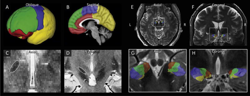
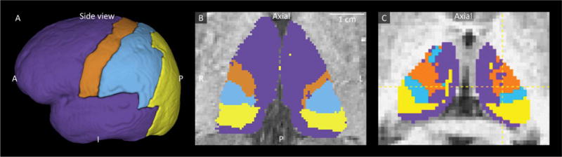
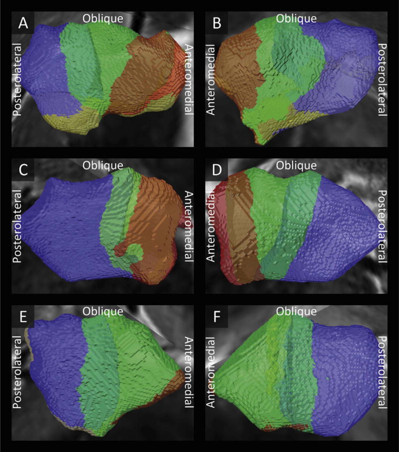
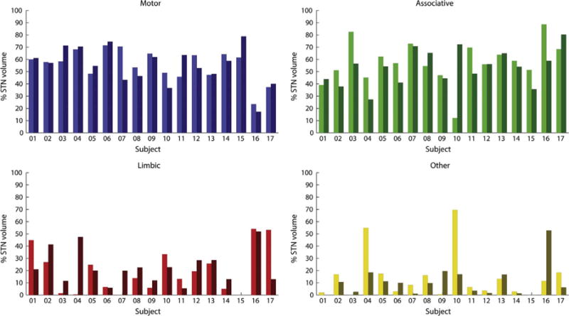
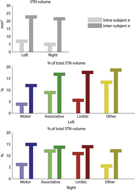
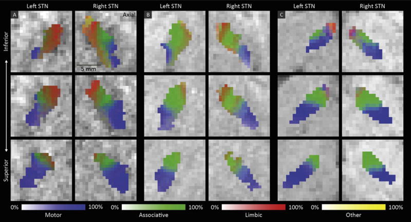
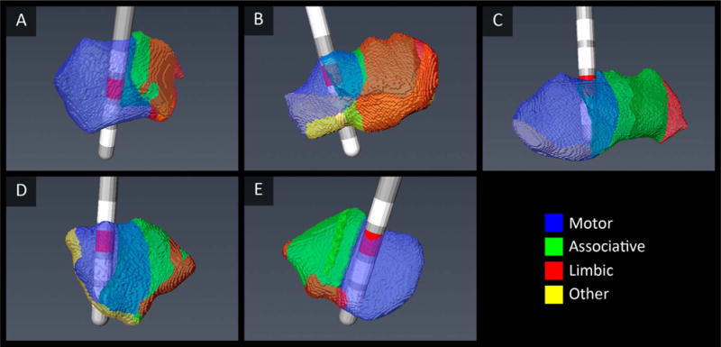
Deep brain stimulation of the subthalamic nucleus (STN) is a widely performed surgical treatment for patients with Parkinson's disease. The goal of the surgery is to place an electrode centered in the motor region of the STN while lowering the effects of electrical stimulation on the non-motor regions. However, distinguishing the motor region from the neighboring associative and limbic areas in individual patients using imaging modalities was until recently difficult to obtain in vivo. Here, using ultra-high field MR imaging, we have performed a dissection of the subdivisions of the STN of individual Parkinson's disease patients. We have acquired 7T diffusion-weighted images of seventeen patients with Parkinson's disease scheduled for deep brain stimulation surgery. Using a structural connectivity-based parcellation protocol, the STN's connections to the motor, limbic, and associative cortical areas were used to map the individual subdivisions of the nucleus. A reproducible patient-specific parcellation of the STN into a posterolateral motor and gradually overlapping central associative area was found in all STNs, taking up on average 55.3% and 55.6% of the total nucleus volume. The limbic area was found in the anteromedial part of the nucleus. Our results suggest that 7T MR imaging may facilitate individualized and highly specific planning of deep brain stimulation surgery of the STN.
Keywords: Deep brain stimulation; Parcellation; Parkinson's disease; Patient-specific; Subthalamic nucleus; Ultra-high field MRI.
Copyright © 2016 Elsevier Inc. All rights reserved.
Figures
References
-
- Alkemade A, Forstmann BU. Do we need to revise the tripartite subdivision hypothesis of the human subthalamic nucleus (STN)? Neuroimage. 2014;95:326–329. - PubMed
-
- Andersson JL, Skare S, Ashburner J. How to correct susceptibility distortions in spin-echo echo-planar images: application to diffusion tensor imaging. Neuroimage. 2003;20:870–888. - PubMed
-
- Andersson JLR, Jenkinson M, Smith S. Non-Linear Registration Aka Spatial Normalisation FMRIB Centre. Oxford; United Kingdom: 2007.
-
- Avecillas-Chasin JM, Alonso-Frech F, Parras O, Del Prado N, Barcia JA. Assessment of a method to determine deep brain stimulation targets using deterministic tractography in a navigation system. Neurosurg Rev. 2015;38:739–751. - PubMed
Publication types
MeSH terms
Grants and funding
LinkOut - more resources
Full Text Sources
Other Literature Sources
Medical
