. 2015 Oct-Dec;19(4):344-51.

Peripheral blood monocytes can differentiate into efficient insulin-producing cells in vitro

Affiliations

Peripheral blood monocytes can differentiate into efficient insulin-producing cells in vitro

A Kyventidis et al. Hippokratia. 2015 Oct-Dec.

Abstract

Background: Recent studies provide evidence that peripheral blood monocytes have the ability to differentiate into mesenchymal-like cells. The ability of cultured monocytes to differentiate and produce insulin in vitro is analysed in the present study.

Methods: Peripheral blood monocytes were isolated from healthy donors and cultivated for fourteen days. Growth factors and liraglutide were used to induce pancreatic differentiation in most of the cultures. The growth factors were: monocyte colony-stimulating factor, interleukin-3, hepatocyte growth factor and epidermal growth factor. The rest of the cultures were cultivated only with nutrient medium and human serum. Insulin levels were measured by an enzyme-linked immunosorbent assay. Cellular morphology was observed using optical and electron microscopy. Cell membrane receptors were detected by flow cytometry.

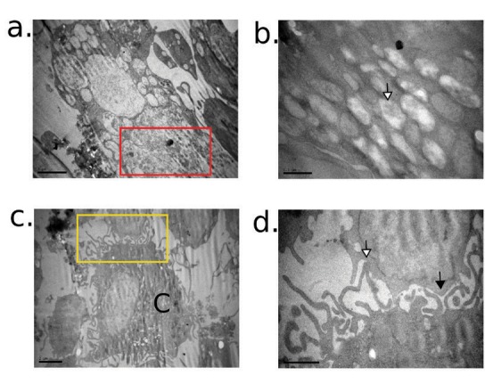
Results: Monocytes were able to synthesize and excrete high levels of insulin after seven days in culture. A further increase in the excretion of insulin was observed after fourteen days. Cells were also able to differentiate and synthesize insulin, even if no growth factors were added to the culture medium. Some of the cultures were able to excrete insulin in a glucose-dependent manner. Differentiated monocytes were connected to neighbouring cells with axons and resembled the morphology of mesenchymal, dendritic and myeloid-progenitor cells. Cells retained their mature receptors and simultaneously developed immature receptors on their membrane.

Conclusions: Monocytes can acquire morphological properties of multipotent cells when they are cultivated under specific conditions in vitro. Differentiated monocytes are able to synthesize and excrete insulin. Hippokratia 2015; 19 (4): 344-351.

Keywords: GLP-1; Monocytes; diabetes; insulin producing cells; liraglutide; mononuclear cells; pancreatic beta cell differentiation.


Figures

Figure 1
Figure 1. Diagrammatic presentation of the cell cultures depending on culture time and glucose concentration. Peripheral blood mononuclear cells from the same donor were divided into four aliquots and cultured in wells. Cells were tested for insulin production after: their isolation from the peripheral blood (Group 1), seven days in culture in the presence of growth factors (Group 2), 14 days in culture - growth factors present in the culture medium (Group 3), and 14 days in culture - growth factors absent in the culture medium (Group 4). The same cultures were tested for insulin excretion either at glucose concentration of 100 mg/dl (A) or 250 mg/dl (B) in the supernatant.
Figure 2
Figure 2. Morphology of the isolated monocytes and lymphocytes on the first day. The black arrows mark one characteristic cell in each picture. a) After isolation, monocytes and lymphocytes were small round cells. b) Afterwards, cells gradually changed shape and developed ellipses. This is the first step to the making of axons. At the same time, they started to attach to the bottom of the well. c) Some cells had larger and darker nucleus and cytoplasm from the first day of the culture. d) Also there were some platelets in the culture, despite the ficoll centrifugation. Some cells were destroyed during the centrifugation process and the cellular debris are evident in the culture well. Platelets and debris reduced each time the supernatant was replaced. Cells were not stained, magnification: 20x.
Figure 3
Figure 3. Morphological changes of the cultivated mononuclear cells observed by optical microscopy. Monocytes constantly changed their morphology and their size during the whole culture period. Lymphocytes remained unchanged. The white arrows show monocytes which enlarged on day seven and they further enlarged on day 14. Their morphology resembled the morphology of myeloid-progenitor cells. Both the nucleus and the cytoplasm became much bigger in comparison with Figure 2. Their morphology resembled the morphology of myeloidprogenitor cells. The black arrows show some cells which exhibited the morphology of dendritic cells on day 14. The red arrows mark cells with morphology of mesenchymal cells. The blue arrow shows a cell which resembled the morphology of myeloblast. The yellow arrows mark the lymphocytes which remained morphologically unchanged. The density of the cells varied on day seven (a), (c), but it became similar on day 14 (b), (d). Cells in pictures (a), (b) (c), (d) were cultured in the presence of the growth factors and cells in pictures (e), (f) were cultured in the absence of the growth factors. Cells were not stained, magnification: 20x.
Figure 4
Figure 4. Optical microscopy images of cell cultures stained by May-Grünwald-Giemsa. When the cells were stained, nuclei enlargement and cellular connections (black arrows) became more obvious. Cellular axons are thinner on day seven in comparison with day 14. The black arrows on day 14 mark the developed cellular axons. The morphology of axon cells resembles the dendritic cells. The white arrows show cells with progenitor morphology. The red arrows show cells with mesenchymal morphology. Cells in pictures (a) and (b) are grown in the presence of growth factors and in (c) and (d) in the absence of them. Magnification: 20x.
Figure 5
Figure 5. Optical microscopy images of cell cultures stained by May-Grünwald-Giemsa on the 14th day. Cells connect to each other with membrane-membrane junctions (red arrow). Some of them develop axons (black arrow). A new type of connection is revealed in this magnification. Cells attach firmly to the bottom of the well, spread their cytoplasmic structures and fuse them together (white arrows). Also cells with two nuclei are revealed (green arrows). Magnification: 40x.
Figure 6
Figure 6. Electron microscopy images of differentiated monocytes after 14 days in culture in the presence of growth factors. a) Differentiated monocytes contain many storing granules (red rectangle). Magnification 10.000x. b) Granules are filled with proteins (white arrow). Magnification 40.000x. c) Monocytic origin cell (C) extends large axons. Magnification 8.000x. d) The yellow rectangle of (c) is magnified. The central cell (C) connects to neighboring cells with gap junctions (black arrow) and tight junctions (white arrow). Magnification 25.000x.

References

    1. Fernandez Pujol B, Lucibello FC, Gehling UM, Lindemann K, Weidner N, Zuzarte ML, et al. Endothelial-like cells derived from human CD14 positive monocytes. Differentiation. 2000;65:287–300. - PubMed
    1. Schmeisser A, Garlichs CD, Zhang H, Escafi S, Graffy C, Ludwiq J, et al. Monocytes coexpress endothelial and macrophagocytic lineage markers and form cord-like structures in Matrigel under angiogenic conditions. Cardiovasc Res. 2001;49:671–680. - PubMed
    1. Heinemann DE, Siggelkow H, Ponce LM, Viereck V, Wiese KG, Peters JH. Alkaline phosphatase expression during monocyte differentiation. Overlapping markers as a link between monocytic cells, dendritic cells, osteoclasts and osteoblasts. Immunobiology. 2000;202:68–81. - PubMed
    1. Tkachenko N, Wojas K, Tabarkiewicz J, Rolinski J. Generation of dendritic cells from human peripheral blood monocytes--comparison of different culture media. Folia Histochem Cytobiol. 2005;43:25–30. - PubMed
    1. Kodama H, Inoue T, Watanabe R, Yasutomi D, Kawakami Y, Oqawa S, et al. Neurogenic potential of progenitors derived from human circulating CD14+ monocytes. Immunol Cell Biol. 2006;84:209–217. - PubMed

LinkOut - more resources