. 2016 Oct 18;113(42):E6496-E6505.
doi: 10.1073/pnas.1607496113. Epub 2016 Sep 29.

Oligodendrocytes contribute to motor neuron death in ALS via SOD1-dependent mechanism

Affiliations

Oligodendrocytes contribute to motor neuron death in ALS via SOD1-dependent mechanism

Laura Ferraiuolo et al. Proc Natl Acad Sci U S A. .

Abstract

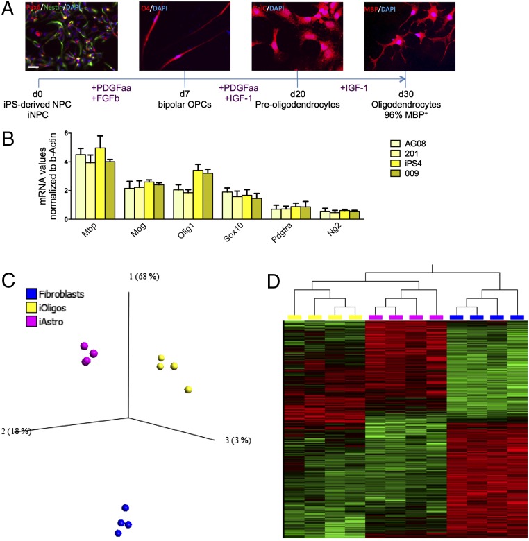
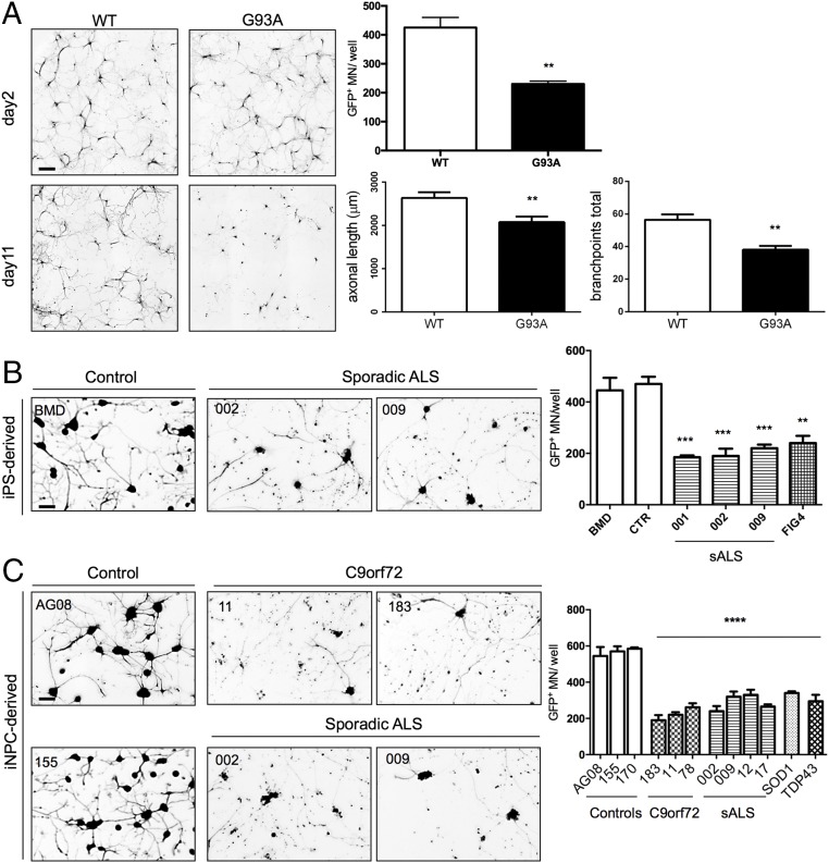
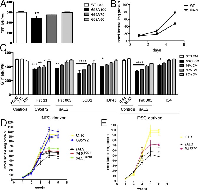
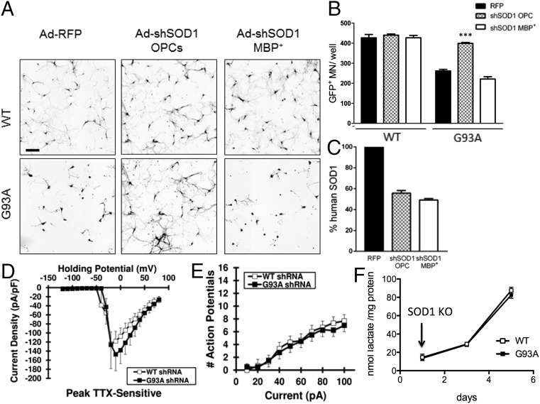
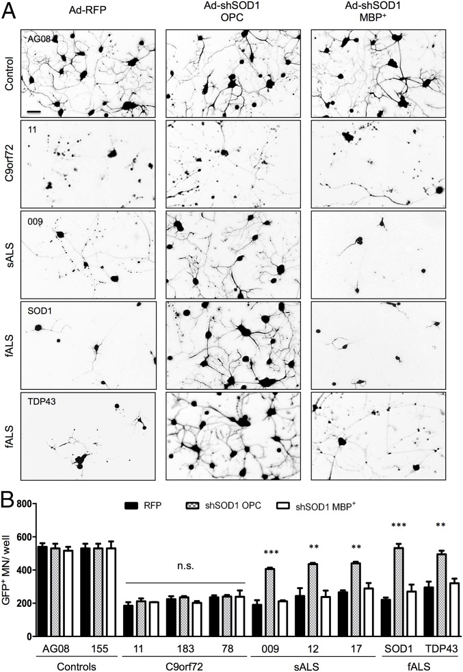
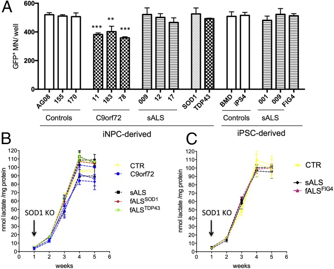
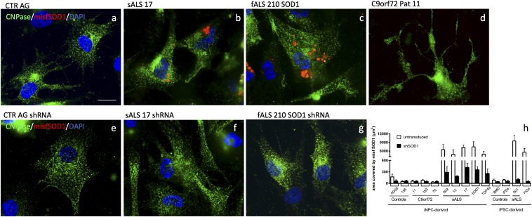
Oligodendrocytes have recently been implicated in the pathophysiology of amyotrophic lateral sclerosis (ALS). Here we show that, in vitro, mutant superoxide dismutase 1 (SOD1) mouse oligodendrocytes induce WT motor neuron (MN) hyperexcitability and death. Moreover, we efficiently derived human oligodendrocytes from a large number of controls and patients with sporadic and familial ALS, using two different reprogramming methods. All ALS oligodendrocyte lines induced MN death through conditioned medium (CM) and in coculture. CM-mediated MN death was associated with decreased lactate production and release, whereas toxicity in coculture was lactate-independent, demonstrating that MN survival is mediated not only by soluble factors. Remarkably, human SOD1 shRNA treatment resulted in MN rescue in both mouse and human cultures when knockdown was achieved in progenitor cells, whereas it was ineffective in differentiated oligodendrocytes. In fact, early SOD1 knockdown rescued lactate impairment and cell toxicity in all lines tested, with the exclusion of samples carrying chromosome 9 ORF 72 (C9orf72) repeat expansions. These did not respond to SOD1 knockdown nor did they show lactate release impairment. Our data indicate that SOD1 is directly or indirectly involved in ALS oligodendrocyte pathology and suggest that in this cell type, some damage might be irreversible. In addition, we demonstrate that patients with C9ORF72 represent an independent patient group that might not respond to the same treatment.

Keywords: C9orf72; SOD1; amyotrophic lateral sclerosis; lactate; oligodendrocytes.


Conflict of interest statement

The authors declare no conflict of interest.

Figures

Fig. 1.
Fig. 1.
Efficient differentiation of human neural progenitors into MBP+ oligodendrocytes. Schematic representation of human NPC differentiation into MBP+ oligodendrocytes (A) and expression of oligodendrocyte markers at the end of differentiation determined by qPCR and normalized to β-actin (B). Expression levels are relative to whole spinal cord homogenates. Transcripts were investigated in four lines, two derived from controls (nos. 155 and 170) and two patients (nos. 12 and 17). Error bar = SD, n = 3 per sample. (Scale bar, 30 μm.) PCA reveals that iOligodendrocytes, iAstrocytes, and fibroblasts are three distinct cell populations (C) based on a two-way ANOVA multigroup comparison analysis (P < 0.001). Differentially expressed transcripts were visualized in a heat map, identifying iOligodendrocytes and iAstrocytes as more closely related cell types than they are to fibroblasts, even if significantly different (D).
Fig. 2.
Fig. 2.
Oligodendrocytes from ALS samples reduce MN survival. Coculture of mouse oligodendrocytes from mSOD1G93A mice and WT Hb9-GFP MNs result in reduced MN survival after 11 d compared with WT oligodendrocyte cocultures. This result is accompanied by reduction in axonal length and branching (A). (Scale bar, 100 μm.) Error bar = SD, n = 6. Coculture of human iPSC- and iNPC-derived oligodendrocytes from sporadic and familial ALS patients results in 50% increased cell death 72 h after plating the MNs (B and C). **P ≤ 0.01, ***P ≤ 0.001, ****P ≤ 0.0001. (Scale bars, 50 μm.) Error bar = SD, n = 3 per line.
Fig. 3.
Fig. 3.
Oligodendrocyte CM from ALS samples induces MN death and is associated with decreased lactate levels. Hb9 GFP+ MN treated with increasing percentages of oligodendrocyte CM from the mSOD1G93A mouse model displayed a significant increase in cell death, while increasing percentages of CM from WT oligodendrocytes to MN medium slightly improved, but did not significantly change MN survival (A), thus we are representing the 100% CM condition. Increased mSOD1G93A oligodendrocyte CM-induced MN death was accompanied by significantly lower levels of lactate in the CM (B). As levels of secreted lactate increase in WT cells as they differentiate into oligodendrocytes, mSOD1G93A cells display lower increments, resulting in a significant difference in secreted lactate at the end of the differentiation protocol. Hb9-GFP+ MN treated with CM from human fully differentiated oligodendrocytes from both iNPCs and iPSCs also display increased cell death when treated with increasing percentages of ALS CM (C). Similarly to the mouse data, human ALS cells secrete progressively less lactate than control cells as they differentiate into oligodendrocytes, with exception of samples carrying C9orf72 repeat expansion (D and E). *P ≤ 0.05, **P ≤ 0.01, ***P ≤ 0.001, ****P ≤ 0.0001. Error bar = SD, n = 3–4 per line.
Fig. 4.
Fig. 4.
Lactate is a major component of CM-mediated toxicity, but not in coculture. Monocultures (mc) (AD) of Hb9 GFP+ MN were treated with 100% WT or SOD1G93A oligodendrocyte CM with addition of 1 or 2 mM lactate, resulting in MN rescue, whereas cocultures (cc) treated with 2 mM lacate (EG) showed only a minimal increase in MN survival. Similarly, monocultures of Hb9-GFP+ MN treated with 100% CM from human control or ALS oligodendrocytes plus 1 or 2 mM lactate showed increase in MN survival with exception of MNs treated with C9orf72 CM (HL). Lactate supplementation only marginally, but significantly, rescued MN survival in coculture (MO). Statistical significance refers to one-way ANOVA with multicomparison test of each treated sample against it own untreated control. (Magnification, 10×.) *P ≤ 0.05, **P ≤ 0.01, ***P ≤ 0.001, ****P ≤ 0.0001.
Fig. 5.
Fig. 5.
Knockdown of the human SOD1 transgene in mSOD1G93A oligodendrocyte progenitors, but not finally differentiated oligodendrocytes, results in MN rescue. Knockdown of SOD1 in oligodendrocyte progenitors (before starting differentiation) results in complete rescue in MN survival (A and B) as well as electrophysiological properties (n = 8) (D and E) and secreted lactate levels (F). No difference between mSOD1G93A oligodendroctes infected with Ad-RFP and Ad-shSOD1 at the end of differentiation was detected in coculture with MNs, i.e., no MN rescue is achieved when mSOD1 in knocked down at the end of differentiation before coculture (A and B). SOD1 protein levels were quantified at the end of coculture (C). ***P ≤ 0.001. Error bar = SD, n = 3 per coculture condition. (Scale bar, 100 μm.)
Fig. 6.
Fig. 6.
Knockdown of human SOD1 in oligodendrocyte progenitors results in MN rescue in patients with sporadic and familial ALS, but not in patients carrying C9orf72 repeat expansions. Knockdown of human WT SOD1 in human oligodendrocyte progenitors obtained from iNPCs results in a significant rescue in MN survival in sporadic and familial ALS cases carrying mutations in SOD1 and TDP43, but not C9orf72 repeat expansion (A and B). SOD1 knockdown was ineffective when performed at the end of differentiation. Error bar = SD, n = 3 per coculture condition per cell line. **P ≤ 0.01, ***P ≤ 0.001. (Scale bar, 50 μm.)
Fig. 7.
Fig. 7.
Knockdown of human SOD1 in oligodendrocyte progenitors results in normal levels of lactate in the growth medium throughout their differentiation. Knockdown of human WT SOD1 in human oligodendrocyte progenitors obtained from iNPCs or iPSCs results in rescue of MN monocultures treated with oligodendrocyte CM from sporadic and familial ALS cases carrying mutations in SOD1 and TDP43, but not C9orf72 repeat expansion (A). This result is accompanied by restoration of normal levels of secreted lactate (B and C). n = 3 per culture condition per cell line (A) and lactate measurements at all time points (B and C). **P ≤ 0.01, ***P ≤ 0.001.
Fig. 8.
Fig. 8.
Oligodendrocytes from patients with sporadic, SOD1, and TDP43-linked ALS display misfolded SOD1. Oligodendrocytes from patients with sporadic, SOD1, and TDP43-linked ALS, but not C9orf72-linked and unaffected individuals, display misfolded SOD1 aggregates (AD). The pattern is mostly perinuclear (B and C). SOD1 knockdown in progenitor cells successfully eliminates such aggregates as shown via immunocytochemistry (EG) and ImageJ analysis software (H). (Scale bar, 10 μm.)

References

    1. Rosen DR. Mutations in Cu/Zn superoxide dismutase gene are associated with familial amyotrophic lateral sclerosis. Nature. 1993;364(6435):362. - PubMed
    1. Kabashi E, et al. TARDBP mutations in individuals with sporadic and familial amyotrophic lateral sclerosis. Nat Genet. 2008;40(5):572–574. - PubMed
    1. Sreedharan J, et al. TDP-43 mutations in familial and sporadic amyotrophic lateral sclerosis. Science. 2008;319(5870):1668–1672. - PMC - PubMed
    1. Kwiatkowski TJ, Jr, et al. Mutations in the FUS/TLS gene on chromosome 16 cause familial amyotrophic lateral sclerosis. Science. 2009;323(5918):1205–1208. - PubMed
    1. Vance C, et al. Mutations in FUS, an RNA processing protein, cause familial amyotrophic lateral sclerosis type 6. Science. 2009;323(5918):1208–1211. - PMC - PubMed

Publication types

MeSH terms