. 2016 Sep 20:4:e2424.
doi: 10.7717/peerj.2424. eCollection 2016.

In situ visualization of bacterial populations in coral tissues: pitfalls and solutions

Affiliations

In situ visualization of bacterial populations in coral tissues: pitfalls and solutions

Naohisa Wada et al. PeerJ. .

Abstract

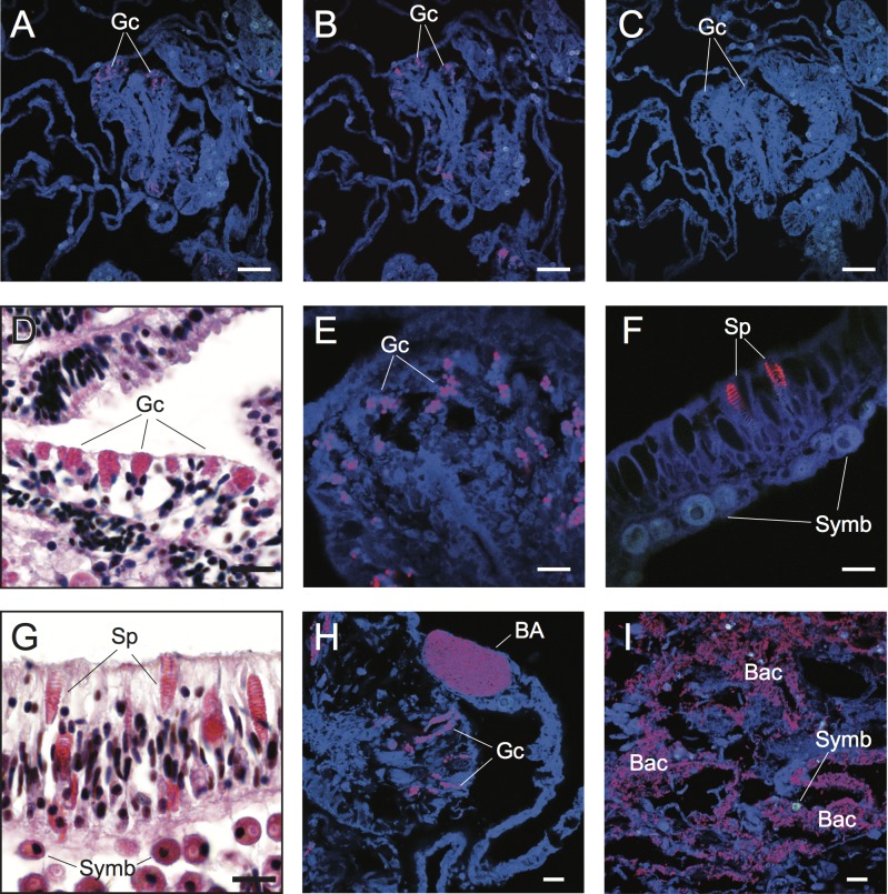
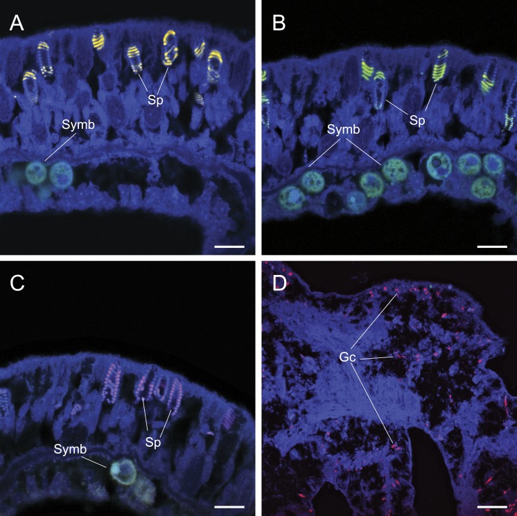
In situ visualization of microbial communities within their natural habitats provides a powerful approach to explore complex interactions between microorganisms and their macroscopic hosts. Specifically, the application of fluorescence in situ hybridization (FISH) to simultaneously identify and visualize diverse microbial taxa associated with coral hosts, including symbiotic algae (Symbiodinium), Bacteria, Archaea, Fungi and protists, could help untangle the structure and function of these diverse taxa within the coral holobiont. However, the application of FISH approaches to coral samples is constrained by non-specific binding of targeted rRNA probes to cellular structures within the coral animal tissues (including nematocysts, spirocysts, granular gland cells within the gastrodermis and cnidoglandular bands of mesenterial filaments). This issue, combined with high auto-fluorescence of both host tissues and endosymbiotic dinoflagellates (Symbiodinium), make FISH approaches for analyses of coral tissues challenging. Here we outline the major pitfalls associated with applying FISH to coral samples and describe approaches to overcome these challenges.

Keywords: Bacteria; Coral; Fluorescence in situ hybridization; Holobiont; In situ visualization.


Conflict of interest statement

The authors declare there are no competing interests.

Figures

Figure 1
Figure 1
Characteristic field appearance of a white syndrome (WS) on a colony of the coral Acropora hyacinthus from reefs near Lizard Island (14°40′S, 145°27′E) in the Northern sector of the Great Barrier Reef (GBR): (A) WS lesion on a coral colony with numbers and arrows displaying (1) dead coral skeleton overgrown with algae, (2) recently exposed coral skeleton prior to algal overgrowth, (3) actively progressing lesion boundary where samples derived for this study were taken, (4) healthy tissue ahead of the lesion and from which samples were also derived. (B) Close up image of lesion boundary displaying diffuse, acute to sub-acute areas of tissue loss revealing white, intact skeleton.
Figure 2
Figure 2. Detection and characterization of specific and non-specific fluorescence in situ hybridization (FISH) probe binding to target bacteria (BA and Bac), granular gland cells (Gc), and spirocysts (Sp) using Cy3-labelled FISH probes (A–C, E, F, H, I) and Hematoxylin and Eosin (H&E) staining of coral tissue sections (D, G).
Non-specific binding of EUB338 (A) and nonEUB338 (B) FISH probes to granular gland cells and lack of auto-fluorescence of these structures in probe-free treatments (C) is demonstrated through serial tissue sections. Detailed granular gland cell morphology within the gastrodermis is shown in H&E stained (D) and EUB338 FISH-hybridized (E) tissue sections. Non-specific binding of EUB338 FISH probes to spirocysts within epidermal cells (F) was detected in tissue sections and was particularly prevalent in coenosarc and tentacle regions of polyps. Detailed spirocyst morphology is shown through H&E staining (G). A bacterial aggregate within the calicoblastic layer is shown near to non-specific binding signals of granular gland cells (H) in healthy coral tissues hybridized with EUB338 FISH probes. Bacteria assemblages were detected within necrotic tissues associated with WS disease using EUB338 FISH probes (I). Scale bars represent 50 µm in (A–C) and 10 µm in (D)–(I). Abbreviations: Gc, granular gland cell; Sp, spirocysts; Symb, Symbiodinium; BA, bacterial aggregation; Bac, bacterial assemblages.
Figure 3
Figure 3. The application of common approaches designed to ameliorate non-specific fluorescence in situ hybridization (FISH) probe binding failed to inhibit hybridization to granular gland cells (Gc) and spirocysts (Sp).
Cy3 (A), Cy5-labelled NonEUB338 (B) and Atto 647-labelled Vib-GV (C) FISH probes all hybridized to spirocysts. Blocking solution also failed to prevent non-specific binding of oligonucleotide probes to granular gland cells (D). Scale bars represent 100 µm in (A)–(C) and 50 µm in (D). Abbreviations: Gc, granular gland cell; Sp, spirocysts; and Symb, Symbiodinium.

References

    1. Ainsworth TD, Fine M, Blackall LL, Hoegh-Guldberg O. Fluorescence in situ hybridization and spectral imaging of coral-associated bacterial communities. Applied and Environmental Microbiology. 2006a;72:3016–3020. doi: 10.1128/AEM.72.4.3016-3020.2006. - DOI - PMC - PubMed
    1. Ainsworth T, Hoegh-Guldberg O. Bacterial communities closely associated with coral tissues vary under experimental and natural reef conditions and thermal stress. Aquatic Biology. 2009;4:289–296. doi: 10.3354/ab00102. - DOI
    1. Ainsworth TD, Hoegh-Guldberg O, Leggat W. Imaging the fluorescence of marine invertebrates and their associated flora. Journal of Microscopy. 2008;232:197–199. doi: 10.1111/j.1365-2818.2008.02089.x. - DOI - PubMed
    1. Ainsworth TD, Kramasky-Winter E, Loya Y, Hoegh-Guldberg O, Fine M. Coral disease diagnostics: what’s between a plague and a band? Applied and Environmental Microbiology. 2007;73:981–992. doi: 10.1128/AEM.02172-06. - DOI - PMC - PubMed
    1. Ainsworth TD, Krause L, Bridge T, Torda G, Raina J-B, Zakrzewski M, Gates RD, Padilla-Gamiño JL, Spalding HL, Smith C, Woolsey ES, Bourne DG, Bongaerts P, Hoegh-Guldberg O, Leggat W. The coral core microbiome identifies rare bacterial taxa as ubiquitous endosymbionts. The ISME Journal. 2015;9:2261–2274. doi: 10.1038/ismej.2015.39. - DOI - PMC - PubMed

LinkOut - more resources