. 2016 Nov 15;7(46):75635-75647.
doi: 10.18632/oncotarget.12314.

Fluorescence-guided surgery of a highly-metastatic variant of human triple-negative breast cancer targeted with a cancer-specific GFP adenovirus prevents recurrence

Affiliations

Fluorescence-guided surgery of a highly-metastatic variant of human triple-negative breast cancer targeted with a cancer-specific GFP adenovirus prevents recurrence

Shuya Yano et al. Oncotarget. .

Abstract

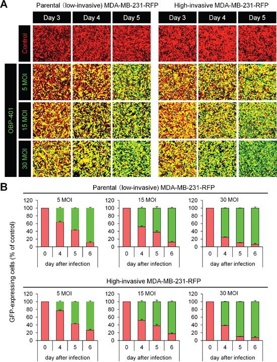
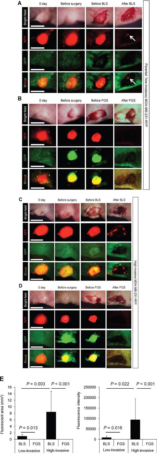
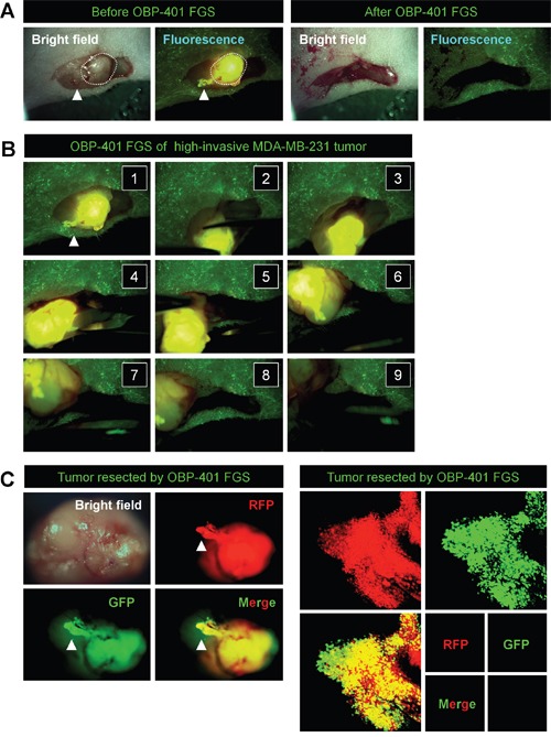
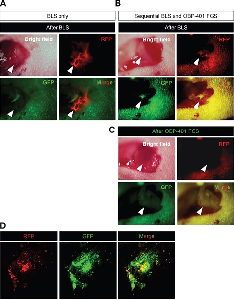
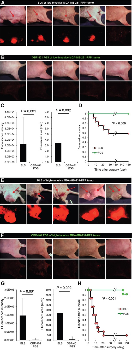
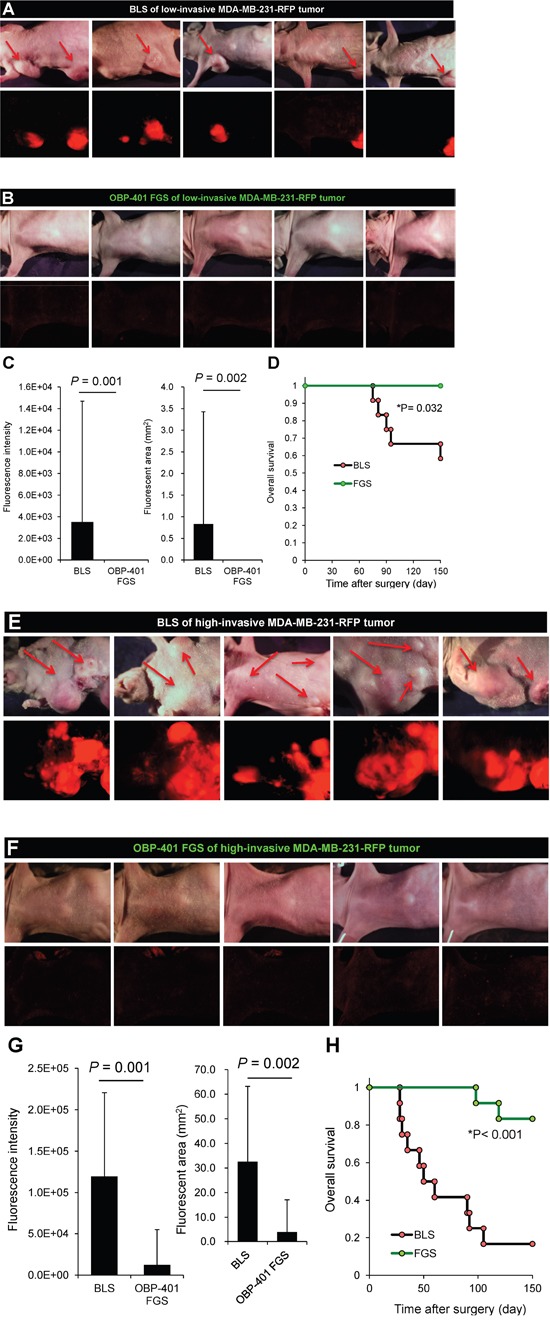
We have previously developed a genetically-engineered GFP-expressing telomerase-dependent adenovirus, OBP-401, which can selectively illuminate cancer cells. In the present report, we demonstrate that targeting a triple-negative high-invasive human breast cancer, orthotopically-growing in nude mice, with OBP-401 enables curative fluorescence-guided surgery (FGS). OBP-401 enabled complete resection and prevented local recurrence and greatly inhibited lymph-node metastasis due to the ability of the virus to selectively label and subsequently kill cancer cells. In contrast, residual breast cancer cells become more aggressive after bright (white)-light surgery (BLS). OBP-401-based FGS also improved the overall survival compared with conventional BLS. Thus, metastasis from a highly-aggressive triple-negative breast cancer can be prevented by FGS in a clinically-relevant mouse model.

Keywords: GFP/RFP; adenovirus; fluorescence-guided surgery (FGS); survival; telomerase dependent.


Conflict of interest statement

CONFLICTS OF INTEREST

Y. Urata is a CEO of Oncolys BioPharma Inc. (the manufacturer of OBP-401). H. Tazawa and T. Fujiwara are consultants of Oncolys BioPharma Inc.

Figures

Figure 1
Figure 1. OBP-401 labels low- and high-invasive MDA-MB-231-RFP breast cancer cells in vitro
Low- and high-invasive variants of MDA-MB-231-RFP cells were seeded in 6-well plates (1 × 10⁵ cells a well). OBP-401 was added at the indicated multiplicity of infection (MOI) 24 hours after seeding. Images were acquired with a confocal laser scanning microscope FV1000 (Olympus, Tokyo, Japan). A. Representative images of low- and high-invasive MDA-MB-231-RFP breast cancer cells 3, 4, and 5 days after infection of OBP-401 at the indicated MOI B. Histogram shows the frequency of GFP-expressing parental and highly metastatic MDA-MB-231-RFP breast cancer cells at indicated days after infection of OBP-401. The number of GFP-expressing cells was counted. Data are shown as average ± SD. N = 5.
Figure 2
Figure 2. Comparison of OBP-401-based fluorescence-guided surgery with bright-light surgery for orthotopic low- and high-invasive MDA-MB-231-RFP
OBP-401 was injected intaratumorally at 1 × 10⁸ PFU when tumors reached approximately 10 mm3 (diameter; 3 mm). A. Representative whole-tumor images of low-invasive MDA-MB-231-RFP cells before and after bright light surgery (BLS). White arrows point to the residual tumor. B. Representative whole-tumor images of low-invasive MDA-MB-231-RFP before injection of OBP-401, before and after OBP-401-based fluorescence-guided surgery (OBP-401 FGS). C. Representative whole-tumor images of high-invasive MDA-MB-231-RFP before and after BLS. D. Representative whole-tumor images of high-invasive MDA-MB-231-RFP before injection of OBP-401 and before and after OBP-401-based fluorescence-guided surgery (OBP-401 FGS). E. Histogram shows the comparison of fluorescent area (left) and fluorescence intensity (right) of residual tumor in the surgical bed after BLS or OBP-401 FGS of low- and high-invasive MDA-MB-231-RFP. Fluorescence intensity and fluorescent area were calculated with ImageJ software. Data are shown as average ± SD. N = 10.
Figure 3
Figure 3. In situ OBP-401 GFP-labeling visualizes invading cancer cells and enables complete resection of high-invasive MDA-MB-231-RFP
A. Representative high-magnification images of high-invasive MDA-MB-231-RFP before and after OBP-401 FGS using the hand-held fluorescence Dino-Lite scope. B. Step-by-step procedure of OBP-401 FGS for high-invasive MDA-MB-231-RFP (see Supplementary Movie S1). C. Representative images of en bloc tumor resected by OBP-401 FGS. Representative images of invading cancer cells resected by OBP-401 FGS visualized at the single-cell level using a confocal laser-scanning microscope (FV1000, Olympus, Tokyo, Japan).
Figure 4
Figure 4. Sequential BLS and OBP-401 FGS of high-invasive MDA-MB-231-RFP
OBP-401 was injected intaratumorally at 1 × 10⁸ PFU when tumors reached approximately 10 mm3 (diameter; 3 mm). The labeled orthotopic tumor was initially resected by BLS and then residual tumor was resected by FGS. A. Representative high-magnification images of invading cancer cells in a lymphatic duct after BLS. B. Representative macroscopic images of incomplete resection of the tumor after BLS (before OBP-401 FGS). C. Representative high-magnification images of surgical area after sequential BLS and OBP-401 FGS. D. Representative microscopic image of resected residual tumor after OBP-401 FGS acquired with confocal laser scanning microscope FV1000 (Olympus, Japan).
Figure 5
Figure 5. OBP-401-based FGS controls local recurrence of high- and low-invasive MDA-MB-231-RFP
A. Representative whole-body images 150 days after BLS of low-invasive MDA-MB-231-RFP. Arrows indicate growing tumors. B. Representative whole body images 150 days after OBP-401 FGS for low-invasive MDA-MB-231-RFP. C. Comparison of fluorescent area of local recurrent tumors after BLS or OBP-401 FGS for low-invasive MDA-MB-231-RFP (right). Comparison of fluorescence intensity of local and metastatic tumors after BLS or OBP-401 FGS for low-invasive MDA-MB-231-RFP (left). D. Kaplan-Meyer shows disease-free survival after BLS or OBP-401 FGS for low-invasive MDA-MB-231-RFP. E. Representative whole-body images 120 days after BLS for high-invasive MDA-MB-231-RFP. Arrows indicate growing tumors. F. Representative whole-body images 120 days after OBP-401 FGS of high-invasive MDA-MB-231-RFP. G. Comparison of fluorescent area of local recurrent tumors after BLS or OBP-401 FGS for high-invasive MDA-MB-231-RFP (right). Comparison of fluorescence intensity of local recurrent tumors after BLS or OBP-401 FGS for high-invasive MDA-MB-231-RFP (left). H. Kaplan-Meyer curve shows disease-free survival after BLS or OBP-401 FGS for high-invasive MDA-MB-231-RFP. Fluorescent area and fluorescence intensity were calculated with ImageJ software. Data are shown as average ± SD.
Figure 6
Figure 6. OBP-401-based FGS inhibits metastatic recurrence
A. Representative whole-body images 150 days after BLS for low-invasive MDA-MB-231-RFP. Arrows indicate growing metastatie tumors. B. Representative whole-body images 150 days after OBP-401 FGS for low-invasive MDA-MB-231-RFP. C. Comparison of fluorescent area of lymph-node metastasis after BLS or OBP-401 FGS for low-invasive MDA-MB-231-RFP (right). Comparison of fluorescence intensity of lymph-node metastasis after BLS or OBP-401 FGS for low-invasive MDA-MB-231-RFP (left). D. Kaplan-Meyer curve shows the overall survival after BLS or OBP-401 FGS of low-invasive breast cancer. E. Representative whole-body images 120 days after BLS for high-invasive MDA-MB-231-RFP. Arrows indicate growing metastatic tumors. F. Representative whole-body images 120 days after OBP-401 FGS for high-invasive MDA-MB-231-RFP. G. Comparison of fluorescent area of lymph node metastasis after BLS or OBP-401 FGS for high-invasive MDA-MB-231-RFP (right). Comparison of fluorescence intensity of l lymph node metastasis after BLS or OBP-401 FGS for high-invasive MDA-MB-231-RFP (left). H. Kaplan-Meyer curve shows the overall survival after BLS or OBP-401 FGS of high-invasive MDA-MB-231-RFP. Fluorescent area and fluorescence intensity were calculated with ImageJ software. Data are shown as average ± SD.

References

    1. Bouvet M, Hoffman RM. Glowing tumors make for better detection and resection. Sci Transl Med. 2011;3:110sf10. - PubMed
    1. Kishimoto H, Zhao M, Hayashi K, Urata Y, Tanaka N, Fujiwara T, Penman S, Hoffman RM. In vivo internal tumor illumination by telomerase-dependent adenoviral GFP for precise surgical navigation. Proc Natl Acad Sci USA. 2009;106:14514–14517. - PMC - PubMed
    1. Kishimoto H, Aki R, Urata Y, Bouvet M, Momiyama M, Tanaka N, Fujiwara T, Hoffman RM. Tumor-selective, adenoviral-mediated GFP genetic labeling of human cancer in the live mouse reports future recurrence after resection. Cell Cycle. 2011;10:2737–2741. - PMC - PubMed
    1. Yano S, Miwa S, Kishimoto H, Uehara F, Tazawa H, Toneri M, Hiroshima Y, Yamamoto M, Urata Y, Kagawa S, Bouvet M, Fujiwara T, Hoffman RM. Curative fluorescence-guided cancer surgery of soft-tissue sarcoma in combination with a GFP-labeling oncolytic adeovirus. Oncotarget. 2015;30:13133–13148. doi: 10.18632/oncotarget.3811. - DOI - PMC - PubMed
    1. Yano S, Miwa S, Kishimoto H, Toneri M, Hiroshima Y, Yamamoto M, Bouvet M, Urata Y, Tazawa H, Kagawa S, Fujiwara T, Hoffman RM. Experimental curative fluorescence-guided surgery of highly invasive glioblastoma multiforme selectively labeled with a killer-reporter adenovirus. Mol Ther. 2015;23:1182–1188. - PMC - PubMed

MeSH terms

Substances