. 2016 Oct 12;138(40):13335-13343.
doi: 10.1021/jacs.6b07830. Epub 2016 Sep 30.

Chemical Proteomic Profiling of Human Methyltransferases

Affiliations

Chemical Proteomic Profiling of Human Methyltransferases

Benjamin D Horning et al. J Am Chem Soc. .

Abstract

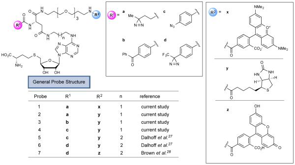
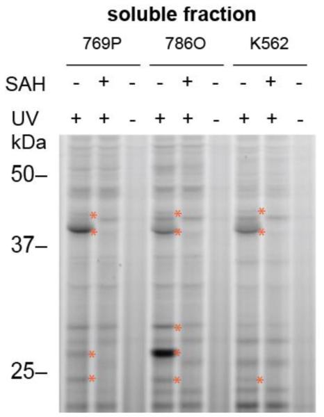
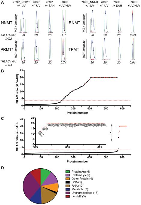
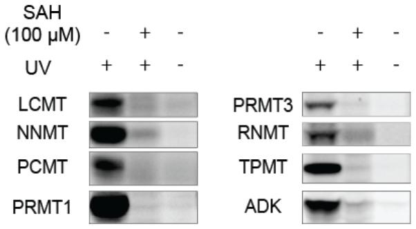
Methylation is a fundamental mechanism used in Nature to modify the structure and function of biomolecules, including proteins, DNA, RNA, and metabolites. Methyl groups are predominantly installed into biomolecules by a large and diverse class of S-adenosyl methionine (SAM)-dependent methyltransferases (MTs), of which there are ∼200 known or putative members in the human proteome. Deregulated MT activity contributes to numerous diseases, including cancer, and several MT inhibitors are in clinical development. Nonetheless, a large fraction of the human MT family remains poorly characterized, underscoring the need for new technologies to characterize MTs and their inhibitors in native biological systems. Here, we describe a suite of S-adenosyl homocysteine (SAH) photoreactive probes and their application in chemical proteomic experiments to profile and enrich a large number of MTs (>50) from human cancer cell lysates with remarkable specificity over other classes of proteins. We further demonstrate that the SAH probes can enrich MT-associated proteins and be used to screen for and assess the selectivity of MT inhibitors, leading to the discovery of a covalent inhibitor of nicotinamide N-methyltransferase (NNMT), an enzyme implicated in cancer and metabolic disorders. The chemical proteomics probes and methods for their utilization reported herein should prove of value for the functional characterization of MTs, MT complexes, and MT inhibitors in mammalian biology and disease.


Figures

Figure 1
Figure 1
S-adenosylhomocysteine (SAH)-based photoreactive probes for chemical proteomic profiling of methyltransferases (MTs). R1 and R2 designate varying photoreactive elements and reporter tags (fluorophore, biotin), respectively.
Figure 2
Figure 2
Gel-based profiling with fluorescent SAH-based probe 1. Probe 1 (25 µM) shows UV-dependent and SAH-competed reactivity with proteins in human cancer cell lysates (1 mg protein/mL). Proteins competed by excess SAH (100 µM) are marked with red asterisks. 769P and 786O are human renal cancer cell lines, and K562 is a human leukemia cell line.
Figure 3
Figure 3
Quantitative MS-based profiling with biotinylated SAH probe 2. (A) Parent ion mass (MS1) traces for representative tryptic peptides for the indicated MTs that were specific (enriched and SAH-competed) targets of probe 2. Blue and red traces represent heavy (+ UV, no SAH) and light (− UV or + UV, SAH-competed) fractions, respectively. For +UV/+UV control group, both heavy and light cell lysates were treated with probe 2 and exposed to UV light. Note that a maximum ratio value of 20 was assigned to all ratios with 20 or greater values. 769P_NNMT cells are a variant of the human renal cancer cell line 769P that has been genetically engineered to overexpress NNMT. (B) SILAC ratio plot for total proteins identified in human cancer cell lysates treated with probe 2 (heavy: 25 µM, + UV; light: 25 µM, − UV). Results are average enrichment values (+ UV/− UV) for 13 independent experiments performed with 769P cells (+/− NNMT expression,), 786O cells, and K562 cells. (C) SILAC ratios plot for total proteins identified in human cancer cell lysates treated with probe 2 (25 µM, + UV) with or without excess SAH (500 µM) competitor. Results are average competition values (− SAH/+ SAH) for 24 independent experiments performed with 769P cells (+/− NNMT expression), 786O cells and K562 cells. Inset shows expanded image of SAH-competed proteins. For (B) and (C), red dashed lines mark four-fold ratio change used to designate UV-dependent and/or SAH-competed proteins enriched by probe 2, and red dots mark MT proteins in the profiles. (D) Categorization of MTs designated as specific (UV-enriched and SAH-competed) targets of probe 2 based on substrate class. Numbers in parentheses refer to the number of probe 2 targets identified in each MT category. MTs without a characterized substrate class are designated as “uncharacterized”.
Figure 4
Figure 4
Probe 1 labeling of recombinantly expressed MTs and SAH-binding protein ADK. Proteins were expressed in HEK-293T cells by transient transfection and assayed in cell lysates, or expressed and purified from bacteria and doped into HEK293T lysates. Samples were treated with probe 1 (25 µM) ± UV light and with or without excess SAH (100 µM). For complete gels, including mock-transfected HEK-293T cells, see supporting Figures S6.
Figure 5
Figure 5
Distribution of MTs enriched by different photoreactive probes. Data are from K562 soluble lysates; 7-8 experiments for each probe that represent a combination of enrichment (UV-vs-no-UV) and SAH competition experiments. See Supporting Table S1, tab “Figure 5 data” for further details.
Figure 6
Figure 6
Discovery of an inhibitor of NNMT that reacts with active-site cysteine C165. (A) Crystal structure (PDB 2IIP) of SAH bound to human NNMT; an active-site cysteine C165 is highlighted. (B) Structure of hit fragment 8 and concentration-dependent blockade of probe1 labeling of endogenous NNMT in 786O soluble lysates (2 mg protein/mL) by 8. (C) Fluorescence polarization screen of a fragment electrophile library for inhibition of probe 1 binding to recombinant, purified human NNMT. See Figure S10 for structures of fragment electrophiles screened. (D) Concentration-dependent effects of 26-30 on probe 1 labeling of NNMT. (E) Structure and characterization of the more potent NNMT inhibitor RS004 (31) and concentration-dependent blockade of probe1 labeling of NNMT by 31. (F) RS004 (indicated concentrations, 60 min) blocks binding of probe 1 to NNMT, but not the C165A-NNMT mutant as measured by fluorescence polarization. Data represent mean values +/− standard deviation; n = 4 per group. (G) RS004 (25 µM, 30 min) blocks probe 2 labeling of NNMT, but not other MTs in cancer cell lysates as measured by quantitative MS-based proteomics. Reported SILAC ratios are average competition values (DMSO/RS004) for three independent experiments performed with 786O cells. Representative MS1 traces for NNMT and other RS004-insensitive MTs are shown.

References

    1. Fontecave M, Atta M, Mulliez E. Trends Biochem Sci. 2004;29:243. - PubMed
    1. Chen K, Zhao BS, He C. Cell Chem Biol. 2016;23:74. - PMC - PubMed
    1. Greer EL, Shi Y. Nat Rev Genet. 2012;13:343. - PMC - PubMed
    1. Fu Y, Dominissini D, Rechavi G, He C. Nat Rev Genet. 2014;15:293. - PubMed
    1. Xing Y, Li Z, Chen Y, Stock JB, Jeffrey PD, Shi Y. Cell. 2008;133:154. - PubMed

Publication types