. 2016 Oct 27;59(20):9390-9408.
doi: 10.1021/acs.jmedchem.6b00948. Epub 2016 Oct 14.

Antivirulence C-Mannosides as Antibiotic-Sparing, Oral Therapeutics for Urinary Tract Infections

Affiliations

Antivirulence C-Mannosides as Antibiotic-Sparing, Oral Therapeutics for Urinary Tract Infections

Laurel Mydock-McGrane et al. J Med Chem. .

Abstract

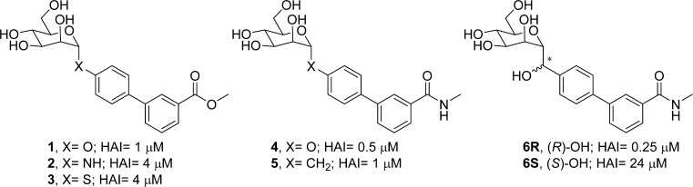
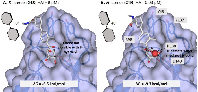
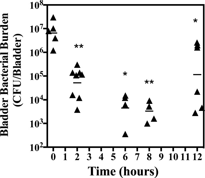
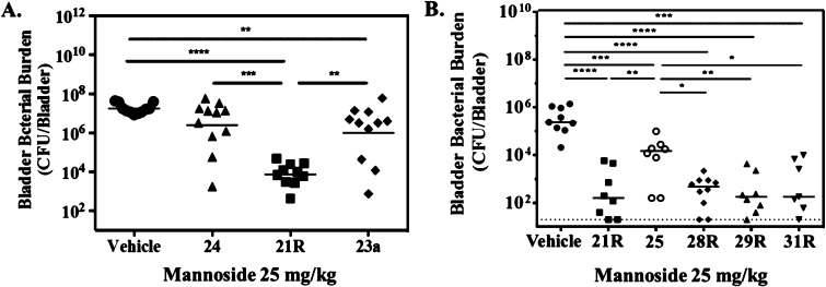
Gram-negative uropathogenic Escherichia coli (UPEC) bacteria are a causative pathogen of urinary tract infections (UTIs). Previously developed antivirulence inhibitors of the type 1 pilus adhesin, FimH, demonstrated oral activity in animal models of UTI but were found to have limited compound exposure due to the metabolic instability of the O-glycosidic bond (O-mannosides). Herein, we disclose that compounds having the O-glycosidic bond replaced with carbon linkages had improved stability and inhibitory activity against FimH. We report on the design, synthesis, and in vivo evaluation of this promising new class of carbon-linked C-mannosides that show improved pharmacokinetic (PK) properties relative to O-mannosides. Interestingly, we found that FimH binding is stereospecifically modulated by hydroxyl substitution on the methylene linker, where the R-hydroxy isomer has a 60-fold increase in potency. This new class of C-mannoside antagonists have significantly increased compound exposure and, as a result, enhanced efficacy in mouse models of acute and chronic UTI.


Conflict of interest statement

The authors declare the following competing financial interest(s): Scott Hultgren and James Janetka are co-founders and stockholders in Fimbrion Therapeutics, Inc.

Figures

Figure 1
Figure 1
Examples of simple glycosidic bond replacement of early lead O-mannosides 1 and 4, with alternate linkers to the biphenyl aglycone.
Scheme 1
Scheme 1. Synthesis of N-, S-, and Triazole-Linked Biphenyl Mannosides
Reagents and conditions: (a) methyl 4′-aminobiphenyl-3-carboxylate, EtOH, 55 °C; (b) 4-bromobenzenethiol, BF3–OEt2, DCM, 0 °C to rt; (c) 3-methoxycarbonylphenyl boronic acid, Pd(PPh3)4, Cs2CO3, dioxane/water (5/1), 80 °C; (d) NaOMe/MeOH, 0 °C to rt; (e) phenylacetylene, CuSO4, Na ascorbate, EtOH/H2O (4/1), rt.
Scheme 2
Scheme 2. Synthesis of C-Linked Amide Mannosides
Reagents and conditions: (a) 25% HCl aq, 50 °C; (b) HATU, DIPEA, 0 °C to rt, DMF; (c) 4-bromobenzoyl chloride, pyridine, rt; (d) Pd(PPh3)4, Cs2CO3, dioxane/water(5/1), 80 °C.
Scheme 3
Scheme 3. Synthesis of C-Linked Methylene and Hydroxy-Methylene Mannosides
Reagents and conditions: (a) DIBAL, CH2Cl2, −78 °C; (b) diethyl ether, −78 °C to −20 °C; (c) Dess–Martin periodinane, pyridine, CH2Cl2, 0 °C; (d) Li(tOBu)3AlH, THF, −40 to 0 °C; (e) 3-(N-methyaminocarbonyl)phenylboronic acid pinacol ester, Pd(PPh3)4, Cs2CO3, dioxane/water (5/1), 80 °C; (f) 10% Pd/C, H2, MeOH, rt.
Figure 2
Figure 2
Direct comparison of the potencies of C-linked methylene mannosides 21R, 21S, and 22 to those of similar O-mannoside analogues 23, 24, and 25.
Figure 3
Figure 3
In vitro analysis of mannoside potency. The difference in the conformational stability of the FimH lectin domain in the presence of mannoside compared to that in the absence of mannoside, as determined by differential scanning fluorimetry, is presented on the left axis. Measurement of the melting temperature is described in the methods. The hemagglutination inhibition data is presented relative to d-mannose on the right axis and is described in the methods.
Scheme 4
Scheme 4. Structures and Synthesis of Prodrug Analogues of O-Mannoside 23
Reagents and conditions: (a) Ac2O, pyridine, rt; (b) POCl3 trimethylphosphate, H2O, 0 °C; (c) TMSCl, Et3N, DMF, 0 °C; (d) AcOH, acetone/MeOH, 0 °C to rt; (e) N,N-dimethylglycine hydrochloride, DMAP, DIC, CH2Cl2/DIPEA, rt; (f) TFA, CH3CN, 0 °C.
Figure 4
Figure 4
(A) X-ray structure of 23 bound to FimH (PDB 5F3F). Water-mediated H-bond of amide carbonyl to Y48. (B) Overlay of 23 with 24 (PDB 5F2F) and 1 (PBD 3MCY). Altered biphenyl ring conformation and amide carbonyl orientation of ortho-substituted mannosides 23 and 24 compared to 1.
Figure 5
Figure 5
Computational docking models of isomeric C-mannosides: (A) S-hydroxy (21S) and (B) R-hydroxy (21R) bound to the FimH mannose-binding pocket. The accepted model of 21R is bound to FimH through water-mediated H-bond to D140 and N135.
Figure 6
Figure 6
(A) Derivatization of key building block 19R (precursor to 21R). (B) Small molecule X-ray structure of acetylated intermediate 27, confirming the R-stereochemistry of the 21R benzylic hydroxyl group.
Figure 7
Figure 7
(A) Mannoside levels were quantified in the urines of mice over a period of 8 h following oral gavage of drug as described in the methods. Data is presented as the mean and standard deviation from at least three independent experiments. Differences in concentration at the 8 h time point were tested for significance using Mann–Whitney U test. (* P < 0.05). (B) Measurement of mannoside in the plasma of rats following either a 10 mg/kg oral dose (dashed lines) or a 3 mg/kg intravenous dose (solid lines). Details regarding the measurement of mannoside are described in the methods.
Figure 8
Figure 8
(A) Prophylactic treatment of an acute UTI. Mannoside was dosed orally at 25 mg/kg in 10% cyclodextrin, 30 min prior to infection with 107 CFUs UPEC. Bladders were harvested 6 h postinfection and bacterial burdens enumerated. (B) Mannoside treatment of chronic UTI. Mice were treated orally with 50 mg/kg of mannoside in 10% cyclodextrin, after 14 days of chronic UPEC infection. Bladders were harvested 6 h following oral dosing and bacterial burdens enumerated. Data from at least two independent experiments are presented in each panel; bars indicate geometric means. Differences between treated groups and vehicle were tested for significance using Mann–Whitney U test (*** P = 0.0001, **** P < 0.0001).
Figure 9
Figure 9
Pharmacodynamics of O-linked mannoside. Chronically infected mice were treated orally with 50 mg/kg of mannoside 24. Bladders were harvested at the designated time points following oral dosing and bacterial burden enumerated. Bars indicate geometric means. Differences between the designated time point and the 0 h time point were tested for significance using Mann–Whitney U test (* P < 0.05, ** P < 0.001).
Figure 10
Figure 10
Improved efficacy of C-linked mannosides. (A) Chronically infected mice were treated orally with 25 mg/kg of mannoside. Bladders were harvested 12 hours following oral dosing and bacterial burdens enumerated. (B) Mice were treated orally with 25 mg/kg of mannoside 30 min prior to infection. Bladders were harvested 6 h following infection and bacterial burdens enumerated. Data from at least two independent experiments are presented; bars indicate geometric means. Differences between treated groups and vehicle were tested for significance using Mann–Whitney U test (**P < 0.01, *** P = 0.0001, **** P < 0.0001).

Similar articles

Cited by

References

    1. Boyle E. C.; Finlay B. B. Bacterial pathogenesis: exploiting cellular adherence. Curr. Opin. Cell Biol. 2003, 15, 633–639. 10.1016/S0955-0674(03)00099-1. - DOI - PubMed
    2. Chagnot C.; Listrat A.; Astruc T.; Desvaux M. Bacterial adhesion to animal tissues: protein determinants for recognition of extracellular matrix components. Cell. Microbiol. 2012, 14, 1687–1696. 10.1111/cmi.12002. - DOI - PubMed
    1. Ofek I.; Beachey E. H.. General concepts and principles of bacterial adherence. In Bacterial Adherence, Receptors and Recognition, Beachey E. H., Ed.; Chapman and Hall: London, 1980; Vol. 6, pp 1–29.
    2. Ofek I.; Doyle R. J.. Common Themes in Bacterial Adhesion. In Bacterial Adhesion to Cells and Tissues; Chapman and Hall: New York, 1994; pp 513–561.
    1. Aminov R. I. A brief history of the antibiotic era: lessons learned and challenges for the future. Front. Microbiol. 2010, 1, 134.10.3389/fmicb.2010.00134. - DOI - PMC - PubMed
    1. Cole S. T. Who will develop new antibacterial agents?. Philos. Trans. R. Soc., B 2014, 369, 20130430.10.1098/rstb.2013.0430. - DOI - PMC - PubMed
    2. Walsh C. Where will new antibiotics come from?. Nat. Rev. Microbiol. 2003, 1, 65–70. 10.1038/nrmicro727. - DOI - PubMed
    1. Lee Y. M.; Almqvist F.; Hultgren S. J. Targeting virulence for antimicrobial chemotherapy. Curr. Opin. Pharmacol. 2003, 3, 513–519. 10.1016/j.coph.2003.04.001. - DOI - PubMed
    2. Rasko D. A.; Sperandio V. Anti-virulence strategies to combat bacteria-mediated disease. Nat. Rev. Drug Discovery 2010, 9, 117–128. 10.1038/nrd3013. - DOI - PubMed

MeSH terms