. 2016 Sep 28;17(10):1598.
doi: 10.3390/ijms17101598.

Decreased Expression of SRSF2 Splicing Factor Inhibits Apoptotic Pathways in Renal Cancer

Affiliations

Decreased Expression of SRSF2 Splicing Factor Inhibits Apoptotic Pathways in Renal Cancer

Hanna Kędzierska et al. Int J Mol Sci. .

Erratum in

Abstract

Serine and arginine rich splicing factor 2(SRSF2) belongs to the serine/arginine (SR)-rich family of proteins that regulate alternative splicing. Previous studies suggested that SRSF2 can contribute to carcinogenic processes. Clear cell renal cell carcinoma (ccRCC) is the most common subtype of kidney cancer, highly aggressive and difficult to treat, mainly due to resistance to apoptosis. In this study we hypothesized that SRSF2 contributes to the regulation of apoptosis in ccRCC. Using tissue samples obtained from ccRCC patients, as well as independent validation on The Cancer Genome Atlas (TCGA) data, we demonstrate for the first time that expression of SRSF2 is decreased in ccRCC tumours when compared to non-tumorous control tissues. Furthermore, by employing a panel of ccRCC-derived cell lines with silenced SRSF2 expression and qPCR arrays we show that SRSF2 contributes not only to splicing patterns but also to expression of multiple apoptotic genes, including new SRSF2 targets: DIABLO, BIRC5/survivin, TRAIL, BIM, MCL1, TNFRSF9, TNFRSF1B, CRADD, BCL2L2, BCL2A1, and TP53. We also identified a new splice variant of CFLAR, an inhibitor of caspase activity. These changes culminate in diminished caspase-9 activity and inhibition of apoptosis. In summary, we show for the first time that decreased expression of SRSF2 in ccRCC contributes to protection of cancer cells viability.

Keywords: CFLAR; SRSF2; TCGA; alternative splicing; apoptosis; caspase-9; ccRCC; mRNA; renal cancer.


Conflict of interest statement

The authors declare no conflict of interest.

Figures

Figure 1
Figure 1
Decreased SRSF2 expression in renal tumours. (A) Western-blot analysis of SRSF2 expression in matched-paired control and tumours samples from eight patients; (B) Analysis of immunohistochemical staining intensity. The plot shows mean staining intensity of immunohistochemical (IHC) performed on C—Control samples: n = 3, T—Tumour samples: n = 11. The intensity scale used for statistical calculations was as follows: strong intensity: 3, moderate intensity: 2, weak intensity: 1. For the IHC images see Figure S2; (C) Analysis of TCGA data. Left panel: decreased SRSF2 expression in tumour samples (n = 72) when compared with controls (n = 72); Right panel: Principal component analysis of TCGA data. Control samples: red circles, tumour samples: blue circles. Statistical analysis was performed using t-test. ** p < 0.01.
Figure 2
Figure 2
The effect of SRSF2 silencing on apoptosis and cell viability. (A) Silencing of SRSF2 in Caki-2 cells illustrated by the results of Western-blot analysis; (B) results of annexin V/PI staining and flow cytometry. The bars show percentage of early apoptotic cells in Caki-2 cells with silenced SRSF2 expression (siSRSF2) when compared with cells transfected with non-specific scrambled control siRNA (siControl). The plot shows mean from three independent biological experiments performed in triplicates; and (C) viability of UV-irradiated Caki-2 cells with silenced SRSF2 expression (siSRSF2) when compared with cells transfected with non-specific scrambled control siRNA (siControl) analysed with 3-[4,5-dimethylthiazol-2-yl]-2,5-diphenyltetrazolium bromide; thiazolyl blue (MTT) assay. The bars show mean results of three independent biological experiments. Statistical analysis was performed using t-test. * p < 0.05, **** p < 0.0001.
Figure 3
Figure 3
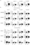
The effect of SRSF2 silencing on the expression of genes involved in the regulation of apoptotic pathways in four renal cancer-derived cell lines. (A) Caki-2, UOK171, KIJ-265T, and KIJ-308T cells were transfected with SRSF2-specific (siSRSF2) or control (siControl) siRNA. The graphs show mean results of qPCR analysis of SRSF2 expression performed on cDNA from three independent biological experiments executed in triplicates; and (B) the expression of apoptotic genes in cells with silenced SRSF2 expression. Only genes that were positively validated in manual qPCR verification are shown. Statistical analysis was performed using t-test. The results for BCL2L2 in Caki-2 cells were analysed using Mann–Whitney test due to lack of normal distribution. * p < 0.05, ** p < 0.01, *** p < 0.001, **** p < 0.0001. NS: Lack of statistical significance.
Figure 4
Figure 4
The effect of SRSF2 silencing on splicing patterns of apoptotic genes. Electrophoretic analysis of PCR-amplified splicing variants of apoptotic genes in four renal cancer-derived cell lines transfected with SRSF2-specific (siSRSF2) or control (siControl) siRNA. CFLAR NEW designates a new CFLAR splice variant, identified in this study. Primers used for amplification of BIM isoforms detected three major variants (BimEL, BimL, and BimS), as well as minor variants (Bimα1, Bimα2, and Bimα3). HPRT1—Internal RT-PCR control.
Figure 5
Figure 5
Decreased expression of SRSF2 affects activity of caspase-9. The activity of caspase-9 in UV-irradiated Caki-2 cells with silenced SRSF2 expression (siSRSF2) when compared with cells transfected with non-specific scrambled control siRNA (siControl). The graph shows results of three independent biological experiments performed in seven replicates. Statistical analysis was performed using t-test. ** p < 0.01.
Figure 6
Figure 6
The model showing how decreased expression of SRSF2 affects apoptotic pathways in renal cancer cells. The changes introduced by the depletion of SRSF2 are shown with the following colours: green, decreased expression; and red, increased expression. The arrows indicate activation, bar-headed lines indicate inhibition. Grey dotted arrow indicates release of cytochrome c. Black dotted arrows indicate final effect on apoptosis. In the extrinsic apoptotic pathway (left side of the drawing) depletion of SRSF2 results in downregulated expression of genes coding for death receptors (TNFRSF1B and TNFRSF9) and CRADD that recruits caspase-2 to complex with death receptors. Additionally, the expression of TRAIL-β, a non-active splice variant of a ligand of the TNFRSF10 receptor [48] is decreased. Overexpressed caspase-8L interferes with the binding of caspase-8a to FADD (Fas-associated death domain-containing protein) and counteracts its activation [49]. Likewise, caspase-8 activation is probably hindered by overexpression of the new splice variant of CFLAR, coding for Usurpin β [18]. In the intrinsic apoptotic pathway (right side of the drawing) BAX and BAK oligomerize, trigger permeabilization of the outer mitochondrial membrane and release of cytochrome c. The proapototic activity of BAX and BAK is counteracted by antiapoptotic proteins of BCL-2 family (MCL-1L, BCL-XL, BCL-2, BCL2L2, and BCL2A1) [14]. In cells with lowered expression of SRSF2, the concurrent decrease of antiapoptotic BCL2A1 and BCL2L2 might be overcome by diminished expression of proapoptotic BimS (the most powerful pro-apoptotic isoform of BIM variants) that inhibits the activity of antiapoptotic BCL2 proteins and directly activates BAX [22,23,24]. A decrease of BimS may, thus, lead to, on one hand, diminished activation of BAX, and on the other hand, to inefficient inhibition of BCL-2 proteins, allowing them to inhibit BAX and BAK. A similar effect is exerted by decreased expression of proapoptotic MCL-1S that acts as an inhibitor of antiapoptotic MCL-1L [21]. The antiapoptotic effect of SRSF2 depletion is possibly executed by diminished activity of caspase-9, resulting from: (1) decreased expression of caspase-9a; (2) increased expression of caspase-9b that inhibits Apaf-1-mediated activation of caspase-9a [50,51]; (3) decreased expression of Smac3 which inactivates XIAP that acts as caspases’ inhibitor; and (4) decreased expression of proapoptotic Surv-2B that acts as an activator of caspase-9 [20]. Notably, the expression of antiapoptotic survivin is not changed by SRSF2 silencing. Finally, decreased expression of SRSF2 results in upregulation of TP53, which inhibits apoptosis in renal cancer cells [38,39,40,41,52].

Similar articles

Cited by

References

    1. Long J.C., Caceres J.F. The SR protein family of splicing factors: Master regulators of gene expression. Biochem. J. 2009;417:15–27. doi: 10.1042/BJ20081501. - DOI - PubMed
    1. Edmond V., Merdzhanova G., Gout S., Brambilla E., Gazzeri S., Eymin B. A new function of the splicing factor SRSF2 in the control of E2F1-mediated cell cycle progression in neuroendocrine lung tumors. Cell Cycle. 2013;12:1267–1278. doi: 10.4161/cc.24363. - DOI - PMC - PubMed
    1. Lin S., Coutinho-Mansfield G., Wang D., Pandit S., Fu X.D. The splicing factor SC35 has an active role in transcriptional elongation. Nat. Struct. Mol. Biol. 2008;15:819–826. doi: 10.1038/nsmb.1461. - DOI - PMC - PubMed
    1. Ji X., Zhou Y., Pandit S., Huang J., Li H., Lin C.Y., Xiao R., Burge C.B., Fu X.D. SR proteins collaborate with 7SK and promoter-associated nascent RNA to release paused polymerase. Cell. 2013;153:855–868. doi: 10.1016/j.cell.2013.04.028. - DOI - PMC - PubMed
    1. McFarlane M., MacDonald A.I., Stevenson A., Graham S.V. Human papillomavirus 16 oncoprotein expression is controlled by the cellular splicing factor SRSF2 (SC35) J. Virol. 2015;89:5276–5287. doi: 10.1128/JVI.03434-14. - DOI - PMC - PubMed

LinkOut - more resources