Review
. 2017 Jan 10;8(2):3628-3639.
doi: 10.18632/oncotarget.12275.

Immune cells in liver regeneration

Affiliations
Review

Immune cells in liver regeneration

Na Li et al. Oncotarget. .

Abstract

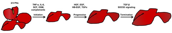
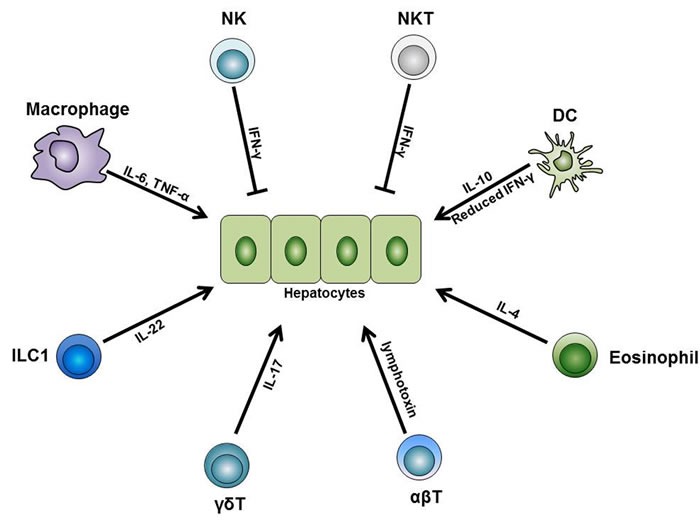
After partial hepatectomy, hepatocytes proliferate to restore mass and function of the liver. Macrophages, natural killer (NK) cells, natural killer T (NKT) cells, dendritic cells (DC), eosinophils, gamma delta T (γδT) cells, and conventional T cells, as well as other subsets of the immune cells residing in the liver control liver regeneration, either through direct interactions with hepatocytes or indirectly by releasing inflammatory cytokines. Here, we review recent progress regarding the immune cells in the liver and their functions during liver regeneration.

Keywords: adaptive immune system; innate immune system; liver; partial hepatectomy; regeneration.


Conflict of interest statement

Conflicts of interest

The authors declare no conflict of interest.

Figures

Figure 1
Figure 1. Three phases of liver regeneration after 2/3 partial hepatectomy
After 2/3 partial hepatectomy, an acute phase response initiates the liver regeneration process. In this process, the complement system in the liver is activated, which triggers different cytokines needed for regeneration priming. Among these cytokines, TNF-α and IL-6 are the most important. In addition, SCF and OSM are beneficial for enhancing the effects of these regeneration-promoting cytokines. In response to this, quiescent hepatocytes enter the cell cycle (Go to G1 phase). Then, various growth factors, such as HGF, EGF, HB-EGF, and TGF-α further drive the cell cycle to S phase, which is the progression phase of liver regeneration. When the liver re-establishes its normal mass and function, signals terminating the regeneration process, such as TGF-β and SOCS3 signals, brakes the regeneration process, and the liver accomplishes the regeneration process after 2/3 partial hepatectomy. Abbreviations: TNF-α, tumor necrosis factor-α; IL-6, interlukin-6; SCF, stem cell factor; OSM, Oncostatin M; HGF, hepatocyte growth factor; EGF, epidermal growth factor; HB-EGF, heparin-binding epidermal growth factor; TGF-α, transforming growth factor-α; TGF-β, transforming growth factor-β; SOCS3, Suppressor Of Cytokine Signaling 3.
Figure 2
Figure 2. The relationship of macrophage activation, cytokine secretion, and liver regeneration
In response to partial hepatectomy, the liver shows an acute phase response, during which the liver is enriched in several chemotaxis mediators for macrophages, such as osteopontin (mainly secreted by biliary epithelial cells [47]), MCP-1 (mainly secreted by hepatic stellate cells and biliary epithelial cells [130, 131] ), and ICAM-1 (mainly secreted by sinusoidal endothelial cells [40]). In the liver, the increased levels of LPS as well as reactive oxygen species (ROS) activate the complement system and the TLR/MyD88 pathway in macrophages. This leads to the activation of NFκB and results in the release of inflammatory cytokines TNF-α and IL-6. TNF-α could function in an autocrine manner and further activate NFκB. IL-6 binds to its receptors on hepatocytes and activate STAT3 signaling, thus promote the proliferation of hepatocytes. Abbreviations: MCP-1, monocyte chemoattractant protein-1; ICAM-1, intercellular adhesion molecule 1; ROS, reactive oxygen species; TNF-α, tumor necrosis factor-α; IL-6, interlukin-6; STAT3, Signal transducer and activator of transcription 3.
Figure 3
Figure 3. The role of immune cells during liver regeneration
Different subsets of the innate and adaptive immune cells are indispensable for normal liver regeneration after partial hepatectomy. Among these cells, liver macrophages produce IL-6 and TNF-α and initiate the regeneration process after partial hepatectomy. Besides, liver DCs upregulate their IL-10 expression level while downregulate their IFN-γ level, thus facilitate liver regeneration. In addition, liver eosinophil-derived IL-4 also promotes the regeneration process. Furthermore, γδT cell-derived IL-17 and ILC1-derived IL-22 are both necessary for normal regeneration. On the other side, NK and NKT cells play inhibitory roles in liver regeneration, and this is mainly dependent on the IFN-γ they secrete. Besides these innate immune cells, conventional αβT cells can secrete lymphotoxin and stimulate liver regeneration. Abbreviations: TNF-α, tumor necrosis factor-α; IFN-γ, interferon-γ; ILC, innate lymphoid cell.

References

    1. Stapleton GN, Hickman R, Terblanche J. Blood supply of the right and left hepatic ducts. Brit J Surg. 1998;85(2):202–207. - PubMed
    1. Kopka L, Rodenwaldt J, Vosshenrich R, Fischer U, Renner B, Lorf T, Graessner J, Ringe B, Grabbe E. Hepatic blood supply: Comparison of optimized dual phase contrast-enhanced three-dimensional MR angiography and digital subtraction angiography. Radiology. 1999;211(1):51–58. - PubMed
    1. Jenne CN, Kubes P. Immune surveillance by the liver. Nature immunology. 2013;14(10):996–1006. - PubMed
    1. Wiest R, Lawson M, Geuking M. Pathological bacterial translocation in liver cirrhosis. J Hepatol. 2014;60(1):197–209. - PubMed
    1. Hackstein CP, Assmus LM, Welz M, Klein S, Schwandt T, Schultze J, Forster I, Gondorf F, Beyer M, Kroy D, Kurts C, Trebicka J, Kastenmuller W, Knolle PA, Abdullah Z. Gut microbial translocation corrupts myeloid cell function to control bacterial infection during liver cirrhosis. Gut. 2016;18 pii: gutjnl-2015-311224. [Epub ahead of print] - PubMed