Review
. 2017 Jan 10;8(2):3649-3665.
doi: 10.18632/oncotarget.12278.

On the origin of myeloid-derived suppressor cells

Affiliations
Review

On the origin of myeloid-derived suppressor cells

Camilla Rydberg Millrud et al. Oncotarget. .

Abstract

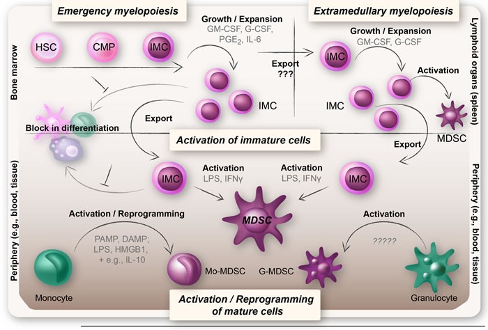
Myeloid-derived suppressor cells (MDSCs) have a strong immunosuppressive character that allows them to regulate immune responses and hinder overt inflammatory responses. In cancer, this leads to tumor immune evasion and disease progression. MDSCs come in at least two forms: monocytic (Mo-MDSCs) and granulocytic (G-MDSCs). The classical definition of MDSCs as immature myeloid cells blocked from differentiating has been challenged by recent studies suggesting that Mo-MDSCs and G-MDSCs may represent monocytes and granulocytes that have acquired immunosuppressive properties. The molecular mechanism behind their generation and their true origins are now widely debated. In this review we discuss the different proposed mechanisms of the generation of both types of MDSCs, with a special focus on human MDSCs in cancer.

Keywords: MDSC origin; emergency myelopoiesis; extramedullary; myelopoiesis; reprogramming.


Conflict of interest statement

CONFLICTs OF INTEREST

The authors declare that there are no conflicts of interest.

Figures

Figure 1
Figure 1. Classical MDSC functions
MDSCs have a strong immunosuppressive character that allows them to limit inflammation so that wound healing and recovery can take place. One of the hallmarks of MDSCs is their ability to suppress T-cell responses. MDSCs have also been described to induce regulatory T-cells (Tregs), modulate the cytokine production of macrophages, and promote other non-immune functions such as tumor angiogenesis and eventually metastasis.
Figure 2
Figure 2. Overview of the theories on the origin of MDSCs
MDSCs arise under pathological conditions when there is an excessive inflammation. Upper left panel: Upon an increased demand for myeloid cells, IMCs expand in the bone marrow and migrate into the periphery, a process known as emergency myelopoiesis. The IMCs are believed to be blocked in their differentiation and to become functionally active MDSCs when exposed to inflammatory mediators (upper and lower panel). Upper right panel: IMCs may also expand and become functionally active MDSCs extramedullary (i.e. in organs outside of the bone marrow, foremost the spleen), a feature often seen in chronic inflammatory diseases such as cancer. Lower panel: More recent hypotheses suggest that Mo-MDSCs and G-MDSCs may represent reprogrammed or activated monocytes and granulocytes. Reprogramming of monocytes into Mo-MDSCs is believed to rely on a repeated TLR-signaling (triggered by PAMPs or DAMPs) in combination with certain cytokines and mediators (e.g., IL-10, Wnt5a, and PGE2), whereas G-MDSCs are thought to represent an activation state of neutrophils.
Figure 3
Figure 3. A schematic illustration of the involvement of CSFs in the generation of MDSCs
A: MDSC development is likely governed by the same growth factors that control normal myelopoiesis e.g., CSFs (GM-CSF, G-CSF, and M-CSF). CSFs are essential survival factors for myeloid cells, in the bone marrow as well as in the periphery. They are also involved in many aspects of MDSC generation such as expansion of IMCs in the bone marrow, migration of myeloid cells into the periphery, and in some instances also for activation of MDSCs. B: All MDSCs undoubtedly derive from common myeloid progenitors in the bone marrow. The IMCs either expand in and migrate from the bone marrow into the periphery where they attain their MDSC phenotype (1), or into the spleen where they expand and become activated MDSCs that further migrate into the periphery (2). IMCs or MDSCs derived from hematopoietic progenitor cells found in the spleen may also directly migrate into the periphery (3). Whether peripheral MDSCs also can migrate into the spleen is currently unknown (4).
Figure 4
Figure 4. The two-signal model of MDSC generation
The classical hypothesis regarding the generation of MDSCs is the “two-signal model”. This model states that an expansion signal induced by e.g., CSFs (such as GM-CSF and G-CSF), IL-6, and PGE2, and mediated by STAT3, expands and mobilizes immature myeloid cells from the bone marrow. This is followed by an activation signal induced by pro-inflammatory stimuli e.g., LPS, PGE2 and S100A8/A9, and mediated by NFκB. According to this hypothesis it is not until the IMCs acquire an activation signal that they obtain a suppressive MDSC-phenotype.
Figure 5
Figure 5. Reprogramming of monocytes into Mo-MDSCs
The molecular mechanism behind reprogramming of monocytes into Mo-MDSCs is suggested to rely on a repeated TLR-signaling in combination with the right cytokine milieu (e.g., IL-10). Upon a first encounter with PAMPs or DAMPs, the monocytes will attain an activated pro-inflammatory phenotype with formation of pro-inflammatory NFκB p65:p50 heterodimers. A subsequent exposure to PAMPs/DAMPs, in combination with certain cytokines (e.g., IL-10), results in the formation of immunosuppressive NFκB p50:p50 homodimers and a reprogramming of monocytes from a pro-inflammatory state to an anti-inflammatory state with down-regulation of TLR, HLA-DR, and co-receptors. These anti-inflammatory monocytes have the same surface phenotype and function as Mo-MDSCs.

References

    1. Gabrilovich DI, Bronte V, Chen SH, Colombo MP, Ochoa A, Ostrand-Rosenberg S, Schreiber H. The terminology issue for myeloid-derived suppressor cells. Cancer Res. 2007;67:425. doi: 10.1158/0008-5472.CAN-06-3037. author reply 6. - DOI - PMC - PubMed
    1. Ostrand-Rosenberg S, Sinha P. Myeloid-derived suppressor cells: linking inflammation and cancer. J Immunol. 2009;182:4499–506. doi: 10.4049/jimmunol.0802740. - DOI - PMC - PubMed
    1. Cuenca AG, Delano MJ, Kelly-Scumpia KM, Moreno C, Scumpia PO, Laface DM, Heyworth PG, Efron PA, Moldawer LL. A paradoxical role for myeloid-derived suppressor cells in sepsis and trauma. Mol Med. 2011;17:281–92. doi: 10.2119/molmed.2010.00178. - DOI - PMC - PubMed
    1. Gabrilovich DI, Nagaraj S. Myeloid-derived suppressor cells as regulators of the immune system. Nat Rev Immunol. 2009;9:162–74. doi: 10.1038/nri2506. - DOI - PMC - PubMed
    1. Poschke I, Kiessling R. On the armament and appearances of human myeloid-derived suppressor cells. Clin Immunol. 2012;144:250–68. doi: 10.1016/j.clim.2012.06.003. - DOI - PubMed

MeSH terms

Substances