

Heterogeneity in Dopamine Neuron Synaptic Actions Across the Striatum and Its Relevance for Schizophrenia
- PMID: 27692238
- PMCID: PMC5121049
- DOI: 10.1016/j.biopsych.2016.07.002
Heterogeneity in Dopamine Neuron Synaptic Actions Across the Striatum and Its Relevance for Schizophrenia
Abstract
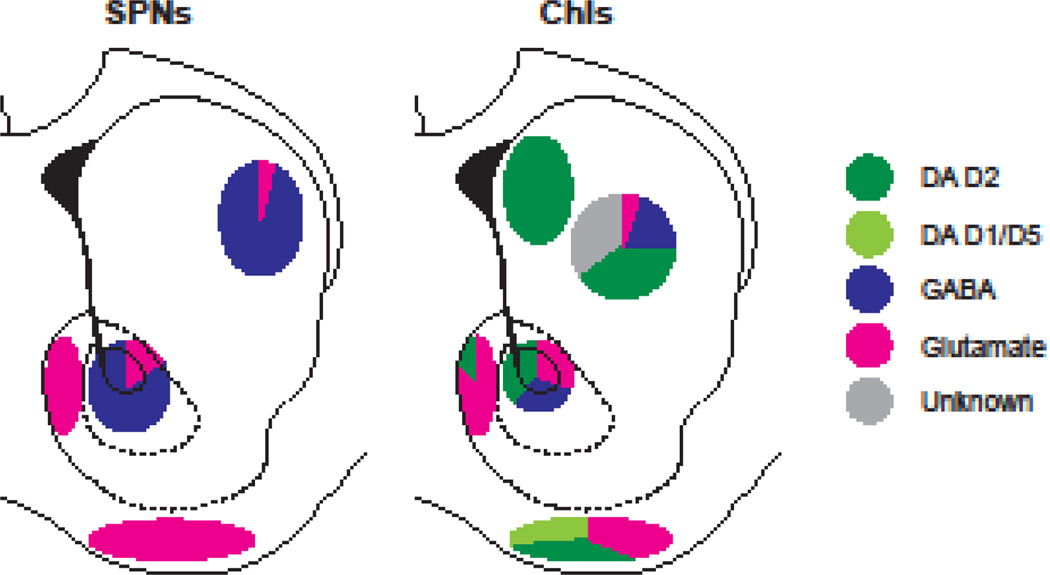
Brain imaging has revealed alterations in dopamine uptake, release, and receptor levels in patients with schizophrenia that have been resolved on the scale of striatal subregions. However, the underlying synaptic mechanisms are on a finer scale. Dopamine neuron synaptic actions vary across the striatum, involving variations not only in dopamine release but also in dopamine neuron connectivity, cotransmission, modulation, and activity. Optogenetic studies have revealed that dopamine neurons release dopamine in a synaptic signal mode, and that the neurons also release glutamate and gamma-aminobutyric acid as cotransmitters, with striking regional variation. Fast glutamate and gamma-aminobutyric acid cotransmission convey discrete patterns of dopamine neuron activity to striatal neurons. Glutamate may function not only in a signaling role at a subset of dopamine neuron synapses, but also in mediating vesicular synergy, contributing to regional differences in loading of dopamine into synaptic vesicles. Regional differences in dopamine neuron signaling are likely to be differentially involved in the schizophrenia disease process and likely determine the subregional specificity of the action of psychostimulants that exacerbate the disorder, and antipsychotics that ameliorate the disorder. Elucidating dopamine neuron synaptic signaling offers the potential for achieving greater pharmacological specificity through intersectional pharmacological actions targeting subsets of dopamine neuron synapses.
Keywords: Corelease; Cotransmission; GABA; Glutamate; Nucleus accumbens; Optogenetics; Vesicular synergy.
Copyright © 2016 Society of Biological Psychiatry. Published by Elsevier Inc. All rights reserved.
Conflict of interest statement
Financial disclosures The authors report no biomedical financial interests or potential conflicts of interest.
Figures


References
-
- Drevets WC, Price JC, Kupfer DJ, Kinahan PE, Lopresti B, Holt D, et al. PET measures of amphetamine-induced dopamine release in ventral versus dorsal striatum. Neuropsychopharmacology. 1999;21:694–709. - PubMed
-
- Colpaert FC. Discovering risperidone: the LSD model of psychopathology. Nat Rev Drug Discov. 2003;2:315–320. - PubMed
Publication types
MeSH terms
Substances
Grants and funding
LinkOut - more resources
Full Text Sources
Other Literature Sources
Medical
