. 2016 Oct 12;20(4):423-428.
doi: 10.1016/j.chom.2016.09.006. Epub 2016 Sep 29.

Zika Virus Infection Induces Cranial Neural Crest Cells to Produce Cytokines at Levels Detrimental for Neurogenesis

Affiliations

Zika Virus Infection Induces Cranial Neural Crest Cells to Produce Cytokines at Levels Detrimental for Neurogenesis

Nicholas L Bayless et al. Cell Host Microbe. .

Abstract

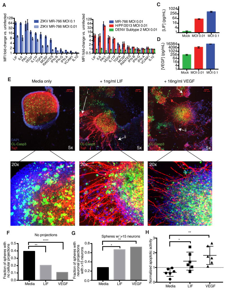
Zika virus (ZIKV) infection during pregnancy is linked to microcephaly, which is attributed to infection of developing brain structures. ZIKV infects neural progenitor cells in vitro, though its effects on other developmentally relevant stem cell populations, including cranial neural crest cells (CNCCs), have not been assessed. CNCCs give rise to most cranial bones and exert paracrine effects on the developing brain. Here, we report that CNCCs are productively infected by ZIKV, but not by the related dengue virus. ZIKV-infected CNCCs undergo limited apoptosis but secrete cytokines that promote death and drive aberrant differentiation of neural progenitor cultures. Addition of two such cytokines, LIF or VEGF, at levels comparable to those secreted by ZIKV-infected CNCCs is sufficient to recapitulate premature neuronal differentiation and apoptotic death of neural progenitors. Thus, our results suggest that CNCC infection by ZIKV may contribute to associated embryopathies through signaling crosstalk between developing face and brain structures.

Keywords: Zika virus; cytokines; growth factors; microcephaly; neural development; stem cells.


Figures

Figure 1
Figure 1. Zika virus (ZIKV) productively infects cranial neural crest cells (CNCCs)
A. Model of in vitro human neural development. B. Embryonic stem cells (ESCs), neurospheres, and CNCCs were infected with ZIKV at MOI 0.1 (or mock infected in PBS) for 1 hour, and incubated for 23 hours at 37C. Infection was detected by intracellular staining for ZIKV with anti-flavivirus group surface antigen and analyzed by flow cytometry. See also Figure S1A. C. Viral titers in culture supernatants as determined by plaque assay on Vero cells. Bars represent mean titer from two technical replicates of supernatants from two independent biological experiments. Plaque formation was not detected (N.D.) in supernatants from mock-infected controls. Error bars are S.D. D. CNCCs were infected with ZIKV or DENV at matched MOI (or mock infected in PBS) for 1 hour, followed by 23 hours of incubation at 37°C. Infection was detected by intracellular staining for ZIKV with anti-flavivirus group surface antigen and flow cytometry. Data is from a representative experiment. E. Summary of infections of CNCCs. Points represent independent biological experiments. ZIKV vs. mock, p = 0.0078, Wilcoxon paired signed-rank test. ZIKV vs. DENV, p = 0.044, Mann-Whitney test. (Mock and ZIKV n=10, DENV n=2). *p < 0.05; **p < 0.01. F–G. Quantifications of flow cytometry analysis of apoptotic death at 24 hours (F) and 72 hours (G) after infection. Cells were stained with Annexin V and propidium iodide to mark early apoptosis (Annexin V+/PI−), late apoptosis (Annexin V+/PI+) and non-apoptotic dead cells (Annexin V−/PI+). See also Figure S1E. H. Confocal immunofluorescent images of neurospheres after 72 hours of transwell culture. Neurospheres were incubated alone (left), or with uninfected (center) and ZIKV-infected (right) CNCCs on the transwell membrane. Neurospheres were stained for cleaved caspase-3 (green), Tuj1 (red), and DAPI (blue). See Figure S1F for ZIKV-infected neurospheres alone.
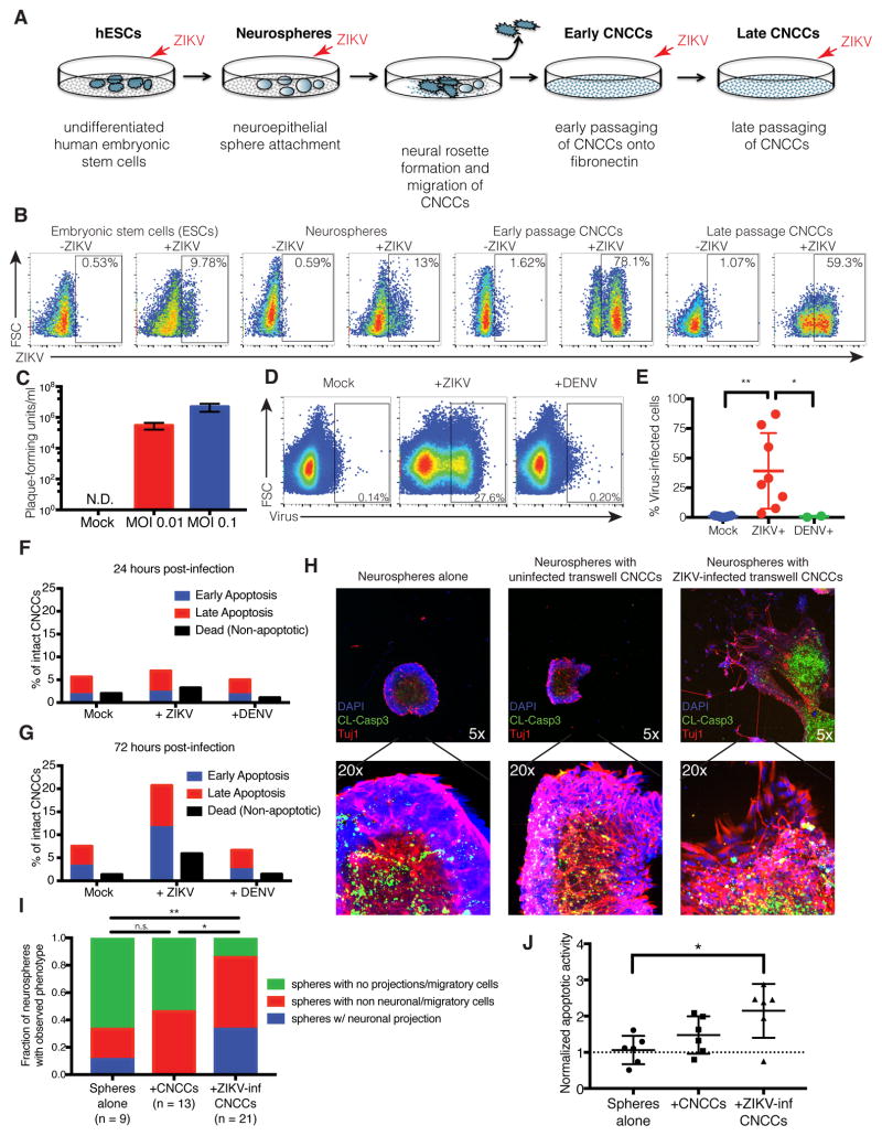
  1. Example of non-neuronal migratory cellular outgrowth

  2. Example of neuronal projections

I. Quantification of neurosphere morphology after exposure to transwell CNCCs. Spheres alone vs. +ZIKV-inf CNCCs, p = 0.0041, chi-squared test of equal proportions; uninfected CNCCs vs. ZIKV-inf CNCCs, p = 0.0139, chi-squared test of equal proportions. *p < 0.05; **p < 0.01. J. Quantification of apoptotic activity in neurospheres. Confocal image z-stacks were analyzed for the intensity of Tuj1 and caspase signals. Caspase-3 intensities were normalized by Tuj1 signal (in the central bolus of the neurosphere) to account for differences in neurosphere size. Spheres alone vs. +ZIKV-inf CNCCs, p = 0.0260, Mann-Whitney test; uninfected CNCCs vs. ZIKV-inf CNCCs, p = 0.1320, Mann-Whitney test. *p < 0.05.
Figure 2
Figure 2. ZIKV-Infected CNCCs secrete LIF and VEGF at high concentrations, promoting apoptosis and premature neuronal formation in neurospheres
A. Cell culture supernatants were collected from CNCCs infected with ZIKV at MOI 0.1 or 0.01 or mock infected for 24 hours. Secreted cytokine levels were measured by a 63-plex Luminex assay. Shown here are analytes with >5-fold induction, IFNs, and VCAM1 and IL-10 for comparison. Data is represented as mean fluorescent intensity (MFI) fold-change compared to uninfected sample. Each bar represents mean MFI from two technical replicates from each of two independent biological experiments. Error bars represent S.D. See also Figure S2A. B. Luminex fold-changes 24 hours after infection with MOI 0.01 ZIKV or DENV or mock infection. Data is represented as mean fluorescent intensity (MFI) fold-change compared to uninfected sample. Each bar represents mean MFI from two technical replicates from each of two independent biological experiments. Error bars represent S.D. See also Figure S2F. C–D. Concentrations of (C) LIF and (D) VEGF in culture supernatants after 24 hour ZIKV infection at MOI 0.1, 0.01 or mock infection. Values represent mean concentration as determined from standard curves for each analyte. Error bars represent S.E.M. of two independent biological experiments with two technical replicates each. See also Figure S2B–E. E. Neural precursor cells were differentiated from hESCs and grown as neurospheres. Seven days after sphere formation, exogenous LIF or VEGF was added to culture media, and spheres were observed for 3 days, followed by fixation and staining for DNA (DAPI, blue), apoptosis (cleaved caspase-3, green), and β-tubulin to mark neurons (Tuj1, red). Representative images are shown of neurospheres grown in culture media alone (left panel), with 1 ng/ml LIF (center panel), and with 16 ng/ml VEGF (right panel). See also Figure S2G.
  1. Examples of non-neuronal migratory cellular outgrowth

  2. Examples of neuronal outgrowth

F. Images were scored by an objective observer for morphology. Data represents proportions of spheres showing no migratory cell projections or neuronal projections under normal media conditions and after the addition of LIF or VEGF. Chi-squared test of equal proportions. See also Figure S2H-I. G. Images scored positive for neurons were grouped into spheres with low (<15) and high (<15) numbers of neurons. Chi-squared test of equal proportions. See also Figure S2J. H. Quantification of apoptotic activity in neurospheres. Confocal image z-stacks were analyzed for the intensity of Tuj1 and caspase signals. Caspase-3 intensities were normalized by Tuj1 signal (in the central bolus of the neurosphere) to account for differences in neurosphere size. Mann-Whitney test. *p < 0.05; **p < 0.01; ***p <0.001; ****p < 0.0001.

References

    1. Bajpai R, Chen DA, Rada-Iglesias A, Zhang J, Xiong Y, Helms J, Chang CP, Zhao Y, Swigut T, Wysocka J. CHD7 cooperates with PBAF to control multipotent neural crest formation. Nature. 2010;463:958–962. doi: 10.1038/nature08733. - DOI - PMC - PubMed
    1. Bauer S, Patterson PH. Leukemia inhibitory factor promotes neural stem cell self-renewal in the adult brain. J Neurosci. 2006;26:12089–12099. doi: 10.1523/JNEUROSCI.3047-06.2006. - DOI - PMC - PubMed
    1. Brasil P, Pereira JP, Raja Gabaglia C, Damasceno L, Wakimoto M, Ribeiro Nogueira RM, Carvalho de Sequeira P, Machado Siqueira A, Abreu de Carvalho LM, Cotrim da Cunha D, Calvet GA, Neves ES, Moreira ME, Rodrigues Baião AE, Nassar de Carvalho PR, Janzen C, Valderramos SG, Cherry JD, Bispo de Filippis AM, Nielsen-Saines K. Zika Virus Infection in Pregnant Women in Rio de Janeiro - Preliminary Report. new england Journal of Medicine. 2016 doi: 10.1056/NEJMoa1602412. - DOI - PMC - PubMed
    1. Cruz-Oliveira C, Freire JM, Conceição TM, Higa LM, Castanho MARB, Da Poian AT. Receptors and routes of dengue virus entry into the host cells. FEMS Microbiol Rev. 2015;39:155–170. doi: 10.1093/femsre/fuu004. - DOI - PubMed
    1. de Fatima Vasco Aragao M, van der Linden V, Brainer-Lima AM, Coeli RR, Rocha MA, Sobral da Silva P, Durce Costa Gomes de Carvalho M, van der Linden A, Cesario de Holanda A, Valenca MM. Clinical features and neuroimaging (CT and MRI) findings in presumed Zika virus related congenital infection and microcephaly: retrospective case series study. BMJ. 2016;353:i1901. doi: 10.1136/bmj.i1901. - DOI - PMC - PubMed