. 2016 Oct 6;167(2):498-511.e14.
doi: 10.1016/j.cell.2016.09.008. Epub 2016 Sep 29.

Replication-Dependent Unhooking of DNA Interstrand Cross-Links by the NEIL3 Glycosylase

Affiliations

Replication-Dependent Unhooking of DNA Interstrand Cross-Links by the NEIL3 Glycosylase

Daniel R Semlow et al. Cell. .

Abstract

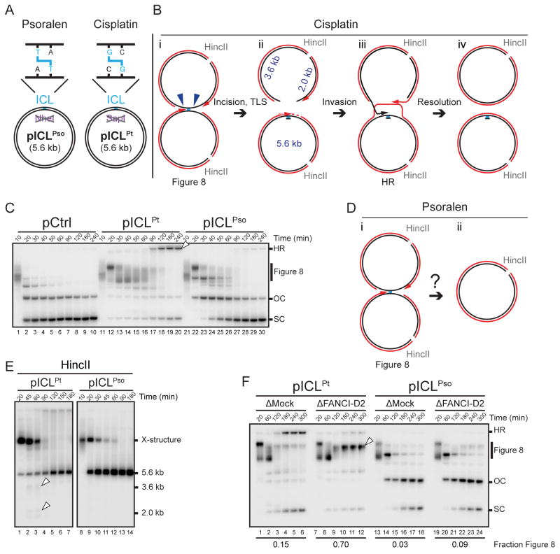
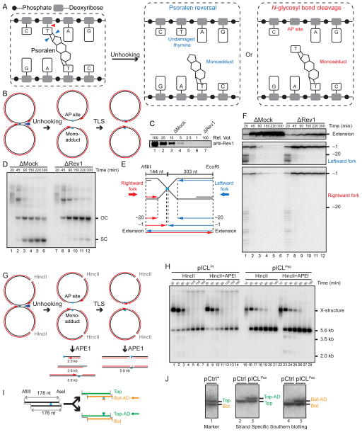
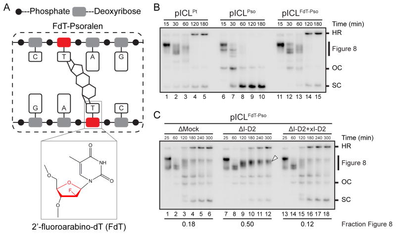
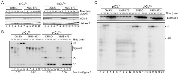
During eukaryotic DNA interstrand cross-link (ICL) repair, cross-links are resolved ("unhooked") by nucleolytic incisions surrounding the lesion. In vertebrates, ICL repair is triggered when replication forks collide with the lesion, leading to FANCI-FANCD2-dependent unhooking and formation of a double-strand break (DSB) intermediate. Using Xenopus egg extracts, we describe here a replication-coupled ICL repair pathway that does not require incisions or FANCI-FANCD2. Instead, the ICL is unhooked when one of the two N-glycosyl bonds forming the cross-link is cleaved by the DNA glycosylase NEIL3. Cleavage by NEIL3 is the primary unhooking mechanism for psoralen and abasic site ICLs. When N-glycosyl bond cleavage is prevented, unhooking occurs via FANCI-FANCD2-dependent incisions. In summary, we identify an incision-independent unhooking mechanism that avoids DSB formation and represents the preferred pathway of ICL repair in a vertebrate cell-free system.

Keywords: DNA interstrand cross-link; FANCD2; FANCI; Fanconi anemia; ICL; NEIL3; abasic site; psoralen.


Figures

Figure 1
Figure 1. A psoralen-ICL is unhooked by an incision-independent pathway that does not require FANCI-D2
(A) Cartoons of pICLPso and pICLPt. NheI and SapI restriction sites coincide with the psoralen-and cisplatin-ICLs, respectively. (B) Cartoon of pICLPt replication intermediates digested with HincII. (C) pCtrl, pICLPt, or pICLPso was replicated in egg extracts with [α-32P]dATP. Replication intermediates were separated on a native agarose gel and visualized by autoradiography. SC, supercoiled. OC, open circular. HR, homologous recombination intermediate, also denoted by white arrowhead. (D) Cartoon of pICLPso replication intermediates digested with HincII. (E) pICLPt or pICLPso was replicated in egg extracts with [α-32P]dATP. Replication intermediates were digested with HincII, separated on a native agarose gel, and visualized by autoradiography. White arrowheads, 3.6 kb and 2.0 kb incision products depicted in (B). (F) pICLPt or pICLPso was replicated in mock or FANCI-D2 depleted egg extracts in the presence of [α-32P]dATP, and the repair intermediates were analyzed as in (C). White arrowhead, Figure 8 DNA structure. The fraction Figure 8 indicates the proportion of Figure 8 structures relative to total species at 300 min.
Figure 2
Figure 2. Psoralen-ICL is unhooked via the cleavage of a DNA N-glycosyl bond
(A) Two possible incision-independent unhooking pathways for a psoralen-ICL. Blue arrowheads (left panel) show unhooking of a psoralen-ICL via breakage of the cyclobutane ring (psoralen reversal), resulting in an undamaged thymine and a monoadduct (middle panel). The red arrowhead (left panel) shows psoralen-ICL unhooking via cleavage of a DNA N-glycosyl bond, resulting in an AP site and a monoadduct (right panel). (B) Model for pICLPso unhooking via cleavage of a DNA N-glycosyl bond, which necessitates gap filling of both daughter molecules by TLS. (C) Mock-depleted and Rev1-depleted NPE were analyzed by Rev1 Western blotting. A relative volume of 100 corresponds to 0.25 μl NPE. (D) pICLPso was replicated in mock or Rev1 depleted egg extracts in the presence of [α-32P]dATP. Repair intermediates were analyzed as in Figure 1C. (E) Schematic illustration of nascent leading strands liberated after digestion of pICLPso with AflIII and EcoRI. (F) pICLPso was replicated in mock- or Rev1-depleted egg extracts in the presence of [α-32P]dATP. Samples were purified and digested with AflIII and EcoRI before separation on a denaturing polyacrylamide gel. Three portions of the autoradiograph are shown with different contrasts for optimal display. (G) Expected species after digestion of pICLPso replication intermediates with HincII and APE1, including 3.6 kb and 2.0 kb fragments. (H) pICLPt or pICLPso was replicated in egg extracts with [α-32P]dATP. Replication intermediates were digested with HincII and APE1, separated on a native agarose gel, and visualized by autoradiography. Quantification of 3.6 kb and 2.0 kb fragments is presented in Figure S1J. (I) Depiction of final repair products after AflIII and AseI digestion. AflIII and AseI generate different sized overhangs, allowing separation of top (178 nt) and bottom (176 nt) strands. Depending on which N-glycosyl bond is cleaved, the mono-adduct is present on the bottom (Bot-AD) or top (Top-AD) strands. (J) Detection of pICLPso mono-adducts. pCtrl or pICLPso was replicated for 3 hours in egg extract. DNA was isolated, digested with AflIII and AseI, separated on a denaturing sequencing gel, and analyzed by strand-specific Southern blotting to visualize the top strand (middle panel) or bottom strand (right panel). The absence of bottom (lane 2) or top strands (lane 4) in the Southern blot of pCtrl indicates the strand specificity of the blotting protocol. To generate size markers for the top (178 nt) and bottom (176 nt) strands (left panel, lane 1), pCtrl was replicated in the presence of [α-32P]dATP (pCtrl*) and analyzed on the same sequencing gel after AflIII and AseI digestion.
Figure 3
Figure 3. Psoralen-ICL undergoes FANCI-D2 dependent processing when the N-glycosyl bond cannot be cleaved
(A) Schematic of a psoralen-ICL formed between two 2′-fluoroarabino-dT (FdT) nucleosides (inset). (B) pICLPt, pICLPso, or pICLFdT-Pso was replicated in egg extracts in the presence of [α-32P]dATP. Repair intermediates were analyzed as in Figure 1C. (C) pICLFdT-Pso was replicated in mock-depleted egg extract, FANCI-D2 depleted extract (ΔI-D2), or ΔI-D2 extract supplemented with xFANCI-D2 (ΔI-D2 + xI-D2) in the presence of [α-32P]dATP, and the repair intermediates were analyzed as in Figure 1C. White arrowhead, Figure 8 DNA structure that persists in the absence of FANCI-D2. The fraction Figure 8 indicates the proportion of Figure 8 structures relative to total species at 300 min.
Figure 4
Figure 4. CMG unloading is not required for psoralen-ICL unhooking
(A) pICLPt or pICLPso (containing 48 lacO repeats) was replicated in the presence or absence of NMS-873 and, at the indicated times, pulled down using LacI-coated beads. Western blotting was performed on chromatin samples with the indicated antibodies. (B) pICLPt and pICLPso were replicated in the presence or absence of NMS-873 with [α-32P]dATP, and repair intermediates were analyzed as in Figure 1C. The fraction Figure 8 indicates the proportion of Figure 8 structures relative to total species at 180 min. (C) DNA samples from (B) were digested with AflIII and EcoRI before separation on a denaturing polyacrylamide gel. Extension products and rightward fork products (see Figure 2E) are shown with different contrast for optimal display.
Figure 5
Figure 5. An AP-ICL is repaired by an incision-independent pathway that does not require FANCI-D2 or CMG unloading
(A) Mechanism of AP-ICL formation. (B) Cartoon of an AP-ICL containing plasmid (pICLAP), in which the SapI restriction site coincides with the ICL. (C) pCtrl, pICLPt, pICLAP, or pICLPso was replicated in egg extracts and analyzed as in Figure 1C. White arrowhead, homologous recombination intermediate that accumulates for pICLAP. (D) pICLAP was replicated in mock-depleted egg extract, FANCI-D2-depleted extract (ΔI-D2), or ΔI-D2 extract supplemented with xFANCI-D2 (ΔI-D2+xI-D2), and analyzed as in Figure 1C. White arrowhead, Figure 8 structures that persist in the absence of FANCI-D2. The fraction Figure 8 indicates the proportion of Figure 8 structures relative to total species at 300 min. (E) Schematic illustration of nascent leading strands liberated by digestion of pICLAP with AflIII. (F) pICLPt and pICLAP were replicated with [α-32P]dATP in the presence or absence of NMS-873. The nascent strands were purified and digested with AflIII before separation on a denaturing polyacrylamide gel. Two portions of the autoradiograph are shown with different contrasts for optimal display. White arrowhead, leftward -1 products. Black arrowhead, rightward -1 products.
Figure 6
Figure 6. An AP-ICL is unhooked via cleavage of the non-native N-glycosyl bond
(A) Expected species after digestion of pICLAP with HincII and APE1, including 3.3 kb and 2.3 kb fragments. (B) pICLAP was replicated in egg extracts and then digested with HincII and APE1 as in Figure 2H. (C) The 3.3 kb and 2.3 kb fragments in (B) were quantified and plotted. (D) Two possible incision-independent unhooking pathways for an AP-ICL. Cleavage of the native N-glycosyl bond (blue arrowhead) yields a non-native adenosine and an AP site (middle panel), whereas cleavage of the non-native N-glycosyl bond (red arrowhead) yields an AP site and a normal adenosine (right panel). (E) Mock-depleted and Rev1-depleted NPE were analyzed by Rev1 Western blotting. A relative volume of 100 corresponds to 0.66 μl NPE. (F) pICLAP was replicated in mock- or Rev1-depleted egg extract in the presence of [α-32P]dATP and NMS-873 and analyzed as in Figure 1C. (G) pICLAP was replicated in mock or Rev1 depleted egg extract in the presence of [α-32P]dATP and NMS-873. Samples were purified and digested with AflIII before separation on a denaturing polyacrylamide gel. Two portions of the autoradiograph are displayed with different contrast for optimal display.
Figure 7
Figure 7. NEIL3 unhooks psoralen- and AP-ICLs
(A) pICLPso or pICLAP was replicated in mock-depleted-, NEIL3-depleted-, or NEIL3-depleted egg extract supplemented with 300 nM recombinant NEIL3 in the presence of [α-32P]dATP. Repair intermediates were analyzed as in Figure 1C. As shown in Figure S7E, 15 nM recombinant NEIL3 also rescued pICLAP unhooking in NEIL3-depleted egg extract. (B) Oligonucleotide substrates containing an AP-ICL were incubated with recombinant wild-type (WT) or K60A mutated (mut) NEIL3, separated on a denaturing polyacrylamide gel, and visualized by autoradiography. Black lines, DNA. Red lines, RNA. Cyan, AP-ICL. Asterisks indicate the 32P radiolabel. Black arrowheads, unhooked ssDNA. Blue arrowheads, β-elimination cleavage product. Bands marked by white arrowheads likely represent substrate that was not denatured during electrophoresis. (C) Model for AP-ICL repair by incision-independent (left branch) or incision-dependent (right branch) pathways. Black lines, parental DNA. Red lines, nascent leading strands. Cyan, AP-ICL. Green ovals, CMG helicase. Pathway utilization is illustrated for unmodified extract (black arrows), FANCI-D2 depleted extract (blue arrows), extract supplemented with NMS-873 (green arrows), and NEIL3-depleted extract (pink arrows). Closed circles indicate point mutations that arise due to bypass of an AP site. Open circles indicate potential point mutations that may be introduced due to bypass of an AP-ICL-derived adenine monoadduct.

References

    1. Alt A, Lammens K, Chiocchini C, Lammens A, Pieck JC, Kuch D, Hopfner KP, Carell T. Bypass of DNA lesions generated during anticancer treatment with cisplatin by DNA polymerase eta. Science. 2007;318:967–970. - PubMed
    1. Brooks SC, Adhikary S, Rubinson EH, Eichman BF. Recent advances in the structural mechanisms of DNA glycosylases. Biochim Biophys Acta. 2013;1834:247–271. - PMC - PubMed
    1. Budzowska M, Graham TG, Sobeck A, Waga S, Walter JC. Regulation of the Rev1-pol zeta complex during bypass of a DNA interstrand cross-link. EMBO J. 2015;34:1971–1985. - PMC - PubMed
    1. Cimino GD, Shi YB, Hearst JE. Wavelength dependence for the photoreversal of a psoralen-DNA cross-link. Biochemistry. 1986;25:3013–3020. - PubMed
    1. Clauson C, Scharer OD, Niedernhofer L. Advances in understanding the complex mechanisms of DNA interstrand cross-link repair. Cold Spring Harb Perspect Biol. 2013;5:a012732. - PMC - PubMed

Publication types

MeSH terms