Review
. 2017 Jan 10;25(1):11-26.
doi: 10.1016/j.cmet.2016.08.016. Epub 2016 Sep 29.

Tissue Immunometabolism: Development, Physiology, and Pathobiology

Affiliations
Review

Tissue Immunometabolism: Development, Physiology, and Pathobiology

Kevin Man et al. Cell Metab. .

Abstract

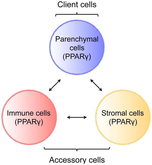
Evolution of metazoans resulted in the specialization of cellular and tissue function. This was accomplished by division of labor, which allowed tissue parenchymal cells to prioritize their core functions while ancillary functions were delegated to tissue accessory cells, such as immune, stromal, and endothelial cells. In metabolic organs, the accessory cells communicate with their clients, the tissue parenchymal cells, to optimize cellular processes, allowing organisms to adapt to changes in their environment. Here, we discuss tissue immunometabolism from this vantage point and use examples from adipose tissues (white, beige, and brown) and liver to outline the general principles by which accessory cells support metabolic homeostasis in parenchymal cells. A corollary of this model is that disruption of communication between client and accessory cells might predispose metabolic organs to the development of disease.


Figures

Figure 1
Figure 1. The core functional unit of metabolic organs
Metabolic organs, such as WAT, BAT, beige fat, and liver, are seeded by immune and stromal cells during development. These accessory cells support their clients, the tissue parenchymal cells. Communication between client and accessory cells is mediated by chemokines, cytokines, growth factors, hormones, small molecules, and direct contact. In addition, expression of common sensors of the metabolic state allows for coordinated sensing of metabolites across the three cell types. For example, in adipose tissue (designated by parentheses), communication between parenchymal, immune, and stromal cells is mediated by PPARγ, which coordinates cellular responses by sensing changes in fatty acids and fatty acid metabolites.
Figure 2
Figure 2. Tissue-specific programming of immune cells in WAT
(A) Local signals (the “inducers”) induce expression of transcription factors (the “effectors”) that confer tissue-specific identity to immune cells. (B) Maturation of adipose tissue macrophages (ATMs) is dependent of type 2 cytokines, IL-4 and IL-13, and PPARγ. ILC2-derived IL-13 and eosinophil-derived IL-4 signal via the IL-4Rα in ATMs to induce PPARγ, which cooperates with lineage-specific transcription factor PU.1 to direct expression of adipose tissue-specific gene expression in ATMs. (C) Maturation of adipose tissue (AT) Tregs is dependent on IL-33 and PPARγ. Stromal cell-derived IL-33 signals via ST2, the receptor for IL-33, to induced expression of PPARγ in Tregs, which cooperates with lineage-specific transcription factor FOXP3 to direct transcriptional programs in adipose tissue Tregs. (D) Maturation of ILC2s in adipose tissue. ILC2s are known to express PPARγ, however, it is not known whether its expression is regulated by IL-33. IL-33 and ST2 are required for the maintenance of adipose tissue ILC2s. PPARγ might cooperate with GATA3, which is necessary for the development of ILC2s, to regulate adipose tissue-specific gene expression in ILC2s.
Figure 3
Figure 3. Interactions between immune and stromal cells regulate thermogenesis
(A) A model of how interactions between immune and stromal cells regulate biogenesis of beige fat. Type 2 immune cells, including ILC2s, eosinophils (Eos), and alternatively activated macrophages (Macs), communicate with stromal cells and beige adipocytes to regulate remodeling of inguinal WAT into thermogenic beige fat. Signaling via the IL-4Rα in macrophages regulates catecholamine biosynthesis and secretion. IL-4/13 signaling in PDGFRα+ cells regulates the numbers and fate of adipocyte precursors in WAT. Differentiation and activation of beige adipocytes is stimulated by myeloid-cell derived norepinephrine (NE) and ILC2-derived methionine-enkephalin (Met-Enk). (B) IL-33 and ST2 regulate licensing of beige and brown adipocytes for uncoupled respiration. IL-33 is an atypical cytokine that is normally located in the nucleus. Its release from cells results in extracellular cleavage by proteases derived from mast cells and neutrophils. Cleaved IL-33 can bind to cell surface receptor ST2 to initiate downstream responses via the adaptor protein MyD88. In BAT, IL-33 and ST2 are required for perinatal licensing of uncoupled respiration by controlling the splicing of Ucp1 mRNA. Although it is not known how IL-33 and ST2 regulate the splicing of Ucp1 mRNA, it is likely to be a non-canonical function of IL-33 because it does not require the adaptor protein MyD88. While it is likely that stromal cells are the cellular source of IL-33, this remains to be determined.
Figure 4
Figure 4. Metabolic and immune zonation of liver
(A) Model of how metabolic and immune functions might be zonated in the liver. (B) Metabolic zonation of liver. In the periportal region, portal vein and hepatic artery provide nutrients and oxygen, respectively, whereas the pericentral region is oxygen-poor. In general, energy consuming catabolic functions are localized to the periportal region, whereas anabolic functions are performed by hepatocytes in the pericentral region. Pericentral zonation is regulated by the Wnt-β-catenin/Tcf pathway, whereas the HRas pathway controls the zonation program around the periportal region. Enzymes or enzymatic activities that show preferential localization to the periportal or pericentral zones are listed next to the metabolic programs they participate in. PEPCK- posphoenolpyruvate carboxy- kinase, G6PC- glucose-6- phosphatase, FBP1- fructosel,6-bisphosphatase, GK-glucokinase, CPT1- carnitine palmitoyltransferase I, FAS-fatty acid synthase, ACC- acetyl-CoA carboxylase, ACL- ATP-dependent citrate lyase, CPS- Carbamoylphosphate synthetase, GS- Glutamine synthetase.

References

    1. Agarwal P, Khan SR, Verma SC, Beg M, Singh K, Mitra K, Gaikwad AN, Akhtar MS, Krishnan MY. Mycobacterium tuberculosis persistence in various adipose depots of infected mice and the effect of anti-tubercular therapy. Microbes Infect. 2014;16:571–580. - PubMed
    1. Andrews NC. Forging a field: the golden age of iron biology. Blood. 2008;112:219–230. - PMC - PubMed
    1. Ardawi MS, Newsholme EA. Glutamine metabolism in lymphocytes of the rat. Biochem J. 1983;212:835–842. - PMC - PubMed
    1. Arrese EL, Soulages JL. Insect fat body: energy, metabolism, and regulation. Annu Rev Entomol. 2010;55:207–225. - PMC - PubMed
    1. Bapat SP, Myoung Suh J, Fang S, Liu S, Zhang Y, Cheng A, Zhou C, Liang Y, LeBlanc M, Liddle C, et al. Depletion of fat-resident Treg cells prevents age-associated insulin resistance. Nature. 2015;528:137–141. - PMC - PubMed

Publication types

LinkOut - more resources