Review
. 2017 Oct:173:42-56.
doi: 10.1016/j.jsbmb.2016.09.024. Epub 2016 Sep 28.

Endogenously produced nonclassical vitamin D hydroxy-metabolites act as "biased" agonists on VDR and inverse agonists on RORα and RORγ

Affiliations
Review

Endogenously produced nonclassical vitamin D hydroxy-metabolites act as "biased" agonists on VDR and inverse agonists on RORα and RORγ

Andrzej T Slominski et al. J Steroid Biochem Mol Biol. 2017 Oct.

Abstract

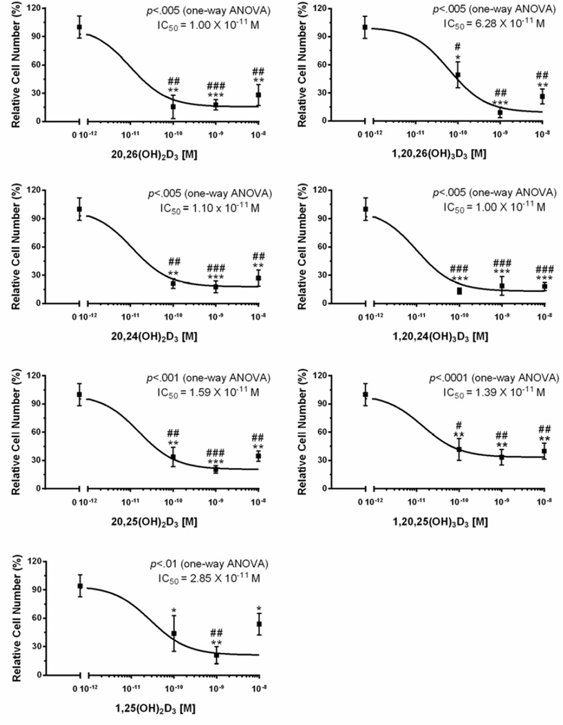
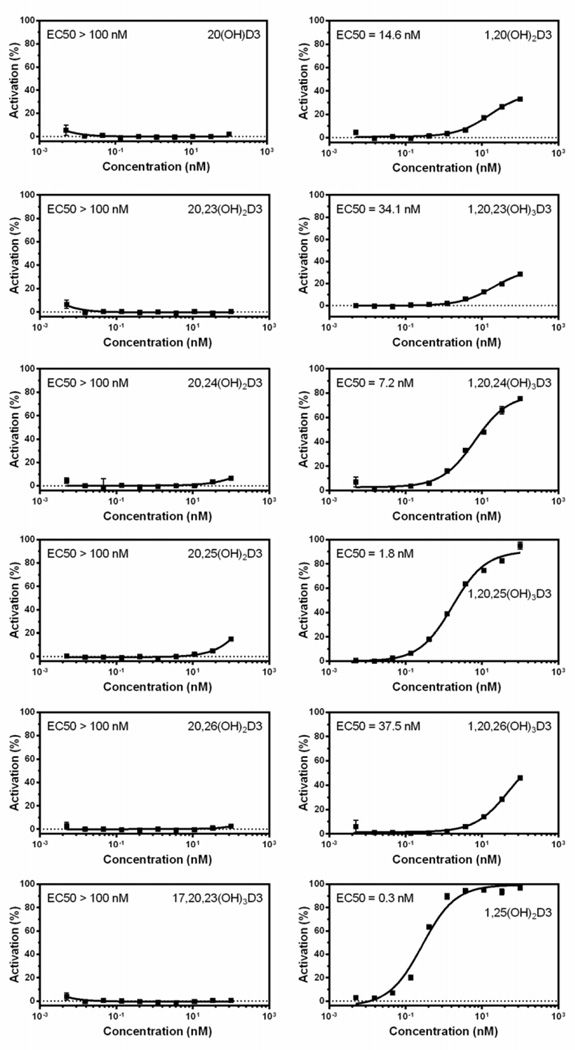
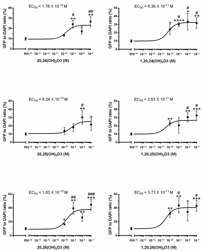
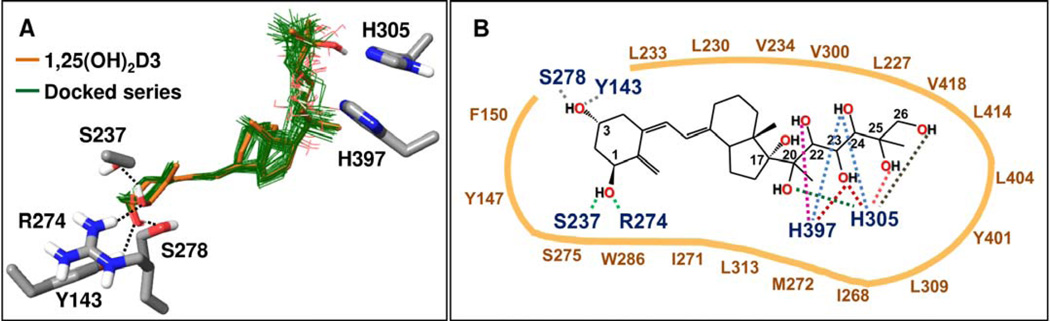
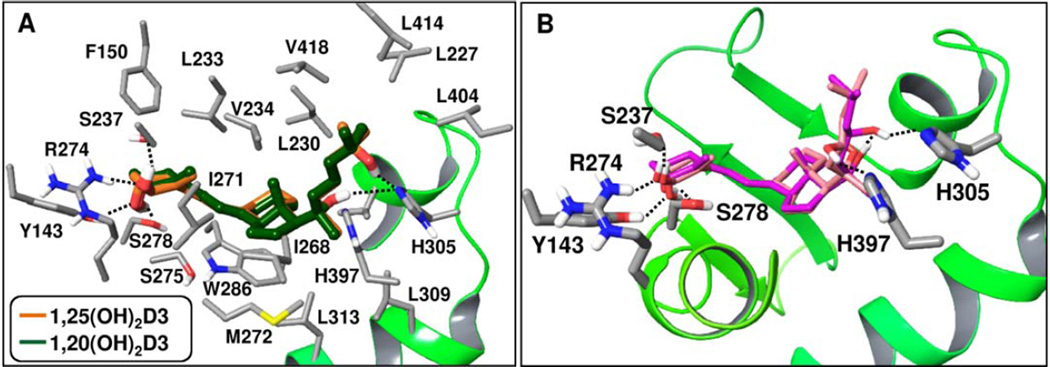
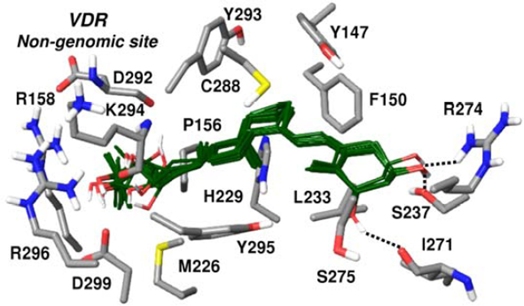
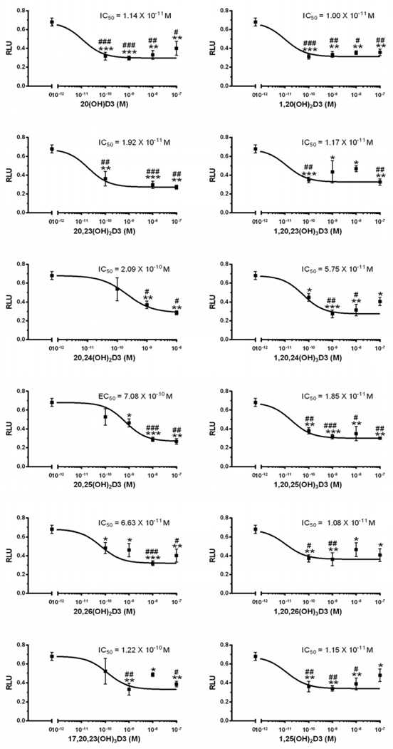
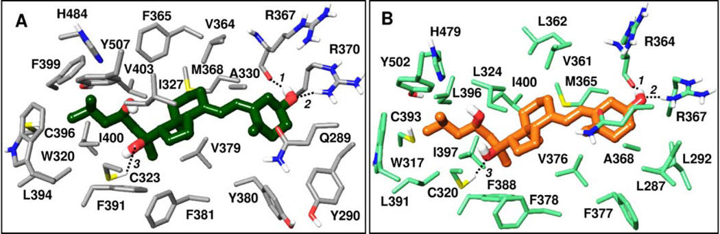
The classical pathway of vitamin D activation follows the sequence D3→25(OH)D3→1,25(OH)2D3 with the final product acting on the receptor for vitamin D (VDR). An alternative pathway can be started by the action of CYP11A1 on the side chain of D3, primarily producing 20(OH)D3, 22(OH)D3, 20,23(OH)2D3, 20,22(OH)2D3 and 17,20,23(OH)3D3. Some of these metabolites are hydroxylated by CYP27B1 at C1α, by CYP24A1 at C24 and C25, and by CYP27A1 at C25 and C26. The products of these pathways are biologically active. In the epidermis and/or serum or adrenals we detected 20(OH)D3, 22(OH)D3, 20,22(OH)2D3, 20,23(OH)2D3, 17,20,23(OH)3D3, 1,20(OH)2D3, 1,20,23(OH)3D3, 1,20,22(OH)3D3, 20,24(OH)2D3, 1,20,24(OH)3D3, 20,25(OH)2D3, 1,20,25(OH)3D3, 20,26(OH)2D3 and 1,20,26(OH)3D3. 20(OH)D3 and 20,23(OH)2D3 are non-calcemic, while the addition of an OH at C1α confers some calcemic activity. Molecular modeling and functional assays show that the major products of the pathway can act as "biased" agonists for the VDR with high docking scores to the ligand binding domain (LBD), but lower than that of 1,25(OH)2D3. Importantly, cell based functional receptor studies and molecular modeling have identified the novel secosteroids as inverse agonists of both RORα and RORγ receptors. Specifically, they have high docking scores using crystal structures of RORα and RORγ LBDs. Furthermore, 20(OH)D3 and 20,23(OH)2D3 have been tested in a cell model that expresses a Tet-on RORα or RORγ vector and a RORE-LUC reporter (ROR-responsive element), and in a mammalian 2-hybrid model that test interactions between an LBD-interacting LXXLL-peptide and the LBD of RORα/γ. These assays demonstrated that the novel secosteroids have ROR-antagonist activities that were further confirmed by the inhibition of IL17 promoter activity in cells overexpressing RORα/γ. In conclusion, endogenously produced novel D3 hydroxy-derivatives can act both as "biased" agonists of the VDR and/or inverse agonists of RORα/γ. We suggest that the identification of large number of endogenously produced alternative hydroxy-metabolites of D3 that are biologically active, and of possible alternative receptors, may offer an explanation for the pleiotropic and diverse activities of vitamin D, previously assigned solely to 1,25(OH)2D3 and VDR.

Keywords: 1,25D3-MARRS; CYP11A1; Hydroxyvitamin D; RORα; RORγ; VDR.


Conflict of interest statement

Conflict of Interest:

The authors declare no conflict of interest

Figures

Figure 1
Figure 1
Hydroxymetabolites of 20(OH)D3 inhibit keratinocytes proliferation. The cells were cultured for 48 h in the presence or absence of 1α,25(OH)2D3, 20S,25(OH)2D3, 1α,20S,25(OH)3D3, 20S,26(OH)2D3, 1α,20S,26(OH)3D3, 20S,24R(OH)2D3, or 1α,20S,24R(OH)3D3 and cell proliferation was measured by the MTS assay as described in the supplemental file and in [90, 92]. Data represent means ± SE (n=3–4). *, p<0.05; **, p<0.01; ***, p<0.001 at student t-test. #, p<0.05; ##, p<0.01; ###, p<0.001 at one-way ANOVA test.
Figure 2
Figure 2
Only the 1α(OH)-derivatives of 20(OH)D3 hydroxymetabolites increase the affinity of the co-activator peptide to the VDR-LBD in the LanthaScreen TR-FRET Vitamin D receptor Coactivator kit assay (Thermo Fisher Scientific, Inc., Waltham, MA) (for details see supplemental file).
Figure 3
Figure 3
Hydroxymetabolites of 20(OH)D3 stimulate VDR-GFP translocation from the cytoplasm to the nucleus in the SKMEL-188 melanoma line stably overexpressing VDR-GFP [53]. The cells were incubated for 90 min in the presence of 20S,25(OH)2D3, 1α,20S,25(OH)3D3, 20S,26(OH)2D3,1α,20S,26(OH)3D3, 20S,24R(OH)2D3 or 1α,20S,24R(OH)3D3, or vehicle (ethanol) and translocation to the nucleus was measured as described in [92] with modifications listed in the supplemental file Data represent means ± SE (n=3–4). *, p<0.05; **, p<0.01; ***, p<0.001 at student t-test. #, p<0.05; ##, p<0.01; ###, p<0.001 at one-way ANOVA test.
Figure 4
Figure 4
Common binding mode predicted for hydroxy-D3 metabolites in the genomic site of the VDR crystal structure (PDB code 1DB1). (A) Docked poses of hydroxy-D3 metabolites (listed in Table 2) are shown with thin bonds and green colored carbons. For comparison, the co-crystallized 1,25(OH)2D3 is shown with thick bonds and light brown colored carbons. All other atoms are colored by atom type (O: red, N: blue, S: yellow, H: white). Only VDR residues involved in polar interactions with the ligands are included. Hydrogen bonding interactions of 1α(OH) and 3(OH) groups common in docked poses are indicated with dashed lines. (B) Schematic summary of interactions formed by docked analogs listed in Table 2 in the genomic site of VDR. Interactions formed between VDR residues and OH substituents in docked poses of the analog series are indicated with dashed lines, colored distinctly for each OH group. For example, 24(OH) in poses of some analogs interact with H397 while in case of other analogs with H305 (light blue dashed lines). VDR residue labels are color coded: those involved in polar interactions are dark blue; those contributing to non-polar interactions are dark brown.
Figure 5
Figure 5
Poses of selected hydroxy-D3 metabolites are shown in the genomic site of VDR (PDB code 1DB1). (A) Docked pose of 1α,20S(OH)2D3 shown in comparison with the co-crystallized 1,25(OH)2D3 in the VDR crystal structure (PDB code 1DB1). Carbon atoms in the docked pose are colored dark green, in the co-crystallized ligand light brown; all other atoms are colored by atom types (as in Fig. 4). Hydrogen bonding interactions are shown with dashed lines. In addition to residues involved in hydrogen bonding with shown ligands, all VDR residues that form non-polar contacts with the docked 1α,20S(OH)2D3 are also displayed. (B) Two possible docked poses obtained for 1α,20S,23R(OH)3D3 are shown simultaneously in the VDR genomic pocket, with their carbon atoms colored distinctly. Hydrogen bonding interactions are indicated with dashed lines. Only VDR residues involved in polar interactions are shown.
Figure 6
Figure 6
Docked poses of hydroxy-D3 metabolites in the non-genomic site of VDR, where the VDR crystal structure (PDB code 1DB1) was refined as described under Computational Methods (Supplemental file). VDR residues shown form polar or non-polar interactions with the docked ligands. Carbons in the docked poses are colored dark green; all other atoms are colored by atom type (as in Fig. 4). Hydrogen bonding interactions involving 1α(OH) and 3(OH) groups shared in docked poses are indicated with dashed lines.
Figure 7
Figure 7
Effect of vitamin D3 hydroxyderivatives on ROR-RE activity in HaCaT cells. The cells were grown on 96 well white plates in DMEM media containing 5% charcoal-treated FBS. After transfection with ROR-RE and a Renilla luciferase construct, the cells were treated with the compounds for 48 h followed by luciferase assay. Methodological details are in supplemental file. *, p<0.05; **, p<0.01; ***, p<0.001 at student t-test. #, p<0.05; ##, p<0.01; ###, p<0.001 at one-way ANOVA test.
Figure 8
Figure 8
20S,23R(OH)2D3 docked in the active site of (A) RORα – dark green colored ligand carbons, and (B) RORγ – light brown colored ligand carbons. Atoms other than carbon are colored by atom type (as in Fig. 4). All residues contributing through polar or non-polar interactions to the binding of the docked ligand are displayed. Distances between hydrogen bonding atoms are as shown via dashed lines between the interacting atoms: (A) Pose at RORα: 1. 2.1 Å, 2. 1.9 Å, 3. 2.5 Å; (B) Pose at RORγ: 1. 1.8 Å, 2. 1.7 Å, 3. 2.0 Å.

References

    1. Holick MF, MacLaughlin JA, Doppelt SH. Regulation of cutaneous previtamin D3 photosynthesis in man: skin pigment is not an essential regulator. Science. 1981;211:590–593. - PubMed
    1. Holick MF. The cutaneous photosynthesis of previtamin D3: a unique photoendocrine system. J Invest Dermatol. 1981;77:51–58. - PubMed
    1. Holick MF. Biological Effects of Sunlight, Ultraviolet Radiation, Visible Light, Infrared Radiation and Vitamin D for Health. Anticancer Res. 2016;36:1345–1356. - PubMed
    1. Holick MF. Vitamin D: A millenium perspective. J Cell Biochem. 2003;88:296–307. - PubMed
    1. Bikle DD. Vitamin D: an ancient hormone. Experimental Dermatology. 2011;20:7–13. - PubMed

Publication types

MeSH terms

Substances

LinkOut - more resources