Clinical Trial
. 2017 Sep;66(9):1542-1554.
doi: 10.1136/gutjnl-2016-312595. Epub 2016 Sep 30.

Hypoxia-inducible factor-2α plays a role in mediating oesophagitis in GORD

Affiliations
Clinical Trial

Hypoxia-inducible factor-2α plays a role in mediating oesophagitis in GORD

Xiaofang Huo et al. Gut. 2017 Sep.

Abstract

Objective: In an earlier study wherein we induced acute reflux by interrupting proton pump inhibitor (PPI) therapy in patients with reflux oesophagitis (RO) healed by PPIs, we refuted the traditional concept that RO develops as an acid burn. The present study explored our alternative hypothesis that RO results from reflux-stimulated production of pro-inflammatory molecules mediated by hypoxia-inducible factors (HIFs).

Design: Using oesophageal biopsies taken from patients in our earlier study at baseline and at 1 and 2 weeks off PPIs, we immunostained for HIF-1α, HIF-2α and phospho-p65, and measured pro-inflammatory molecule mRNAs. We exposed human oesophageal squamous cell lines to acidic bile salts, and evaluated effects on HIF activation, p65 function, pro-inflammatory molecule production and immune cell migration.

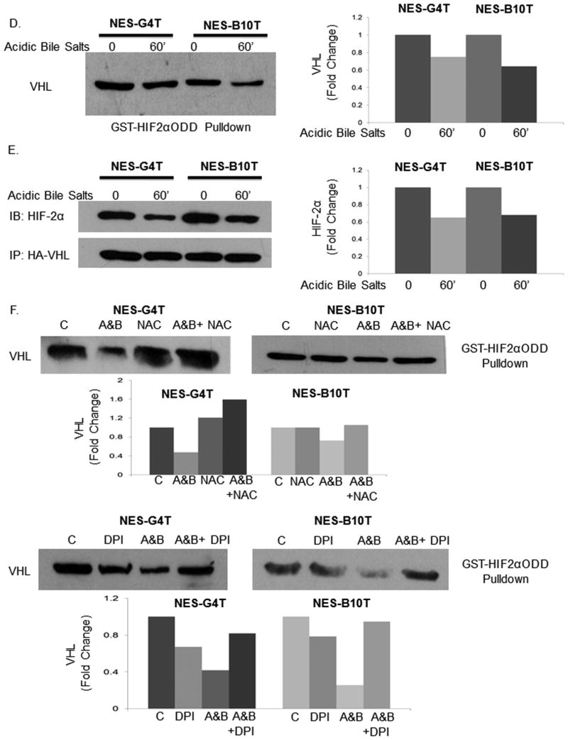
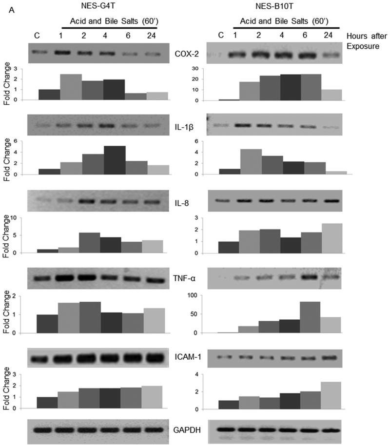
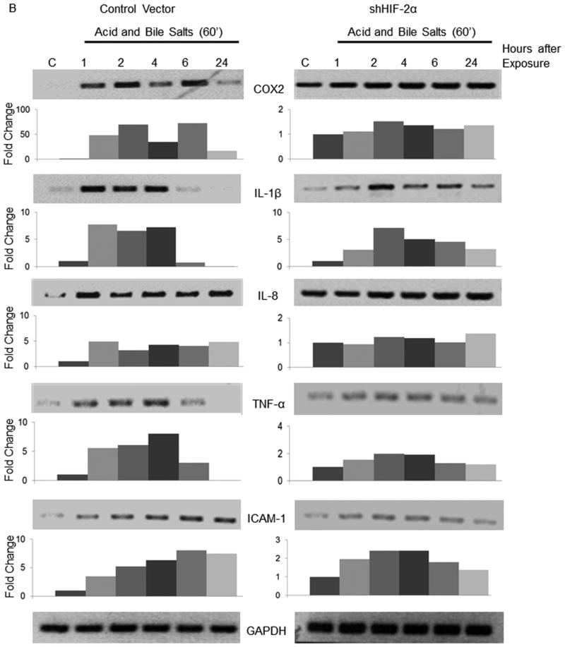
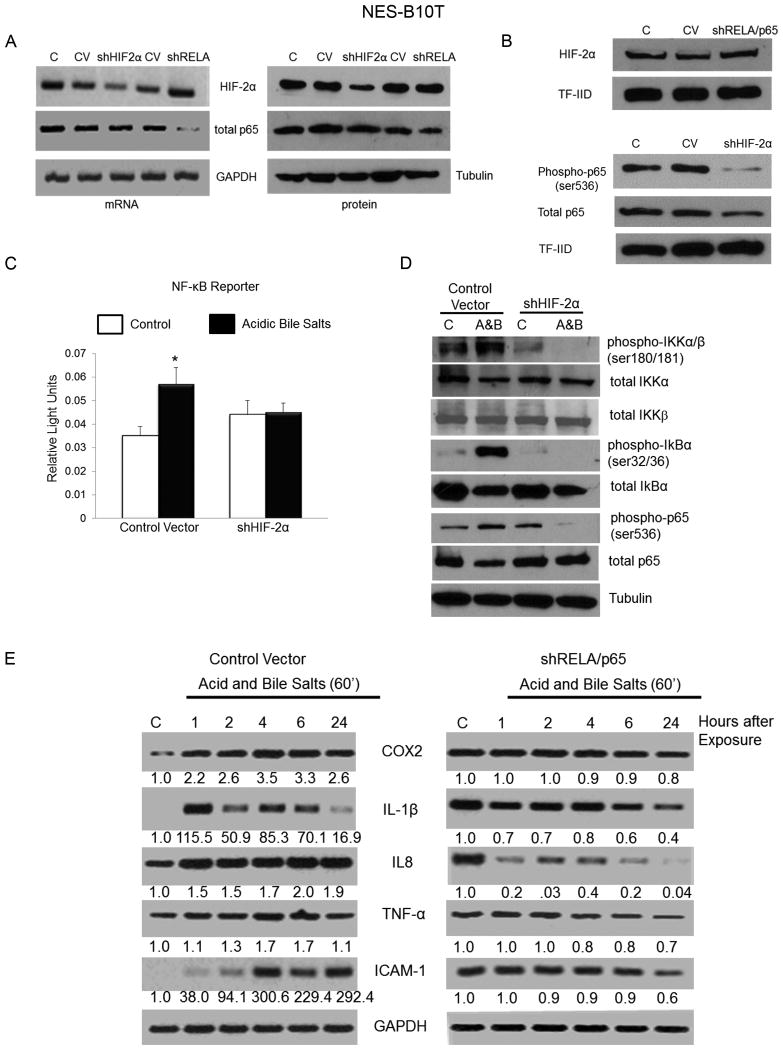
Results: In patient biopsies, increased immunostaining for HIF-2α and phospho-p65, and increased pro-inflammatory molecule mRNA levels were seen when RO redeveloped 1 or 2 weeks after stopping PPIs. In oesophageal cells, exposure to acidic bile salts increased intracellular reactive oxygen species, which decreased prolyl hydroxylase function and stabilised HIF-2α, causing a p65-dependent increase in pro-inflammatory molecules; conditioned media from these cells increased T cell migration rates. HIF-2α inhibition by small hairpin RNA or selective small molecule antagonist blocked the increases in pro-inflammatory molecule expression and T cell migration induced by acidic bile salts.

Conclusions: In patients developing RO, increases in oesophageal HIF-2α correlate with increased pro-inflammatory molecule expression. In oesophageal epithelial cells, acidic bile salts stabilise HIF-2α, which mediates expression of pro-inflammatory molecules. HIF-2α appears to have a role in RO pathogenesis.

Trial registration number: NCT01733810; Results.

Keywords: CELL SIGNALLING; CYTOKINES; INFLAMMATORY DISEASES; OESOPHAGEAL DISEASE.


Figures

Figure 1
Figure 1
Development of acute reflux oesophagitis is associated with increased levels of HIF-2α in patient oesophageal biopsy specimens. (A) Patient flow chart. (B) HIF-2α and HIF-1α immunohistochemistry in biopsies of squamous oesophagus from patients with healed reflux oesophagitis at baseline (on PPIs), and at 1 and 2 weeks after stopping PPIs. Scale bar: 200 μM (C) Quantitation of HIF-1α and HIF-2α staining in patient biopsies using H scores. Bar graphs depict mean ± SEM. NS, not significant. *, p≤0.05 compared with baseline
Figure 2
Figure 2
Induction of acute reflux oesophagitis increases pro-inflammatory mediator mRNA expression that correlates with increased HIF-2α protein in patient oesophageal biopsies. Representative PCR and semi-quantitative analyses for (A) IL-8, (B) IL-1β, (C) TNF-α, (D) COX-2 and (E) ICAM-1 expression at baseline (on PPIs), and at 1 and 2 weeks after stopping PPIs. Bar graphs depict mean ± SEM values for all 12 patients. *, p≤0.05; **, p≤0.01 compared with baseline (F) Table demonstrating eta scores (n=12) for associations between HIF-2α protein levels and pro-inflammatory molecule mRNA levels in patients' oesophageal biopsies. Eta2 × 100% indicates percent of variance in mRNA changes explained by changes in HIF-2α; values ≥14% indicate a large effect.
Figure 3
Figure 3
In NES-G4T and NES-B10T cells, acidic bile salts (A&B) increase nuclear levels of HIF-2α protein by reducing PHD function through ROS generated via the NADPH oxidase system. Representative Western blot and semi-quantitative analysis of (A) total HIF-2α, (B) nuclear HIF-2α, (C) PHD1&2, (D) VHL binding to hydroxylated HIF-2αODD, (E) HIF-2α binding to HA-tagged VHL, (F) VHL binding to hydroxylated HIF-2αODD in the presence of NAC or in the presence of DPI. Note that the densitometry scales vary among samples. Western blots shown are representative of the results from 2 independent experiments.
Figure 3
Figure 3
In NES-G4T and NES-B10T cells, acidic bile salts (A&B) increase nuclear levels of HIF-2α protein by reducing PHD function through ROS generated via the NADPH oxidase system. Representative Western blot and semi-quantitative analysis of (A) total HIF-2α, (B) nuclear HIF-2α, (C) PHD1&2, (D) VHL binding to hydroxylated HIF-2αODD, (E) HIF-2α binding to HA-tagged VHL, (F) VHL binding to hydroxylated HIF-2αODD in the presence of NAC or in the presence of DPI. Note that the densitometry scales vary among samples. Western blots shown are representative of the results from 2 independent experiments.
Figure 4
Figure 4
Acidic bile salts induce HIF-2α and increase expression of pro-inflammatory mediators by oesophageal squamous epithelial cells. PCR and semi-quantitative analysis (depicted beneath individual gels) of expression of pro-inflammatory mediators after (A) acidic bile salts, (B) stable infection with a control vector or shHIF-2α, and (C) treatment with active or inactive enantiomers of a small molecule HIF-2α antagonist and acidic bile salts. Note that the densitometry scales vary among samples. PCR gels shown are representative of the results from 2 independent experiments.
Figure 4
Figure 4
Acidic bile salts induce HIF-2α and increase expression of pro-inflammatory mediators by oesophageal squamous epithelial cells. PCR and semi-quantitative analysis (depicted beneath individual gels) of expression of pro-inflammatory mediators after (A) acidic bile salts, (B) stable infection with a control vector or shHIF-2α, and (C) treatment with active or inactive enantiomers of a small molecule HIF-2α antagonist and acidic bile salts. Note that the densitometry scales vary among samples. PCR gels shown are representative of the results from 2 independent experiments.
Figure 5
Figure 5
Crosstalk between HIF-2α and RELA/p65 regulates expression of pro-inflammatory mediators in oesophageal squamous cells. (A) PCR and Western blot analyses of whole cell lysates demonstrating efficiency of HIF-2α and RELA/p65 knockdown by shRNA in NES-B10T cells. Western blot analysis of (B) nuclear HIF-2α in cells after stable infection with either a control vector (CV) or shRELA/p65 or nuclear phospho-and total p65 in cells after stable infection with either a control vector or shHIF-2α. (C) Luciferase reporter assay for NF-κB activity in response to acidic bile salts in cells stably infected with either a control vector or shHIF-2α. (D) Western blot analysis of NF-κB signaling proteins after exposure to acidic bile salts in cells stably infected with either a control vector or shHIF-2α. (E) PCR and semi-quantitative analyses of pro-inflammatory mediator expression after exposure to acidic bile salts in cells stably infected with shRELA/p65 or a control vector. Bar graphs depict mean ± SEM (n=3). *, p≤0.05 compared with control vector. PCR gels and Western blots shown are representative of the results from 2 independent experiments.
Figure 6
Figure 6
Development of acute reflux oesophagitis is associated with increased expression of phospho-p65 in patient oesophageal biopsies. (A) Immunohistochemical staining for phospho-p65 and total p65 in squamous oesophagus from reflux oesophagitis patients at baseline (on PPIs) and at 1 and 2 weeks off PPIs. Scale bar = 200 μm (B) Quantitation of phospho-p65/total p65 in patient oesophageal biopsies using an H score. (C) Table demonstrating eta scores for associations between phospho-p65/total p65 protein levels and pro-inflammatory mediator mRNA levels in patients' oesophageal biopsies. Eta2 × 100% indicates percent of variance in mRNA changes explained by changes in phospho-p65/total p65; values ≥14% indicate a large effect. Bar graphs depict mean ± SEM. *, p≤0.05 compared with baseline. NS; not significant
Figure 7
Figure 7
HIF-2α induced by exposing oesophageal squamous epithelial cells to acidic bile salts mediates their secretion of T cell chemokines. Migration rate of T cells treated with conditioned media (CM) collected from NES-B10T cells exposed to acidic bile salts (A) using cells stably infected with a control vector or shHIF-2α, or (B) using cells treated with active or inactive enantiomers of a HIF-2α small molecule antagonist. Bar graphs depict mean ± SEM (n=3). **, p≤0.01, ***, p≤0.001 compared to indicated control. (C) Schematic model demonstrating mechanisms of acid and bile salt-induced HIF-2α stabilisation and induction of pro-inflammatory mediators that might contribute to development of reflux oesophagitis.

References

    1. Winkelstein A. Peptic esophagitis: a new clinical entity. Jama. 1935;104:906–909.
    1. Ismail-Beigi F, Horton PF, Pope CE., 2nd Histological consequences of gastroesophageal reflux in man. Gastroenterology. 1970;58:163–74. - PubMed
    1. Frierson HF., Jr Histology in the diagnosis of reflux esophagitis. Gastroenterol Clin North Am. 1990;19:631–44. - PubMed
    1. Souza RF, Huo X, Mittal V, et al. Gastroesophageal reflux might cause esophagitis through a cytokine-mediated mechanism rather than caustic acid injury. Gastroenterology. 2009;137:1776–84. - PubMed
    1. Dunbar KB, Agoston AT, Odze RD, et al. Association of Acute Gastroesophageal Reflux Disease With Esophageal Histologic Changes. Jama. 2016;315:2104–12. - PMC - PubMed

Publication types

MeSH terms

Substances

Associated data