. 2017 Jan;7(1):72-85.
doi: 10.1158/2159-8290.CD-16-0502. Epub 2016 Sep 30.

Tumor Cell-Independent Estrogen Signaling Drives Disease Progression through Mobilization of Myeloid-Derived Suppressor Cells

Affiliations

Tumor Cell-Independent Estrogen Signaling Drives Disease Progression through Mobilization of Myeloid-Derived Suppressor Cells

Nikolaos Svoronos et al. Cancer Discov. 2017 Jan.

Abstract

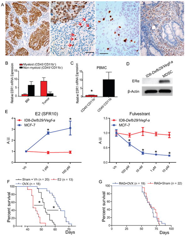
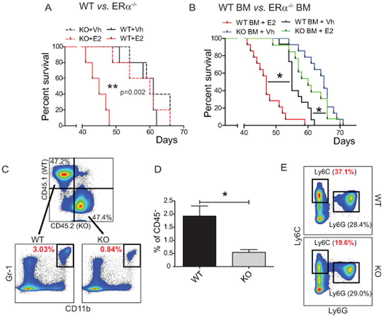
The role of estrogens in antitumor immunity remains poorly understood. Here, we show that estrogen signaling accelerates the progression of different estrogen-insensitive tumor models by contributing to deregulated myelopoiesis by both driving the mobilization of myeloid-derived suppressor cells (MDSC) and enhancing their intrinsic immunosuppressive activity in vivo Differences in tumor growth are dependent on blunted antitumor immunity and, correspondingly, disappear in immunodeficient hosts and upon MDSC depletion. Mechanistically, estrogen receptor alpha activates the STAT3 pathway in human and mouse bone marrow myeloid precursors by enhancing JAK2 and SRC activity. Therefore, estrogen signaling is a crucial mechanism underlying pathologic myelopoiesis in cancer. Our work suggests that new antiestrogen drugs that have no agonistic effects may have benefits in a wide range of cancers, independently of the expression of estrogen receptors in tumor cells, and may synergize with immunotherapies to significantly extend survival.

Significance: Ablating estrogenic activity delays malignant progression independently of the tumor cell responsiveness, owing to a decrease in the mobilization and immunosuppressive activity of MDSCs, which boosts T-cell-dependent antitumor immunity. Our results provide a mechanistic rationale to block estrogen signaling with newer antagonists to boost the effectiveness of anticancer immunotherapies. Cancer Discov; 7(1); 72-85. ©2016 AACR.See related commentary by Welte et al., p. 17This article is highlighted in the In This Issue feature, p. 1.


Conflict of interest statement

COI: The authors declare no conflict of interest

Figures

Figure 1
Figure 1. Estrogen-depletion impairment of ovarian tumor progression is independent of tumor cell signaling and is immune dependent
(A) Frozen human ovarian tumor sections stained for ERα. Scale bar indicates 1 μm. (B-C) Reverse transcription qPCR for ESR1 expression in myeloid (CD45+ CD11b+) and non-myeloid (CD45+ CD11b-) cells isolated from dissociated ovarian tumor or bone marrow (B) or the peripheral blood (C) of 5 ovarian cancer patients. (D) Western-blot for ERα (66 kDa) expression by ID8-Defb29/Vegf-a tumor cells and myeloid-derived suppressor cells isolated from mouse tumors. (E) Proliferation relative to vehicle of ID8-Defb29/Vegf-a and MCF-7 (positive control) cells in response increasing doses of E2 (in steroid-free media) and the ER antagonist fulvestrant as determined by MTS assay. (F) Survival of WT oöphorectomized (OVX) or sham-operated mice challenged with intraperitoneal ID8-Defb29/Vegf-a tumors and supplemented or not with E2. Pooled from 3 independent experiments. Total number of mice per group is depicted. (G) Survival of OVX or sham-operated Rag1 KO mice challenged with i.p. ID8-Defb29/Vegf-a. pooled from 6 independents Total number of mice per group is depicted. *p < 0.05.
Figure 2
Figure 2. Estrogen suppresses anti-tumor T cell responses
(A) Proportion of CD45+ cells isolated from ID8-Defb29/Vegf-a peritoneal tumors that are T cells (CD45+ CD3+ γδ-TCR-). (B) Proportion of activated CD44+ CD69+ double positive cells among CD4+ and CD8+ T cells. (C) ELISpot analysis of T cells isolated from ID8-Defb29/Vegf-a peritoneal tumors stimulated with tumor lysate-pulsed BM-derived dendritic cells. Results shown are representative of multiple independent experiments. *p < 0.05.
Figure 3
Figure 3. Estrogen drives accumulation of myelomonocytic (M-MDSC) and granulocytic (G-MDSC) myeloid-derived suppressor cells and increases the immunosuppressive potential of G-MDSCs
(A) Intraperitoneal LLC lung tumor progression in oöphorectomized vs. sham-treated WT mice (left) or subcutaneous LLC growth in WT mice treated with vehicle (Vh) vs. E2 (right) (5 mice/group in both cases). (B) Flank A7C11 breast tumor growth in oöphorectomized WT mice treated with vehicle (Vh) vs. E2 (5 mice/group). (C) 5×10⁵ B16.F10 cells (ATCC) were injected I.V. into mice (n=5/group). Upon injection, mice were treated with vehicle (0.1% EtOH) or E2 (10 uM) in drinking water. After 15 days, mice were euthanized and the number of lung metastases per mm2 was determined by H&E staining. Representative images of lung metastases are also shown. (D, E) Expression and quantification of M-MDSCs (Ly6Chigh Ly6G-) and G-MDSCs (Ly6C+ Ly6G+) in the spleen of ID8-Defb29/Vegf-a peritoneal tumor-bearing mice. (F, G) Expression and quantification of M-MDSCs (Ly6Chigh Ly6G-) and G-MDSCs (Ly6C+ Ly6G+) in the peritoneal cavity of ID8-Defb29/Vegf-a peritoneal tumor-bearing mice. (H) Flank A7C11 breast tumor growth in oöphorectomized mice treated with vehicle (Vh) vs. E2 and receiving 250 μg of anti-Gr1 (RB6-8C5; BioXCell) vs. control isotype antibodies every other day, starting at day 2 after tumor challenge (5 mice/group). (I) Dilution of Cell Trace Violet in labeled T cells activated with anti-CD3/CD28 beads co-cultured with varying ratios of M- and G-MDSCs isolated from ID8-Defb29/Vegf-a tumors. *p < 0.05; **p < 0.01.
Figure 4
Figure 4. Host estrogen receptor α (ERα) activity is required for E2-driven tumor acceleration and optimal MDSC accumulation
(A) Survival of WT and ERα KO mice treated with Vh or E2 and challenged with i.p. ID8-Defb29/Vegf-a tumors (n=5 mice/group). (B) Survival of WT mice lethally irradiated (10 Gy) and reconstituted with WT or ERα KO bone marrow treated with Vh or E2 and challenged with i.p. ID8-Defb29/Vegf-a tumors. Pooled from 3 independent experiments (n≥9 mice/group). (C, D) Expression and quantification of WT and ERα KO MDSCs (CD45+ CD11b+ Gr-1+) in the spleens of tumor bearing mice lethally irradiated and reconstituted with a 1:1 mix of WT CD45.1+ and ERα KO CD45.2+ bone marrow. (E) Expression of Ly6C and Ly6G by WT and ERα KO CD11b+ MHC-II- cells in the spleens of mixed BM reconstituted mice. *p < 0.05.
Figure 5
Figure 5. Optimal MDSC expansion and suppressive activity is dependent on estrogen signaling
(A) Expression of Ly6C and Ly6G (left) or MHC-II and CD11c (right) by naïve mouse WT bone marrow cultured with GM-CSF+IL6 and treated with Vh or 2 μM anti-estrogen MPP for 3 and 6 days. (B) Total number of M-MDSCs and G-MDSCs after culturing naïve WT mouse BM with GM-CSF+IL6 and treating with 2μM MPP for 6 days. Cumulative results of 3 independent experiments. (C) Dilution of Cell Trace Violet by labeled T cells activated with anti-CD3/CD28 beads co-cultured with varying ratios of G- or M-MDSCs isolated from 6-day bone marrow cultures treated with Vh, 100 ng/mL E2, or 2 μM MPP. (D) Expansion of human M-MDSCs (CD45+ HLA-DR- CD11b+ CD33+ CD14+) and G-MDSCs (CD45+ HLA-DR- CD11b+ CD33+ CD15+) from lung cancer patient bone marrow cultured in GM-CSF+IL6 and treated with Vh, 2 μM, or 10 μM MPP. (E) Total number of human M- and G-MDSCs derived from lung cancer patient bone marrow. *p < 0.05. (F) Scatter plot of the level of CD3E and PRF1 mRNA (measured as FKPM) in 266 serous ovarian cancers from TCGA datasets, separated by CYP19A1 expression above or below the median.
Figure 6
Figure 6. Estrogen increases cytokine-induced STAT3 activation during MDSC expansion
(A) Phosphorylated and total STAT3 protein expression in M- and G-MDSCs sorted from the peritoneal cavity of i.p. ID8-Defb29/Vegf-a tumor-bearing oöphorectomized mice, supplemented or not with E2 during malignant progression (pooled from 5 animals/group). (B) pSTAT3 and total STAT3 protein expression in in vitro BM-derived MDSC cultures treated with Vh, 100 ng/mL E2, or 2 μM MPP. (C) In vitro BM-derived MDSC surface expression of IL6Rα and GP-130 in response to Vh, E2, or MPP treatment. (D) Jak2 RNA expression of in vitro BM-derived MDSC in response to estrogen agonists and antagonists. (E) Expression of total Jak2 protein under the same conditions. (F) Active (phosphorylated) Jak2 protein expression in day 6 in vitro BM-derived MDSCs in response to a 5 h. pulse of 100 ng/mL E2. (G) pSrc protein expression in in vitro BM-derived MDSC cultures treated with Vh, E2, or MPP as in B. (H) Live cell counts in day 6. BM-derived MDSC cultures treated with Vh, 2 μM MPP and/or 1μM of Ruxolitinib. Fresh inhibitors were replaced at day 3. (I) Ly6C/Ly6G differentiation in BM-MDSC cultures in the presence of Vh, 10 nM of Dasatinib or 1μM of Ruxolitinib. (J) Same as in F using Dasatinib instead of Ruxolitinib. *p < 0.05.
Figure 7
Figure 7. T cell-intrinsic ERα activity suppresses anti-tumor response, but is insufficient to abrogate the effectiveness of tumor-primed T cells
(A) Intratumoral T cell expression of CD44 and CD69 in mice lethally irradiated and reconstituted with a 1:1 mix of WT CD45.1+ and ERα KO CD45.2+ bone marrow. (B) ELISpot analysis of intratumoral WT and ERα KO T cells FACS-isolated from tumor-bearing mice stimulated with tumor antigen loaded BMDCs. (C) Survival of tumor-bearing Vh or E2 treated mice following adoptive transfer of tumor antigen-primed WT or ERα KO T cells. Representative survival curves shown for multiple independent experiments. *p < 0.05.

Comment in

References

    1. Gallo MA, Kaufman D. Antagonistic and agonistic effects of tamoxifen: significance in human cancer. Semin Oncol. 1997;24:S1-71–S1-80. - PubMed
    1. Sini V, Cinieri S, Conte P, De Laurentiis M, Leo AD, Tondini C, et al. Endocrine therapy in post-menopausal women with metastatic breast cancer: From literature and guidelines to clinical practice. Crit Rev Oncol Hematol. 2016;100:57–68. - PubMed
    1. Hasan J, Ton N, Mullamitha S, Clamp A, McNeilly A, Marshall E, et al. Phase II trial of tamoxifen and goserelin in recurrent epithelial ovarian cancer. Br J Cancer. 2005;93:647–51. - PMC - PubMed
    1. del Carmen MG, Fuller AF, Matulonis U, Horick NK, Goodman A, Duska LR, et al. Phase II trial of anastrozole in women with asymptomatic mullerian cancer. Gynecol Oncol. 2003;91:596–602. - PubMed
    1. Smyth JF, Gourley C, Walker G, MacKean MJ, Stevenson A, Williams AR, et al. Antiestrogen therapy is active in selected ovarian cancer cases: the use of letrozole in estrogen receptor-positive patients. Clin Cancer Res. 2007;13:3617–22. - PubMed