Review
. 2016 Nov;16(11):661-675.
doi: 10.1038/nri.2016.100. Epub 2016 Oct 3.

Crosstalk between autophagy and inflammatory signalling pathways: balancing defence and homeostasis

Affiliations
Review

Crosstalk between autophagy and inflammatory signalling pathways: balancing defence and homeostasis

Ken Cadwell. Nat Rev Immunol. 2016 Nov.

Abstract

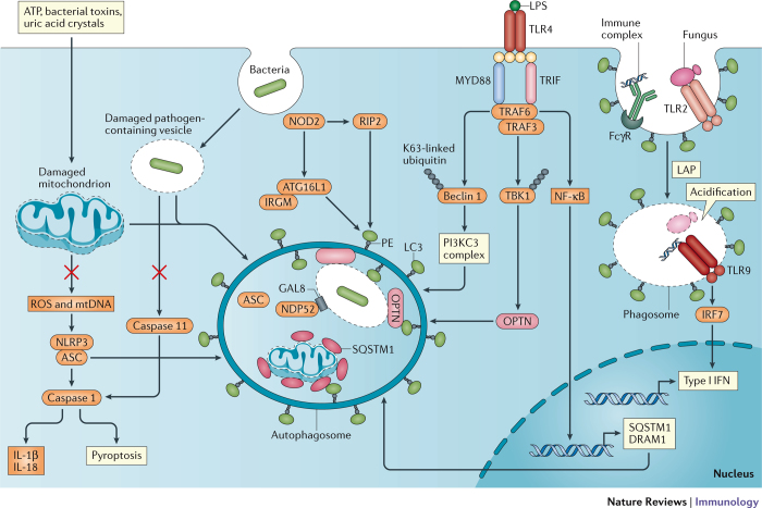
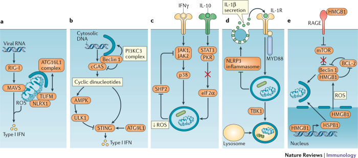
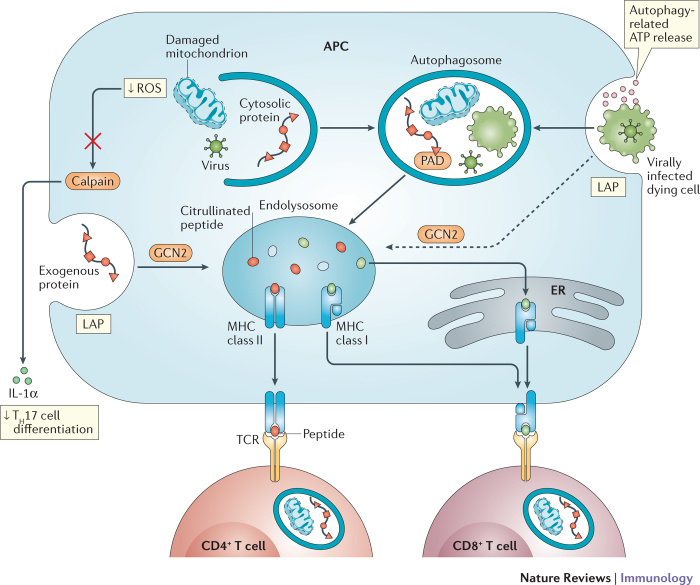
Autophagy has broad functions in immunity, ranging from cell-autonomous defence to coordination of complex multicellular immune responses. The successful resolution of infection and avoidance of autoimmunity necessitates efficient and timely communication between autophagy and pathways that sense the immune environment. The recent literature indicates that a variety of immune mediators induce or repress autophagy. It is also becoming increasingly clear that immune signalling cascades are subject to regulation by autophagy, and that a return to homeostasis following a robust immune response is critically dependent on this pathway. Importantly, examples of non-canonical forms of autophagy in mediating immunity are pervasive. In this article, the progress in elucidating mechanisms of crosstalk between autophagy and inflammatory signalling cascades is reviewed. Improved mechanistic understanding of the autophagy machinery offers hope for treating infectious and inflammatory diseases.


Conflict of interest statement

The author declares no competing financial interests.

Figures

Figure 1
Figure 1. Crosstalk between Toll-like receptor and NOD-like receptor signalling and autophagy.
Exposure to exogenous agents including ATP, bacterial toxins and uric acid crystals damages mitochondria, which results in the release of factors such as reactive oxygen species (ROS) and mitochondrial DNA (mtDNA) that activate the NLRP3 inflammasome. Autophagy inhibits inflammasome activation by sequestering damaged mitochondria and the NLRP3-binding protein ASC to prevent caspase 1-mediated cleavage of pro-interleukin-1β (pro-IL-1β) and pro-IL-18 into the active forms, as well as to prevent pyroptosis. Nucleotide-binding oligomerization domain 2 (NOD2) in the cytosol recognizes bacterial peptidoglycan and viral RNA from internalized pathogens and subsequently induces autophagy, either by signalling through receptor-interacting protein 2 (RIP2) or by directly recruiting ATG16L1 in a complex with immunity-related GTPase family M protein (IRGM). Incorporation of the bacteria into the autophagosome is achieved by LC3-binding proteins, such as sequestosome 1 (SQSTM1), optineurin (OPTN) and NDP52, which recognize the damaged vacuole or mitochondrion tagged by ubiquitin and galectin 8 (GAL8). In addition to inhibiting pathogen replication, this process reduces leakage of bacterial lipopolysaccharide (LPS) into the cytosol to prevent activation of the caspase 11 inflammasome. Following LPS-mediated activation of Toll-like receptor 4 (TLR4), myeloid differentiation primary-response protein 88 (MYD88) and TIR-domain-containing adaptor protein inducing IFNβ (TRIF) signal through TNFR-associated factor 6 (TRAF6) and TRAF3 to facilitate autophagy by inducing the K63-linked ubiquitylation and release of beclin 1 from inhibition, through the phosphorylation and activation of OPTN through TANK-binding kinase 1 (TBK1) ubiquitylation, and by inducing nuclear factor-κB (NF-κB) signalling and transcription of SQSTM1 and the autophagosome maturation protein DNA damage-regulated autophagy modulator protein 1 (DRAM1). Recognition of internalized fungal glycans and immune complexes by TLR2 and the Fcγ receptor (FcγR) triggers LC3-associated phagocytosis (LAP). As the phagosome acidifies, the fungal pathogen is degraded and activates TLR9 to induce signals through interferon regulatory factor 7 (IRF7) and type I interferon (IFN) transcription. PI3KC3, phosphoinositide 3-kinase catalytic subunit type III. PowerPoint slide
Figure 2
Figure 2. Intersection between autophagy and cytokines.
a | Several examples have been reported in which autophagy regulates cytokine production, or is subject to regulation by cytokines. The recognition of viral RNA by retinoic acid inducible gene I (RIG-I) induces mitochondrial reactive oxygen species (ROS) production and mitochondrial antiviral signalling protein (MAVS) signalling, leading to type I interferon (IFN) transcription. The recruitment of the ATG16L1 complex by NLR family member X1 (NLRX1) and elongation factor Tu, mitochondria (TUFM; or COX5B, not shown) to the mitochondrion promotes mitophagy thereby inhibiting type I IFN production by removing the MAVS signalling platform and inhibiting ROS production. b | Cyclic GMP–AMP synthase (cGAS) converts cytosolic DNA (from viruses, bacteria or the host) to cyclic dinucleotides for recognition by stimulator of interferon genes (STING) and subsequent type I IFN production. cGAS activates ULK1, which mediates an inhibitory phosphorylation of STING. Also, the binding of beclin 1 to cGAS allows beclin 1 to induce autophagy through the PI3KC3 complex to remove the cytosolic DNA and simultaneously inhibits cGAS-dependent type I IFN production. c | IFNγ induces autophagy through Janus kinase 1 (JAK1), JAK2 and p38. This pathway is reinforced by mitophogy, which reduces the levels of mitochondrial ROS that activate SHP2, an inhibitor of IFNγ signalling. By contrast, interleukin-10 (IL-10) can inhibit autophagy by promoting binding between signal transducer and activator of transcription 3 (STAT3) and protein kinase R (PKR), which inhibits the activation of the autophagy inducer eukaryotic translation initiation factor 2α (eIF2α). d | In addition to suppressing IL-1β production by inhibiting the NLRP3 inflammasome, autophagy mediates the secretion of IL-1β. Extracellular IL-1β signals through IL-1 receptor (IL-1R) and myeloid differentiation primary-response protein 88 (MYD88) to induce autophagy, and autophagosome maturation and degradation of engulfed material including intracellular bacteria is dependent on TANK-binding kinase 1 (TBK1) and suppresses the NLRP3 inflammasome. e | High mobility group box 1 (HMGB1), both within the cell and upon release by dying cells, promotes autophagy. Binding of extracellular HMGB1 by receptor for advanced glycation end products (RAGE) induces autophagy by inhibiting mechanistic target of rapamycin (mTOR). Nuclear HMGB1 promotes mitophagy by inducing the expression of ROS, it induces autophagy by protecting beclin 1 from an inhibitory interaction with B cell lymphoma 2 (BCL-2) or cleavage by calpain (not shown). HSPB1, heat shock protein B1. PowerPoint slide
Figure 3
Figure 3. Autophagy coordinates a multicellular adaptive immune response.
Autophagy activity in T cells, antigen-presenting cells (APCs) and dying cells affects T cell immunity. Autophagy in APCs such as dendritic cells delivers intracellular and extracellular antigens to endolysosomal vesicles where they are loaded onto MHC class II molecules for presentation to CD4+ T cells. Peptidylarginine deiminases (PADs) in autophagosomes generate citrullinated peptides, thereby affecting the repertoire of antigens being presented. Extracellular antigens can also be delivered to the endosolysosome by LC3-associated phagocytosis (LAP). Autophagy-mediated release of ATP by dying cells, such as those infected by viruses, leads to engulfment by the APC. Internalized antigens from the dying cell can then be cross-presented on MHC class I molecules (in the endolysosome) to stimulate CD8+ T cells, a process mediated by autophagy or, potentially, LAP downstream of GCN2 activation. By inhibiting the release of key inflammatory cytokines, autophagy decreases the differentiation of CD4+ T cells into T helper 17 (TH17) cells. Alternatively, exogenous antigens may be exported into the cytosol and delivered to MHC class I molecules in the endoplasmic reticulum (ER). For instance, the removal of reactive oxygen species (ROS)-producing mitochondria inhibits calpain-mediated processing of interleukin-1α (IL-1α). Naive CD4+ T cells require autophagy for survival and proliferation (and cytokine production once activated). The establishment of memory by CD8+ T cells is also dependent on autophagy. PowerPoint slide

References

    1. Russell RC, et al. ULK1 induces autophagy by phosphorylating Beclin-1 and activating VPS34 lipid kinase. Nat. Cell Biol. 2013;15:741–750. - PMC - PubMed
    1. Ge L, Melville D, Zhang M, Schekman R. The ER-Golgi intermediate compartment is a key membrane source for the LC3 lipidation step of autophagosome biogenesis. eLife. 2013;2:e00947. - PMC - PubMed
    1. Hamasaki M, et al. Autophagosomes form at ER-mitochondria contact sites. Nature. 2013;495:389–393. - PubMed
    1. Dooley HC, et al. WIPI2 links LC3 conjugation with PI3P, autophagosome formation, and pathogen clearance by recruiting Atg12-5-16L1. Mol. Cell. 2014;55:238–252. - PMC - PubMed
    1. Randow F, Youle RJ. Self and nonself: how autophagy targets mitochondria and bacteria. Cell Host Microbe. 2014;15:403–411. - PMC - PubMed

Publication types