. 2016 Nov;22(11):1285-1293.
doi: 10.1038/nm.4192. Epub 2016 Oct 3.

Hyaluronan and TLR4 promote surfactant-protein-C-positive alveolar progenitor cell renewal and prevent severe pulmonary fibrosis in mice

Affiliations

Hyaluronan and TLR4 promote surfactant-protein-C-positive alveolar progenitor cell renewal and prevent severe pulmonary fibrosis in mice

Jiurong Liang et al. Nat Med. 2016 Nov.

Abstract

Successful recovery from lung injury requires the repair and regeneration of alveolar epithelial cells to restore the integrity of gas-exchanging regions within the lung and preserve organ function. Improper regeneration of the alveolar epithelium is often associated with severe pulmonary fibrosis, the latter of which involves the recruitment and activation of fibroblasts, as well as matrix accumulation. Type 2 alveolar epithelial cells (AEC2s) are stem cells in the adult lung that contribute to the lung repair process. The mechanisms that regulate AEC2 renewal are incompletely understood. We provide evidence that expression of the innate immune receptor Toll-like receptor 4 (TLR4) and the extracellular matrix glycosaminoglycan hyaluronan (HA) on AEC2s are important for AEC2 renewal, repair of lung injury and limiting the extent of fibrosis. Either deletion of TLR4 or HA synthase 2 in surfactant-protein-C-positive AEC2s leads to impaired renewal capacity, severe fibrosis and mortality. Furthermore, AEC2s from patients with severe pulmonary fibrosis have reduced cell surface HA and impaired renewal capacity, suggesting that HA and TLR4 are key contributors to lung stem cell renewal and that severe pulmonary fibrosis is the result of distal epithelial stem cell failure.


Conflict of interest statement

COMPETING FINANCIAL INTERESTS

The authors declare no competing financial interests.

Figures
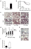
Figure 1
Figure 1
Tlr4−/− mice demonstrate higher mortality and more severe fibrosis after bleomycin-induced lung injury. (a) Tlr4 expression in the lungs of untreated (n = 3) and bleomycin-treated wild-type (WT) mouse (day 7, n = 3) as assessed by RT-PCR (****P < 0.0001 by Student t-test). (b) Percentages of surviving Tlr4−/− mice (n = 35) and WT mice (n = 34) plotted over a 21-day period after intratracheal treatment with bleomycin (2.5 U/kg). *P < 0.05 by log-rank test. (c,d) Representative images (c) of trichrome staining and hydroxyproline contents (d) of lungs from Tlr4−/− and WT mice 21 d after bleomycin injury (1.25 – 5 U/kg) (d, for saline WT n = 5, Tlr4−/− n = 4; for 1.25 U, WT n = 6, Tlr4−/− n = 9; for 2.5 U, WT n = 4, Tlr4−/− n = 4; for 5 U, WT n = 9, Tlr4−/− n = 12; *P < 0.05, **P < 0.01 by Two way analysis of variance (ANOVA) followed by Sidak’s multiple comparison test). (e) Representative images of total 4 images photographed for each group of anti-α-SMA immunostaining of lungs from Tlr4−/− and WT mice 21 days after bleomycin injury (2.5 U/kg). (f) α-SMA (Acta2) expression in lungs from bleomycin-treated Tlr4−/− (n = 4) and WT mice (n = 4) as assessed by qPCR (*P < 0.05 by Student t-test). Scale bars, 200 μm.
Figure 2
Figure 2
Tlr4−/− deficiency leads to loss of AEC2 cells in mouse lungs after bleomycin-induced injury. (a) Percent changes of CD24Sca-1 AEC2 population gated from total lung epithelial cells (EpCAM+Lin) in bleomycin treated WT mice. (b) Numbers of CD24Sca-1 AEC2s recovered from the lungs of bleomycin treated Tlr4−/− and WT mice at indicated time points (for day 0, n = 3 each; for day 3, n = 4 each; for day 7, WT n = 6, Tlr4−/− n = 4; for day 14, n = 5 each; *P < 0.05 by Student t-test). (c) Tlr4 expression of CD24Sca-1 AEC2s flow-sorted from bleomycin treated WT mice at indicated time points by RT-PCR (n = 4 each group; **P < 0.01; ****P < 0.0001 by one-way ANOVA followed by Sidak’s multiple comparison test). (d) Gated CD24Sca-1 AEC2s in Tlr4−/− and WT mouse lungs day 0 and day 14 after bleomycin.
Figure 3
Figure 3
AEC2 differentiation and proliferation requires TLR4 signaling. (a) Representative images of 9 images photographed for each group of co-staining of SFTPC and BrdU of lung sections from WT and Tlr4−/− mice after bleomycin. Arrows indicate overlap staining. (b) Percentage of SFTPC+BrdU+ in total SFTPC+ cells of WT (n = 6) and Tlr4−/− mouse lungs (n = 5). (c,d) Representative images of 9 images photographed for each group of co-staining (c) and percentage (d) of SFTPC+Ki67+ in total SFTPC+ cells of lung sections of WT (n = 3) and Tlr4−/− mice (n = 3) after bleomycin. (e) Representative image of total 6 colonies of WT AEC2s stained with SFTPC and T1α. (f) Replating CFEs of CD24Sca-1 AEC2s from bleomycin-treated WT mice. Passage (P) 0 (n = 8); P1 (n = 3); and P2 (n = 3). (g) CFEs of CD24Sca-1 AEC2s of Tlr4−/− (n = 3) and WT mice (n = 3) 3 d after bleomycin. (h) Colony sizes of CD24Sca-1 AEC2s of Tlr4−/− (n = 44) and WT mice (n = 39). (i) CFEs of CD24Sca-1 AEC2s with and without Healon (WT n = 5 each, Tlr4−/− n = 3 each). (j) CFEs of uninjured WT AEC2s with medium only (n = 4), Healon (n = 5), or Healon and pep-1 (n = 3). Scale bars, 50 μm (a,c), and 20 μm (e). **P < 0.01, ***P < 0.001, ****P < 0.0001 by Student t-test (b,d,g), Mann-Whitney U-test (h), two-way ANOVA followed by Sidak’s multiple comparison test (i), by one-way ANOVA followed by Sidak’s multiple comparison test (j).
Figure 4
Figure 4
Has2 deficient AEC2s have lower colony forming capacity. (a) Cell surface HA of CD24Sca-1 AEC2s of SFTPC-Cre;Has2flox/flox and Has2flox/flox mice. (b) HA concentration of CD24Sca-1 AEC2s from SFTPC-Cre;Has2flox/flox (n = 3), Has2flox/flox (n = 3), and WT mice (n = 3). (c) Numbers of CD24Sca-1 AEC2s recovered from bleomycin day 3 SFTPC-Cre;Has2flox/flox (n = 4) and Has2flox/flox mice (n = 6). (d) Percent of Ki67+ AEC2s in total gated CD24Sca-1 AEC2s from bleomycin-treated day 3 SFTPC-Cre;Has2flox/flox (n = 9) and SFTPC-Cre mice (n = 9). (e) CFEs of CD24Sca-1 AEC2s sorted from SFTPC-Cre;Has2flox/flox (n = 3) and Has2flox/flox (n = 3) 3 days after bleomycin. (f) CFEs of CD24Sca-1 AEC2s sorted from SFTPC-Cre;Has2flox/flox (n = 4 each) and Has2flox/flox (medium n = 5 and Healon n = 4) 3 days after bleomycin. (g) Survival curves of SFTPC-Cre;Has2flox/flox (n = 27) and Has2flox/flox littermates (n = 31) after bleomycin. (h) Hydroxyproline contents in lungs of SFTPC-Cre;Has2flox/flox and Has2flox/flox mice (for day 0 SFTPC-Cre;Has2flox/flox n = 5, Has2flox/flox n = 7; for day 21 SFTPC-Cre;Has2flox/flox n = 6. Has2flox/flox n = 10). (i) Representative images of 6 images photographed for each group of trichrome staining of lung sections of SFTPC-Cre;Has2flox/flox and Has2flox/flox day 21 after bleomycin. Scale bars, 200 μm. *P < 0.05, **P < 0.01, ***P < 0.001, ****P < 0.0001 by one-way ANOVA followed by Sidak’s multiple comparison test (b), two-way ANOVA followed by Holm-Sidak’s (f) or Sidak’s multiple comparison test (h), Student t-test (c,d,e), or by long-rank test (g).
Figure 5
Figure 5
IL-6 promotes AEC renewal and limits lung fibrosis. (a,b) IL-6 concentrations in BALF of Tlr4−/− and WT mice (a) or SFTPC-Cre;Has2flox/flox and littermates (b) at indicated time points (in a, day 0, WT n = 3, Tlr4−/− n = 5; day 1, n = 4 each; day 3, WT n = 4, Tlr4−/− n = 6; day 5, WT n = 5, Tlr4−/− n = 4; day 10, WT n = 8, Tlr4−/− n = 4; in b, day 0, n = 4 each; day 1, Has2flox/flox n = 4, SFTPC-Cre;Has2flox/flox n = 5; day 3, Has2flox/flox n = 4, SFTPC-Cre;Has2flox/flox n = 3). (c,d) CFEs of CD24Sca-1 AEC2s from bleomycin-treated WT mice were treated with (c) anti-IL-6 antibodies and control IgG (μg/ml) or (d) IL-6 protein (in c, n = 3 each; in d, n = 6 except 10 ng/ml group where n = 3). (e,f) CFEs of CD24Sca-1 AEC2s from bleomycin-treated (e) Tlr4−/− and WT mice or (f) SFTPC-Cre;Has2flox/flox and littermates with IL-6 protein (100 ng/ml) (in e, for medium, WT n = 6, Tlr4−/− n = 5, for IL-6 n = 3 each; in f, n = 3 each). (g) Bleomycin-treated Tlr4−/− mice with IL-6 protein or buffer. CD24Sca-1 AEC2s recovered (top panel, buffer n = 8, IL-6 n = 4), Ki67 staining of AEC2s (middle panel, buffer n = 6, IL-6 n = 8), and total BAL protein (bottom panel, n = 4 each). (h,i) Hydroxyproline contents of bleomycin-treated (h) Tlr4−/− mice or (i) SFTPC-Cre;Has2flox/flox mice with IL-6 protein or buffer (in h, buffer n = 4, IL-6 n = 9; in i, buffer n = 9, IL-6 n = 6). *P < 0.05, **P < 0.01, ***P < 0.001, ****P < 0.0001 by two-way ANOVA followed by Sidak’s multiple comparison test (a,b,e,f), one-way ANOVA followed by Sidak’s multiple comparison test (c,d), or by Student t-test (g,h,i).
Figure 6
Figure 6
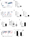
Loss of cell surface HA and impaired renewal capacity of IPF AEC2s. (a) HTII-280+ AEC2s in lungs from healthy donors (left) or from patients with IPF lungs (right). (b) Percentage of HTII-280+ AEC2s in lungs from healthy donors (n = 4) or patients with IPF (n = 7). (c) Cell surface HA on HTII-280+ AEC2s from healthy (left) or diseased lungs (right). (d) Percentages of HABP+ HTII-280+ AEC2s from healthy (n = 6) and diseased lungs (n = 9). (e) Has2 expression between HTII-280+ AEC2s from healthy and diseased lungs were determined using RT-PCR (n = 3 each). The results were repeated with AEC2s from two more subjects with IPF). (f) CFE between HTII-280+ AEC2s from healthy and diseaed lungs (n = 3 each). The results were repeated with AEC2s from three healthy controls and five more subjects with IPF. (g) Colony sizes of HTII-280+ AEC2s from lungs from healthy donors (n = 61) and patients with IPF (n = 75). (h) CFE of diseased HTII-280+ AEC2s treated with or without IL-6 (n = 3 each). The results were repeated with AEC2s from two more individuals with IPF. (i) Flow gated HAhi and HAlow HTII-280+ AEC2s from healthy donors (left) and patients with IPF (right). (j) CFE between HAhi and HAlow AEC2s from lungs of healthy donors (n = 4 each) and patients with IPF (n = 3 each). The results were repeated with cells from two more subjects of each group. *P < 0.05, **P < 0.01, ***P < 0.001, ****P < 0.0001 by Student t-test (b,d,e,f,h), by Mann-Whitney U-test (g), or by two-way ANOVA followed by Sidak’s multiple comparison test (j).

Comment in

References

    1. Rakoff-Nahoum S, Hao L, Medzhitov R. Role of toll-like receptors in spontaneous commensal-dependent colitis. Immunity. 2006;25:319–329. - PubMed
    1. Rakoff-Nahoum S, Paglino J, Eslami-Varzaneh F, Edberg S, Medzhitov R. Recognition of commensal microflora by toll-like receptors is required for intestinal homeostasis. Cell. 2004;118:229–241. - PubMed
    1. Fukata M, et al. Cox-2 is regulated by Toll-like receptor-4 (TLR4) signaling: Role in proliferation and apoptosis in the intestine. Gastroenterology. 2006;131:862–877. - PMC - PubMed
    1. Hogan BL, et al. Repair and regeneration of the respiratory system: complexity, plasticity, and mechanisms of lung stem cell function. Cell Stem Cell. 2014;15:123–138. - PMC - PubMed
    1. Kotton DN, Morrisey EE. Lung regeneration: mechanisms, applications and emerging stem cell populations. Nat Med. 2014;20:822–832. - PMC - PubMed

Publication types

MeSH terms