. 2016 Nov;48(11):1349-1358.
doi: 10.1038/ng.3676. Epub 2016 Oct 3.

Mutations in the HECT domain of NEDD4L lead to AKT-mTOR pathway deregulation and cause periventricular nodular heterotopia

Affiliations

Mutations in the HECT domain of NEDD4L lead to AKT-mTOR pathway deregulation and cause periventricular nodular heterotopia

Loïc Broix et al. Nat Genet. 2016 Nov.

Abstract

Neurodevelopmental disorders with periventricular nodular heterotopia (PNH) are etiologically heterogeneous, and their genetic causes remain in many cases unknown. Here we show that missense mutations in NEDD4L mapping to the HECT domain of the encoded E3 ubiquitin ligase lead to PNH associated with toe syndactyly, cleft palate and neurodevelopmental delay. Cellular and expression data showed sensitivity of PNH-associated mutants to proteasome degradation. Moreover, an in utero electroporation approach showed that PNH-related mutants and excess wild-type NEDD4L affect neurogenesis, neuronal positioning and terminal translocation. Further investigations, including rapamycin-based experiments, found differential deregulation of pathways involved. Excess wild-type NEDD4L leads to disruption of Dab1 and mTORC1 pathways, while PNH-related mutations are associated with deregulation of mTORC1 and AKT activities. Altogether, these data provide insights into the critical role of NEDD4L in the regulation of mTOR pathways and their contributions in cortical development.


Conflict of interest statement

The authors declare no competing financial interests.

Figures
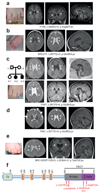
Figure 1
Figure 1
Mutations in NEDD4L cause PNH and syndactyly. (a-e) Photographs and representative sections of brain magnetic resonance imaging (MRI) of affected individuals illustrating the frequent toes syndactyly and the constant PNH. For each patient, two axial sections, or one axial and one coronal section show confluent nodules of heterotopia lining the lateral ventricles, (arrowsheads). Sagittal sections show thin (c) or dysmorphic (c, II-3 and a) corpus callosum. In (d), in addition to the PNH (arrowheads), MRI section shows frontal polymicrogyria (white arrows). MRIs were performed at the age of 8 years (a), 12 months (b), 9 and 12 months (c), 7 months (d) and 8 months (e). For the patient Pnh31124 with the c.2082G>T; p.Gln694His mutation, MR images were not available. (f) Linear representation of NEDD4L polypeptide showing positions in the HECT domain of the heterozygous PNH-associated mutation. NEDD4L protein is characterized by an N-terminal C2 domain known to bind Ca2+ and phospholipids, 2 to 4 WW protein-protein interacting domains responsible for the recognition of the substrate, and the C-terminal catalytic HECT domain.
Figure 2
Figure 2
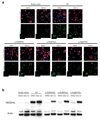
Expression and cellular localization of WT and NEDD4L mutants. (a) Tomato/DAPI and NEDD4L detection in N2A cells transfected with empty vector, WT and mutant NEDD4L cDNA constructs. For each construct, cultured cells were either treated with DMSO or with MG132. NEDD4L immunostaining (green) shows a cytoplasmic distribution, with enrichment in the periphery of N2A cells, of WT NEDD4L, whereas p.Gln694His, p.Glu893Lys and p.Arg897Gln mutants are not detectable. (b) Western blots using protein extracts of N2A cells transfected with WT and NEDD4L mutants and cultured either in presence of DMSO or MG132. They show the lack of expression of PNH-associated mutants, while transfection of WT lead to a significant expression of NEDD4L protein. Note that PNH-associated NEDD4L mutants become detectable upon treatment of N2A cells with MG132.
Figure 3
Figure 3
Ubiquitination activity of NEDD4L mutant. (a) Immunoprecipitation assay (using anti-V5 antibody to precipitate tagged NEDD4L) analyzed by immunoblots using anti-ubiquitin, anti-V5 antibodies to detect ubiquitinated NEDD4L. (b) Analysis of NEDD4L ubiquitination activity in an in vitro assay using WT and NEDD4L mutant immunopurified from transfected N2A cell lysates and incubated with ATP, E1 enzyme and E2 (UbcH7) enzyme with (+) or without (-) ubiquitin. Reaction mixtures were analyzed by immunoblotting using anti-V5 and anti-NEDD4L antibodies. Note that because of the instability of mutant NEDD4L and the resulting unbalanced amounts of WT and NEDD4L mutant and immunoblot signals as shown in Supplementary Figure 7, Western blot analysis was performed using four times less of reaction mixture corresponding to WT NEDD4L than to NEDD4L mutant.
Figure 4
Figure 4
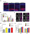
Effect of WT and NEDD4L mutants on neuronal position and progenitors proliferation. (a) Coronal sections of mouse brains at E18.5, 4 days after IUEP with empty vector (EV), WT NEDD4L, or NEDD4L mutant constructs in combination with a Tomato reporter construct. CP, cortical plate; IZ intermediate zone; VZ/SVZ ventricular zone/subventricular zone. Scale bar, 100 μm. (b) Fluorescent neurons were quantified in the regions highlighted in (a): VZ/SVZ: EV vs p.Glu893Lys, P = 0.0078; IZ: EV vs WT, P < 0.0001, EV vs p.Glu893Lys, P<0.0001, EV vs p.Arg897Gln, P < 0.0001; CP: EV vs WT, P < 0.0001, EV vs p.Glu893Lys, P < 0.0001, EV vs p.Arg897Gln, P<0.0001. Bars represent the mean of fluorescent neurons ± s.e.m. of independent brains (EV, n = 4; WT, n=3; p.Glu893Lys, n = 4; p.Arg897Gln, n = 3). (c) Immunofluorescence staining of NEDD4L (magenta) in tomato positive neurons (green) in IZ of E18.5 brains electroporated at E14.5 with NEDD4L constructs. Scale bar 5 µm. (d) Percentage of electroporated neurons positive for PH3 marker against all electroporated cells (mitotic index) in the VZ (Fig. 4a and Supplementary Fig. 9d): EV vs WT, P < 0.0001, EV vs p.Glu893Lys, P = 0.0021, EV vs p.Arg897Gln, P = 0.0001. Number of analyzed brains are as follows: EV, n = 4; WT, n = 3; p.Glu893Lys, n = 4; p.Arg897Gln, n = 3). (e) Quantification of Pax6+/Tomato+ and Tbr2+/Tomato+ cells in the VZ/SVZ (Fig. 4a and Supplementary Fig. 9e) two days after electroporation at E14.5: Pax6: EV vs p.Glu893Lys, P = 0.0003, EV vs p.Arg897Gln, P = 0.0001; Tbr2: EV vs p.Glu893Lys, P = 0.0147, EV vs p.Arg897Gln, P = 0.0014. (EV, n = 4; WT, n = 3; p.Glu893Lys, n = 4; p.Arg897Gln, n = 3). Error bars represent s.e.m.
Figure 5
Figure 5
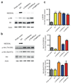
WT NEDD4L and PNH-related mutants induce deregulation of mTORC1 and AKT activities. (a,b) Representative immunoblots using protein extracts of N2A cells transfected with empty vector, WT and NEDD4L mutant constructs, and showing the effect of the WT and NEDD4L mutants on p-S6 level (phosphorylated S6 that reflects mTORC1 activity) and p-Akt (Thr308/Ser473). (c) Histograms of densitometric measurements illustrating S6 and Akt phosphorylation. Data represent mean ± sem of three independent experiments. *p≤0.05, **p≤0.01, ***p≤0.001, ****p≤0.0001
Figure 6
Figure 6
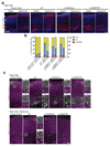
Effect of rapamycin treatment on neuronal positioning and Dab1 localization. (a) Confocal microscope images of coronal sections from E18.5 brains electroporated at E14.5 with either empty vector, WT NEDD4L, or mutants NEDD4L, and the Tomato reporter vector. Following IUEP, pregnant mice were either treated with vehicle (DMSO) or with rapamycin (0.5 mg/kg/day). CP, cortical plate; IZ intermediate zone; VZ/SVZ ventricular zone/subventricular zone. Scale bar 100 µm. (b) Electroporated neurons were quantified in the regions indicate in (a): VZ/SVZ: WT DMSO vs WT Rapamycin, P=0.012, IZ: WT DMSO vs WT Rapamycin, P<0.0001; CP: WT DMSO vs WT Rapamycin, P<0.0001); IZ: p.Glu893Lys DMSO vs p.Glu893Lys Rapamycin, P=0.0377, p.Arg897Gln DMSO vs p.Arg897Gln Rapamycin, P=0.0004; CP : p.Arg897Gln DMSO vs p.Arg897Gln Rapamycin, P=0.0013. Bars represent the mean of electroporated neurons in each regions ± s.e.m. of 3 independent brains. (c) Immunolabelings of Dab1 (magenta) on cortical slices at E18.5 from brain embryos subjected to DMSO or rapamycin treatment. Right panels of each coronal section are higher magnifications of white boxes in the CP (1) and IZ (2) showing the distribution of Dab1. In arrested neurons electroporated with WT NEDD4L of non-treated mice, note the specific pattern of Dab1 distribution and its enrichment in the periphery of the cytoplasm (mainly in arrested neurons of the IZ and to a less extend in neurons of the cortical plate). Scale bar 100 µm; 10 µm (higher magnifications).
Figure 7
Figure 7
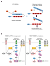
Models depicting consequences of PNH-related mutations on NEDD4L stability and PI3K/Akt/mTOR and TGF-β/Smad pathways. (a) WT NEDD4L (on the left) is shown in its closed and inactive conformation, and NEDD4L mutant (on the right) with an alteration in the HECT domain predicted to lead to conformation changes that favours transition from inactive to open active state and triggers auto-ubiquitination and degradation. (b) Overview of observed deregulations in basal conditions resulting from an excess of WT NEDD4L (on the left) and expression PNH-related NEDD4L mutants (on the right). Red contours indicate deregulated proteins and dotted arrows depict indirect and poorly understood relations between partners of signaling pathways.

References

    1. Caviness VS, Jr, Takahashi T, Nowakowski RS. Numbers, time and neocortical neuronogenesis: a general developmental and evolutionary model. Trends Neurosci. 1995;18:379–83. - PubMed
    1. Rakic P, Caviness VS., Jr Cortical development: view from neurological mutants two decades later. Neuron. 1995;14:1101–4. - PubMed
    1. Barkovich AJ, Guerrini R, Kuzniecky RI, Jackson GD, Dobyns WB. A developmental and genetic classification for malformations of cortical development: update 2012. Brain: a journal of neurology. 2012;135:1348–69. - PMC - PubMed
    1. Francis F, et al. Human disorders of cortical development: from past to present. Eur J Neurosci. 2006;23:877–93. - PubMed
    1. Guerrini R, Dobyns WB. Malformations of cortical development: clinical features and genetic causes. Lancet Neurol. 2014;13:710–26. - PMC - PubMed

Substances