. 2016 Oct 3:6:34459.
doi: 10.1038/srep34459.

Glycine triggers a non-ionotropic activity of GluN2A-containing NMDA receptors to confer neuroprotection

Affiliations

Glycine triggers a non-ionotropic activity of GluN2A-containing NMDA receptors to confer neuroprotection

Rong Hu et al. Sci Rep. .

Abstract

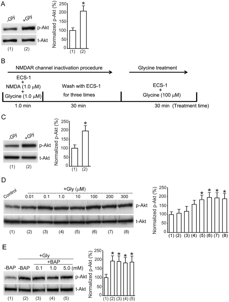
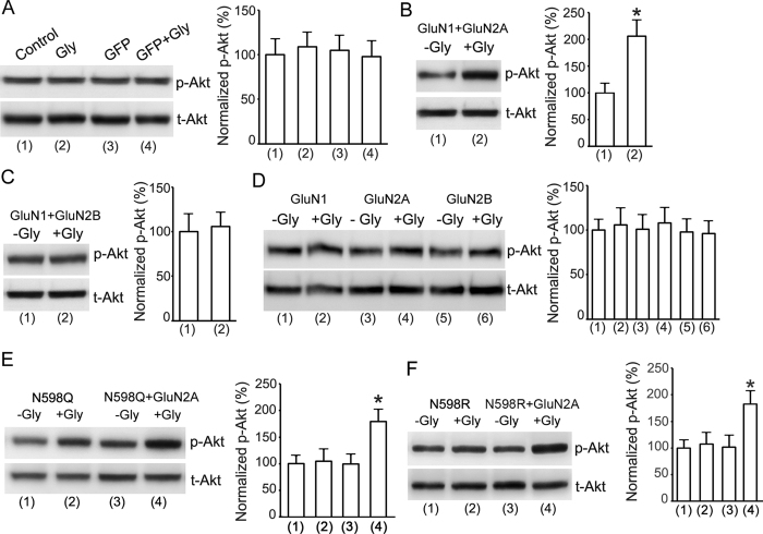
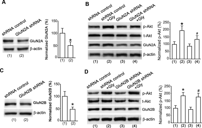
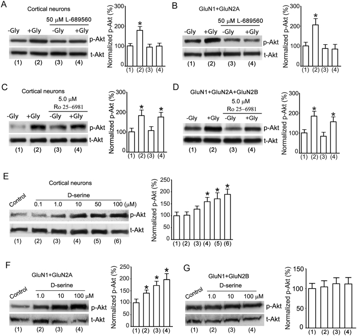
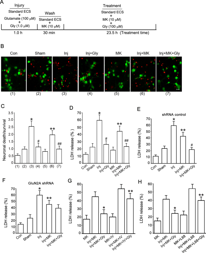
Ionotropic activation of NMDA receptors (NMDARs) requires agonist glutamate and co-agonist glycine. Here we show that glycine enhances the activation of cell survival-promoting kinase Akt in cultured cortical neurons in which both the channel activity of NMDARs and the glycine receptors are pre-inhibited. The effect of glycine is reduced by shRNA-mediated knockdown of GluN2A subunit-containing NMDARs (GluN2ARs), suggesting that a non-ionotropic activity of GluN2ARs mediates glycine-induced Akt activation. In support of this finding, glycine enhances Akt activation in HEK293 cells over-expressing GluN2ARs. The effect of glycine on Akt activation is sensitive to the antagonist of glycine-GluN1 binding site. As a functional consequence, glycine protects against excitotoxicity-induced neuronal death through the non-ionotropic activity of GluN2ARs and the neuroprotective effect is attenuated by Akt inhibition. Thus, this study reveals an unexpected role of glycine in eliciting a non-ionotropic activity of GluN2ARs to confer neuroprotection via Akt activation.


Figures

Figure 1
Figure 1. Enhancement of Akt phosphorylation by glycine in cortical neurons does not require Ca2+-mediated channel activities of NMDARs.
(A) Glycine (100 μM) increases Akt phosphorylation (p-Akt) in neurons treated with ECS without addition of Ca2+ but with addition of 5.0 mM EGTA (n = 7, Student’s t test, *p < 0.05 vs. −Gly). (B) A schematic diagram showing the NMDAR channel inactivation and glycine treatment procedure. (C) Glycine (100 μM) increases p-Akt in neurons where NMDAR channel activities are inhibited (n = 9, Student’s t test, *p < 0.05 vs. −Gly). (D) Glycine-induced increase of p-Akt is dose-dependent in neurons where NMDAR channel activities are inhibited (n = 6, ANOVA test, *p < 0.05 vs. control). (E) The enhancement of p-Akt by glycine (100 μM) is not altered by BAPTA that is included in the ECS-1 (n = 6, ANOVA test, *p < 0.05 vs. -BAP). The p-Akt analyses were normalized to group (1) labeled in the bar graphs. Gly: glycine; BAP: BAPTA.
Figure 2
Figure 2. Enhancement of Akt phosphorylation by glycine in cortical neurons does not require activation of glycine receptors.
(A) Treatment of strychnine (10 μM) does not interfere with the enhancement of p-Akt by glycine (100 μM) in neurons where NMDAR channel activities are pre-inhibited (n = 6, Student’s t test, *p < 0.05 vs. −Gly). (B) Glycine has no significant effect on p38-MAPK phosphorylation (p-p38) in cortical neurons following NMDAR channel inactivation procedure (n = 6; ANOVA test). Gly: glycine; p38: p38-MAPK.
Figure 3
Figure 3. Non-ionotropic activity of GluN2AR mediates glycine-induced enhancement of Akt phosphorylation in HEK293 cells expressing GluN2ARs.
(A) In HEK293 cells without or with GFP transfection, the levels of p-Akt are not altered by glycine (100 μM) treatment after the channel activities of NMDARs are inhibited by the NMDAR channel inactivation procedure (n = 6; ANOVA test). (B) In HEK293 cells transfected with GluN1 + GluN2A cDNAs, glycine (100 μM) increases p-Akt after the channel activities of NMDARs are inhibited (n = 9, Student’s t test, *P < 0.05 vs. -Gly). (C) In HEK293 cells transfected with GluN1 + GluN2B cDNAs, the levels of p-Akt are not altered by glycine (100 μM) after the channel activities of NMDARs are inhibited (n = 6; Student’s t test). (D) In HEK293 cells transfected with GluN1, GluN2A or GluN2B cDNAs, respectively, the levels of p-Akt are not altered by glycine (100 μM) after the channel activities of NMDARs are inhibited (n = 6; ANOVA test). (E) In HEK293 cells transfected with GluN1(N598Q) + GluN2A, but not GluN1(N598Q) alone, glycine enhances Akt phosphorylation after the channel activities of NMDARs are inhibited (n = 6, ANOVA test, *P < 0.05 vs. -Gly). (F) Glycine increases Akt phosphorylation in HEK293 cells transfected with GluN1(N598R) + GluN2A following NMDAR channel inactivation procedure (n = 6; ANOVA test, *P < 0.05 vs. -Gly). Gly: glycine.
Figure 4
Figure 4. Non-ionotropic activity of GluN2AR mediates glycine-induced enhancement of Akt phosphorylation in cortical neurons.
(A) The GluN2A protein expression in cortical neurons is suppressed by GluN2A shRNA (n = 6, Student’s t test, *P < 0.05 vs. shRNA control). (B) GluN2A knockdown by GluN2A shRNA attenuates glycine-induced increase of p-Akt in cortical neurons where NMDAR channels are inhibited (n = 6, ANOVA test, *P < 0.05 vs. shRNA control; #P < 0.05 vs. shRNA control + Gly). (C) The GluN2B protein expression in cortical neurons is suppressed by GluN2B shRNA transduction (n = 6, Student’s t test, *P < 0.05 vs. shRNA control). (D) GluN2B knockdown by GluN2B shRNA does not interfere with glycine-induced increase of p-Akt in cortical neurons where NMDAR channels activities are inhibited (n = 6, ANOVA test, *P < 0.05 vs. shRNA control; #P < 0.05 vs. GluN2B shRNA). Gly: glycine.
Figure 5
Figure 5. The glycine-GluN1 binding is required for glycine-induced non-ionotropic activation of GluN2ARs.
(A) Glycine-GluN1 binding site antagonist L-689560 (50 μM) blocks glycine (100 μM)-induced increase of p-Akt in cultured cortical neurons after the channel activities of NMDARs are inhibited (n = 6, ANOVA test, *P < 0.05 vs.-Gly). (B) L-689560 (50 μM) blocks glycine (100 μM)-induced increase of p-Akt in HEK293 cells transfected with GluN1 + GluN2A after the channel activities of NMDARs are inhibited (n = 6, ANOVA test, *P < 0.05 vs.-Gly). (C) GluN2BR antagonist Ro 25-6981 (5.0 μM) does not interfere with glycine (100 μM)-induced increase of p-Akt in cultured cortical neurons following the NMDAR channel inactivation procedure (n = 6, ANOVA test, *P < 0.05 vs.-Gly). (D) Ro 25-6981 (5.0 μM) does not interfere with glycine (100 μM)-induced increase of p-Akt in HEK293 cells transfected with GluN1 + GluN2A + GluN2B following the NMDAR channel inactivation procedure (n = 6, ANOVA test, *P < 0.05 vs.-Gly). (E) D-serine increases the level of p-Akt in cultured cortical neurons after the channel activities of NMDARs are inhibited (n = 6, ANOVA test, *P < 0.05 vs. Control). (F) D-serine increases the level of p-Akt in HEK293 cells transfected with GluN1 + GluN2A after the channel activities of NMDARs are inhibited (n = 5, ANOVA test, *P < 0.05 vs. Control). (G) D-serine has no effect on the level of p-Akt in HEK293 cells transfected with GluN1 + GluN2B after the channel activities of NMDARs are inhibited (n = 5, ANOVA test). Gly: glycine.
Figure 6
Figure 6. Glycine protects against glutamate neurotoxicity-induced neuronal injury in cortical neurons through non-ionotropic activation of GluN2ARs.
(A) A schematic diagram showing glutamate neurotoxicity injury and glycine treatment procedure. (B) Representative images showing that glycine (100 μM) reduces glutamate neurotoxicity-induced cell death in neurons where NMDAR channel activity is inactivated. Green: FDA; Red: PI. Scale bar = 25 μm. (C) Summarized data of B (n = 5. Total 3136 cells counted for Con group, 2825 cells for Sham group, 3225 cells for Inj group, 3208 cells for Inj + Gly group, 3003 cells for MK group, 3160 cells for Inj + MK group and 3231 cells for Inj + MK + Gly group. ANOVA test, *P < 0.05 vs. Sham; #P < 0.05 vs. Inj; **P < 0.05 vs. Inj; ##P < 0.05 vs. Inj + MK). (D) In neurons where NMDAR channel activities are inhibited, glycine (100 μM) prevents glutamate neurotoxicity-induced increase of LDH release (n = 6, ANOVA test, *P < 0.05 vs. Sham; #P < 0.05 vs. Inj; **P < 0.05 vs. Inj; ##P < 0.05 vs. Inj + MK). (E) Glycine (100 μM) reduces glutamate neurotoxicity-induced increase of LDH release in neurons where shRNA control is transfected and NMDAR channel activity is suppressed (n = 6, ANOVA test, *P < 0.05 vs. Sham; **P < 0.05 vs. Inj; #P < 0.05 vs. Inj + MK). (F) Glycine (100 μM) does not prevent glutamate neurotoxicity-induced increase of LDH release in neurons where GluN2A expression is suppressed by GluN2A shRNA and NMDAR channel activity is inhibited (n = 6, ANOVA test, *P < 0.05 vs. Sham; **P < 0.05 vs. Inj). (G) Akt inhibitor IV (1.0 μM) decreases glycine (100 μM)-induced reduction of LDH release in neurons where NMDAR channel activity is inhibited (n = 6, ANOVA test, *P < 0.05 vs. Inj + MK; **P < 0.05 vs. Inj + MK + Gly). (H) Glycine-GluN1 binding antagonist L-689560 (50 μM) decreases glycine (100 μM)-induced reduction of LDH release in neurons where NMDAR channel activity is inhibited (n = 6, ANOVA test, *P < 0.05 vs. Inj + MK; **P < 0.05 vs. Inj + MK + Gly). Con: control; Inj: injury; Gly: glycine; MK: MK-801; L68: L-689560.

References

    1. Dingledine R., Borges K., Bowie D. & Traynelis S. F. The glutamate receptor ion channels. Pharmacol Rev 51, 7–61 (1999). - PubMed
    1. Monyer H. et al.. Heteromeric NMDA receptors: molecular and functional distinction of subtypes. Science 256, 1217–1221 (1992). - PubMed
    1. Johnson J. W. & Ascher P. Glycine potentiates the NMDA response in cultured mouse brain neurons. Nature 325, 529–531 (1987). - PubMed
    1. Malenka R. C. & Nicoll R. A. Long-term potentiation–a decade of progress? Science 285, 1870–1874 (1999). - PubMed
    1. Barria A. & Malinow R. Subunit-specific NMDA receptor trafficking to synapses. Neuron 35, 345–353 (2002). - PubMed

Publication types

Substances

LinkOut - more resources