. 2016 Dec;17(12):1373-1380.
doi: 10.1038/ni.3580. Epub 2016 Oct 3.

Identification of a role for TRIM29 in the control of innate immunity in the respiratory tract

Affiliations

Identification of a role for TRIM29 in the control of innate immunity in the respiratory tract

Junji Xing et al. Nat Immunol. 2016 Dec.

Erratum in

Abstract

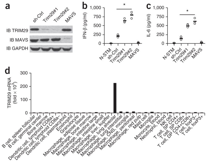
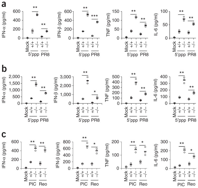
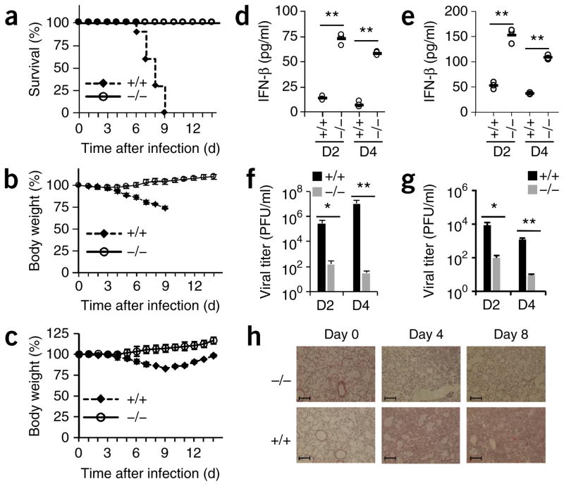
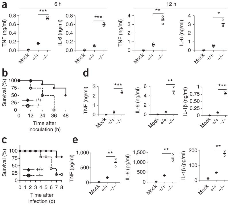
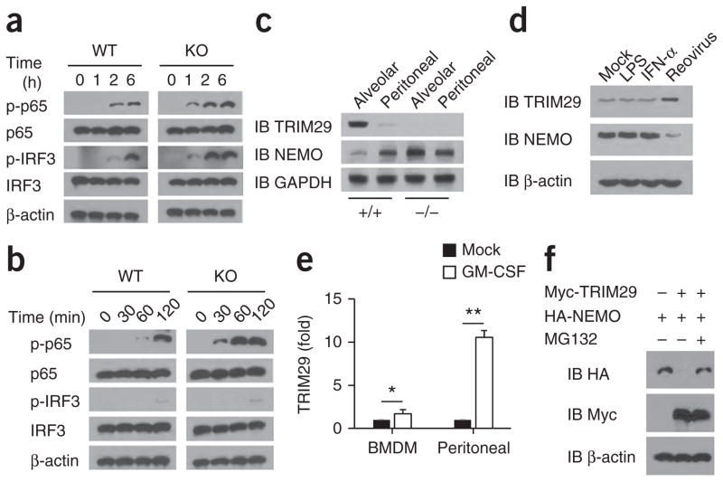
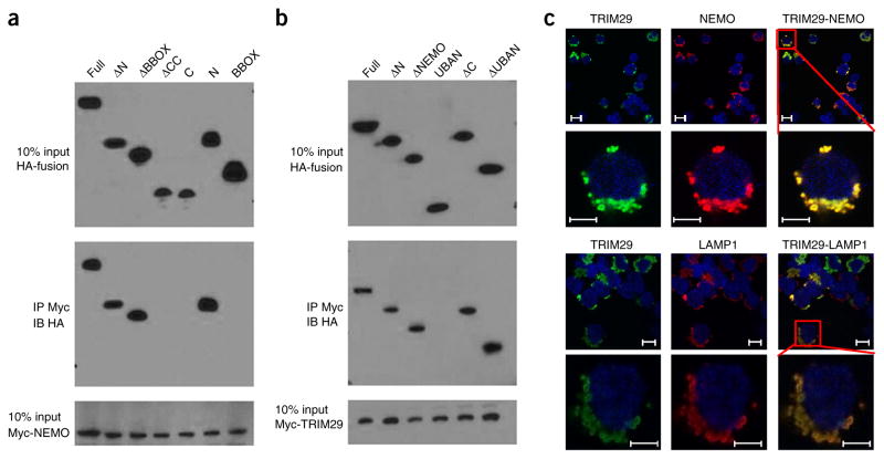
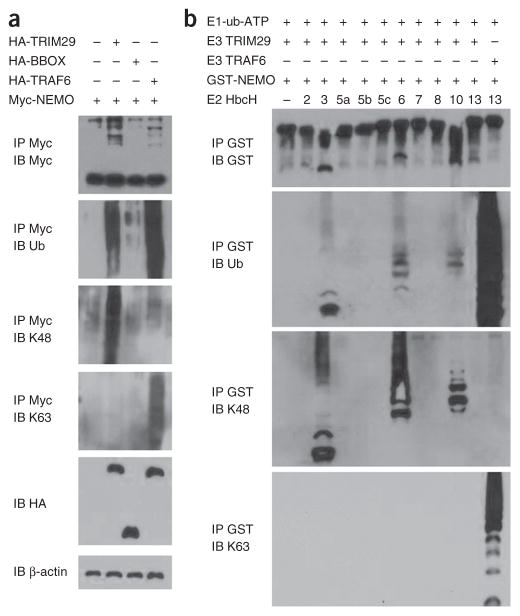
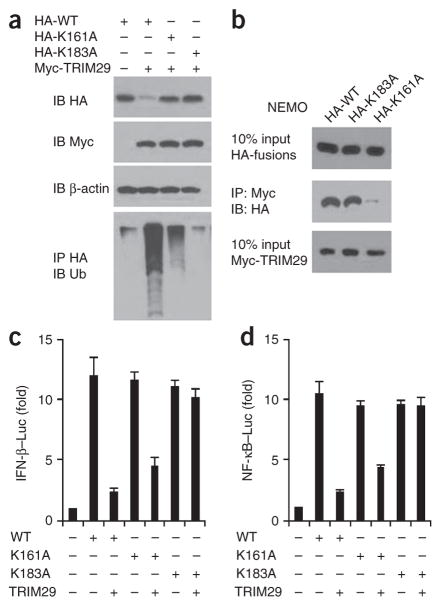
The respiratory tract is heavily populated with innate immune cells, but the mechanisms that control such cells are poorly defined. Here we found that the E3 ubiquitin ligase TRIM29 was a selective regulator of the activation of alveolar macrophages, the expression of type I interferons and the production of proinflammatory cytokines in the lungs. We found that deletion of TRIM29 enhanced macrophage production of type I interferons and protected mice from infection with influenza virus, while challenge of Trim29-/- mice with Haemophilus influenzae resulted in lethal lung inflammation due to massive production of proinflammatory cytokines by macrophages. Mechanistically, we demonstrated that TRIM29 inhibited interferon-regulatory factors and signaling via the transcription factor NF-κB by degrading the adaptor NEMO and that TRIM29 directly bound NEMO and subsequently induced its ubiquitination and proteolytic degradation. These data identify TRIM29 as a key negative regulator of alveolar macrophages and might have important clinical implications for local immunity and immunopathology.


Figures

Figure 1
Figure 1
TRIM29 inhibits cytokine production by AMs in response to 5′ppp RNA. (a) Immunoblot analysis (IB) of TRIM29, MAVS and GAPDH (loading control throughout) in MH-S cells treated with shRNA with a scrambled sequence (as a control (sh-Ctrl)) or shRNA to knock down expression of TRIM29 (Trim29#1 and Trim29#2)) or MAVS (above lanes). (b,c) ELISA of IFN-β (b) and IL-6 (c) in supernatants of MH-S cells left unstimulated and treated with the control shRNA (N-STM) or stimulated for 16 h with 5′pppRNA (1.0 μg/ml) and then treated with shRNA as in a (horizontal axes). Each symbol represents an individual independent experiment; small horizontal lines indicate the average of biological triplicates. (d) Real-time PCR analysis of TRIM29 mRNA in primary cells (horizontal axis) isolated from C57BL/6 mice; results are resented relative to those of GAPDH. *P < 0.0005 (unpaired t-test). Data are representative of three independent experiments with similar results (ac) or two experiments (d).
Figure 2
Figure 2
TRIM29 inhibits the innate immune response to 5′pppRNA, poly(I:C) and RNA viruses in primary AMs. (a,b) ELISA of type I interferons, TNF and IL-6 in supernatants of wild-type AMs without stimulation or infection (Mock) or Trim29+/+ (wild-type) (+/+) and Trim29−/− (−/−) primary AMs stimulated for 6 h (a) or 12 h (b) with 5′pppRNA (5′ppp) (1 μg/ml) or infected for those times with influenza virus at a multiplicity of infection (MOI) of 5 (PR8). (c) ELISA of type I interferons, TNF and IL-6 in supernatants of Trim29+/+ and Trim29−/− primary AMs mock treated (Mock), stimulated for 12 h with high-molecular-weight poly(I:C) (10 μg/ml) (PIC) or infected with reovirus at an MOI of 5 (Reo). Each symbol represents an individual independent experiment; small horizontal lines indicate the average of biological triplicates. *P < 0.005, **P < 0.0005 and ***P < 0.0001 (unpaired t-test). Data are representative of three independent experiments with similar results.
Figure 3
Figure 3
TRIM29 has an important role in host defense against infection with RNA viruses in vivo. (ac) Survival (a) and body weight (b,c) of age- and sex-matched 6-week-old Trim29+/+ and Trim29−/− mice (n = 10 per strain) after intranasal infection with a high dose (1 × 10⁵ PFU per mouse) (a,b) or low dose (1 × 10² PFU per mouse) (c) of influenza virus PR8. (dg) ELISA of IFN-β in BALF (d,e) and pulmonary viral titers (f,g) of 6-week-old Trim29+/+ and Trim29−/− mice (n = 3 per strain) at day 2 (D2) or day 4 (D4) after intranasal infection with a high dose (d,f) or low dose (e,g) (as in ac) of influenza virus PR8. Each symbol (d,e) represents an individual independent experiment; small horizontal lines indicate the average of biological triplicates. (h) Hematoxylin-and-eosin staining of lung sections from Trim29+/+ and Trim29−/− mice on day 0, day 4 or day 8 after infection with influenza virus PR8 (1 × 10⁵ PFU per mouse). Scale bars, 200 μm. *P < 0.0005 and **P < 0.0001 (unpaired t-test). Data are representative of two independent experiments with similar results (mean + s.d. in f,g).
Figure 4
Figure 4
TRIM29 prevents septic shock induced by LPS and infection with H. influenzae in the lungs. (a) ELISA of TNF and IL-6 in supernatants of Trim29+/+ primary AMs left unstimulated (Mock) or Trim29+/+ and Trim29−/− primary AMs stimulated for 6 h or 12 h (above plots) with LPS (10 ng/ml). (b) Survival of age- and sex-matched 6-week-old Trim29+/+ and Trim29−/− mice (n = 8 per strain) inoculated intranasally with LPS (80 mg per kg body weight) and monitored every hour for 48 h. (c) Survival of age- and sex-matched 6-week-old Trim29+/+ mice left uninfected (Mock) or Trim29+/+ and Trim29−/− mice (n = 5 per strain) inoculated intratracheally with H. influenzae (1 × 10⁷ CFU per mouse) and monitored daily for 8 d. (d,e) ELISA of TNF, IL-6 and IL-1β in BALF of Trim29+/+ mice left untreated or uninfected (Mock) or Trim29+/+ and Trim29−/− mice at 12 h after intranasal inoculation with LPS (80 mg per kg body weight) (d) or at day 1 after intratracheal inoculation with H. influenzae (1 × 10⁷ CFU per mouse) (e). Each symbol (a,d,e) represents an individual independent experiment; small horizontal lines indicate the average of biological triplicates. *P < 0.005, **P < 0.0005 and ***P < 0.0001 (unpaired t-test). Data are representative of two independent experiments with similar results.
Figure 5
Figure 5
TRIM29 inhibits the activation of IRF3 and p65 in AMs after viral infection or LPS treatment by degrading NEMO. (a,b) Immunoblot analysis of total and phosphorylated (p−) p65 and IRF3, as well as β-actin (loading control throughout), in lysates of Trim29+/+ (WT) and Trim29−/− (KO) AMs infected for various times (above lanes) with influenza virus PR8 at an MOI of 5 (a) or stimulated for various time (above lanes) with LPS (20 ng/ml) (b). (c) Immunoblot analysis of TRIM29 and NEMO in primary AMs (Alveolar) or peritoneal macrophages (Peritoneal) from Trim29+/+ and Trim29−/− mice. (d) Immunoblot analysis of TRIM29 and NEMO in bone-marrow-derived dendritic cells given mock treatment or treated for 6 h with LPS (20 ng/ml), IFN-μ (25 ng/ml) or reovirus. (e) Real-time PCR analysis of TRIM29 mRNA in wild-type bone-marrow-derived macrophages (BMDM) and peritoneal macrophages l treated for 6 h with GM-CSF (50 ng/ml); results are presented relative to those of untreated cells (Mock), set as 1. (f) Immunoblot analysis HEK293T cells transfected to express various combinations (above lanes) of Myc-tagged TRIM29 and HA-tagged NEMO and treated with MG132 (+) or not (−). *P < 0.05 and **P < 0.001 (unpaired t-test). Data are representative of three independent experiments with similar results (mean + s.d. in e).
Figure 6
Figure 6
TRIM29 binds to and colocalizes with NEMO in the lysosome. (a) Immunoblot analysis of purified Myc-tagged NEMO with anti-Myc (bottom), and immunoblot analysis (with anti-HA) of purified HA-tagged full-length TRIM29 (Full) or TRIM29 with deletion of the N terminus (ΔN), the B-box zinc-finger domain (ΔBBOX) or the coiled-coil domain (ΔCC) or containing only the carboxyl terminus (C), the N terminus (N) or the BBOX domain (above lanes), alone (top) or after incubation with Myc-tagged NEMO and immunoprecipitation (IP) with anti-Myc (middle). (b) Immunoblot analysis of purified Myc-tagged TRIM29 with anti-Myc (bottom), and immunoblot analysis (with anti-HA) of purified HA-tagged full-length NEMO (Full) and NEMO with deletion of the N terminus (ΔN), a fragment of the NEMO domain (ΔNEMO), the carboxyl terminus (ΔC) or the UBAN domain (ΔUBAN) or containing only the UBAN (UBAN), alone (top) or after incubation with Myc-tagged TRIM29 and immunoprecipitation with anti-Myc (middle). (c) Confocal microscopy of HEK293T cells cotransfected with expression plasmids for HA-tagged TRIM29 and Myc-tagged NEMO and stained with Alexa Fluor 488–conjugated antibody to HA (green) and Alexa Fluor 555–conjugated antibody to Myc (red); LAMP1 serves as a marker of lysosome (red), and the DNA-binding dye DAPI serves as a marker of nuclei (blue). Bottom rows are an enlargement of the areas outlined at top right. Scale bars, 10μm (top rows) or 5 μm (bottom rows). Data are representative of three independent experiments with similar results.
Figure 7
Figure 7
TRIM29 induces ubiquitination of NEMO by K48 linkage. (a) Immunoblot analysis with anti-Myc (top blot) or of total ubiquitination (second blot), K48-linked ubiquitination (third blot) or K63-linked ubiquitination (fourth blot) of Myc-tagged NEMO in HEK293T cells transfected with empty vector or expression vector for HA-tagged TRIM29, TRIM29 BBOX or TRAF6 and treated for 3 h with 25 μM MG132, assessed after immunoprecipitation with anti-Myc; and immunoblot analysis of whole-cell lysates with anti-HA (fifth blot) or anti-β-actin (bottom blot). (b) Immunoblot analysis of the degradation and total, K48-mediated or K63-mediated ubiquitination of GST-tagged NEMO, assessed by in vitro ubiquitination assay with various combinations (above lanes) of a mixture of E1 plus ubiquitin plus ATP (E1-ub-ATP), TRIM29 (E3 TRIM29), GST-tagged NEMO (GST-NEMO), GST-tagged TRAF6, and purified E2 enzymes (HbcH; numbers above lanes indicate enzyme), after immunoprecipitation with anti-GST. Data are representative of three independent experiments with similar results.
Figure 8
Figure 8
TRIM29 induces degradation of NEMO after its ubiquitination at Lys183. (a) Immunoblot analysis of HEK293T cells cotransfected an expression vector for HA-tagged wild-type NEMO (HA-WT) or NEMO with substitution of alanine for Lys183 (HA-K183A) or Lys161 (HA-K161A) and with empty vector (−) or an expression vector for Myc-tagged TRIM29 (above lanes). (b) Immunoblot analysis of purified Myc-tagged TRIM29 with anti-Myc (bottom blot), and immunoblot analysis (with anti-HA) of purified HA-tagged NEMO (as in a) alone (top) or after incubation with Myc-tagged TRIM29 and immunoprecipitation with anti-Myc (middle). (c,d) Activation of the promoter of the gene encoding IFN-β (c) or NF-κB (d) in HEK293T cells transfected with luciferase reporters for those genes (Luc), plus expression vector for NEMO (as in a) with or without an expression vector for TRIM29 (below plots); results are presented relative to those of renilla luciferase (cotransfected as an internal control). Data are representative of three independent experiments with similar results (mean + s.d. in c,d).

References

    1. Guilliams M, et al. Alveolar macrophages develop from fetal monocytes that differentiate into long-lived cells in the first week of life via GM-CSF. J Exp Med. 2013;210:1977–1992. - PMC - PubMed
    1. Takeuchi O, Akira S. Pattern recognition receptors and inflammation. Cell. 2010;140:805–820. - PubMed
    1. Vlahos R, Bozinovski S. Role of alveolar macrophages in chronic obstructive pulmonary disease. Front Immunol. 2014;5:435. - PMC - PubMed
    1. Murray PJ, Wynn TA. Protective and pathogenic functions of macrophage subsets. Nat Rev Immunol. 2011;11:723–737. - PMC - PubMed
    1. Kopf M, Schneider C, Nobs SP. The development and function of lung-resident macrophages and dendritic cells. Nat Immunol. 2015;16:36–44. - PubMed

MeSH terms