. 2016 Oct 3;14(10):e1002565.
doi: 10.1371/journal.pbio.1002565. eCollection 2016 Oct.

Diversity Takes Shape: Understanding the Mechanistic and Adaptive Basis of Bacterial Morphology

Affiliations

Diversity Takes Shape: Understanding the Mechanistic and Adaptive Basis of Bacterial Morphology

David T Kysela et al. PLoS Biol. .

Abstract

The modern age of metagenomics has delivered unprecedented volumes of data describing the genetic and metabolic diversity of bacterial communities, but it has failed to provide information about coincident cellular morphologies. Much like metabolic and biosynthetic capabilities, morphology comprises a critical component of bacterial fitness, molded by natural selection into the many elaborate shapes observed across the bacterial domain. In this essay, we discuss the diversity of bacterial morphology and its implications for understanding both the mechanistic and the adaptive basis of morphogenesis. We consider how best to leverage genomic data and recent experimental developments in order to advance our understanding of bacterial shape and its functional importance.


Conflict of interest statement

The authors have declared that no competing interests exist.

Figures

Fig 1
Fig 1. Myriad morphologies have evolved throughout the bacterial domain.
Bacterial phylogeny derived from genome sequence data for selected species, with an emphasis on morphologically and phylogenetically diverse taxa. Sequence data gathered from the Joint Genome Institute [3] and the National Center for Biotechnology Information [4] were searched for reference genes and aligned using Phylosift [5]. FastTree [6] generated an approximate maximum likelihood tree from the resulting concatenated alignment. The final tree was formatted using iTol [7]. Black dots denote ancestral nodes of selected major taxa: DT, Deinococcus-Thermus; Ac, Actinobacteria; Cf, Chloroflexi; Cn, Cyanobacteria; Fi, Firmicutes (inclusive of Mollicutes); Sp, Spirochetes; PVC, Planctomycetes, Verrucomicrobia, Chlamydiae; Cb, Chlorobi; Bd, Bacteroidetes; α, β, γ, δ, ε, Proteobacteria subdivisions. 1. Bifidobacterium longum. 2. Streptomyces coelicolor (mycelial [multicellular] filament with hyphae and spores). 3. Corynebacterium diphtheriae (two cells, dumbbell and club shapes). 4. Herpetosiphon aurantiacus (filament of multiple cylindrical cells). 5. Calothrix (filament of multiple disk-shaped cells). 6. Mycoplasma genitalium. 7. Spiroplasma culicicola. 8. Lactococcus lactis (predivisional cell). 9. Borrelia burgdorferi. 10. Gimesia maris (previously Planctomyces maris, predivisional cell with proteinaceous stalk). 11. Prosthecochloris aestuarii. 12. Pelodictyon phaeoclathratiforme (filament of multiple trapezoidal cells). 13. Spirosoma linguale. 14. Muricauda ruestringensis (appendage includes nonreproductive bulb). 15. Desulfovibrio vulgaris (two cells, helical and curved shapes). 16. Helicobacter pylori. 17. Caulobacter crescentus (predivisional cell). 18. Hyphomonas neptunium (predivisional cell). 19. Rhodomicrobium vannielii (filament of multiple ovoid cells, one is predivisional). 20. Prosthecomicrobium hirschii. 21. Simonsiella muelleri (filament of multiple curved cells). 22. Nevskia ramosa (two cells with bifurcating slime stalk). 23. Beggiatoa leptomitiformis (filament of multiple, giant cylindrical cells). 24. Thiomargarita nelsonii (single, giant cell). 25. Escherichia coli. 26. Mariprofundus ferrooxydans (single cell with metal-encrusted stalk). Bacterial schematics are not to scale. Species names are colored according to morphology as indicated in the key. Colored dots are appended to indicate species with multiple morphologies. Names of species depicted in schematics are emphasized in large, bold font.
Fig 2
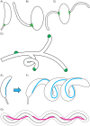
Fig 2. Different mechanisms underlie the evolution of morphogenesis.
(A–C) In prosthecate Alphaproteobacteria, simply repositioning zonal PG synthesis machinery (shown in green) can generate polar (Caulobacter crescentus, A), subpolar (Asticcacaulis excentricus, B), or bilateral (Asticcacaulis biprosthecum, C) prosthecate phenotypes. (D) Similar repositioning of the PG growth zone yields a branching phenotype in Streptomyces coelicolor, which grows from the cell poles. (E) The intermediate filament-like protein crescentin (shown in cyan) of C. crescentus constrains PG synthesis to generate cell wall curvature. (F) When cells filament, this constraint results in long helical shapes. (G) The periplasmic flagella of Borrelia burgdorferi directly deform the cell body into a planar wave shape. Note that the scale of the periplasmic space, relative to the cell membranes, has been modified to highlight this arrangement. For simplicity, details of the periplasmic compartment are shown only for panel G.
Fig 3
Fig 3. The Caulobacterales lineage exhibits diversification of the prosthecate morphology.
(A) Phylogeny of the order Caulobacterales generated as described in Fig 1. Schematics and corresponding colors indicate inferred ancestral morphologies and their subsequent inheritance. Black branches indicate rod-shape, nonappendaged morphology, including several apparent prostheca loss events. Scale bar indicates 0.1 amino acid substitutions per site. (B) Transmission electron micrographs of members of the Caulobacterales, highlighting disparate prosthecate morphologies. For each morphology, a brief description and the name of one representative species is provided, followed by the image source in parentheses. 1. Bilateral prosthecae, Asticcacaulis biprosthecum (Chao Jiang, Stanford University). 2. Subpolar prostheca, Asticcacaulis excentricus (Chao Jiang, Stanford University). 3. Polar prostheca, Caulobacter crescentus (Paul Caccamo, Indiana University). 4. Polar prostheca, Maricaulis maris (Patrick Viollier, University of Geneva). 5. Short polar prostheca, Brevundimonas subvibriodes (Brynn Heckel, Indiana University); note other members of this genus display a much longer prostheca. 6. Polar prostheca through which budding reproduction occurs, Hirschia baltica (Paul Caccamo, Indiana University). Magnification varies between micrographs. All images are reproduced with permission.

References

    1. Pangborn J, Kuhn DA, Woods JR. Dorsal-ventral differentiation in Simonsiella and other aspects of its morphology and ultrastructure. Arch Microbiol. 1977;113(3):197–204. 10.1007/BF00492025 - DOI - PubMed
    1. Whittenbury R, Dow CS. Morphogenesis and differentiation in Rhodomicrobium vannielii and other budding and prosthecate bacteria. Bacteriol Rev. 1977;41(3):754–808. - PMC - PubMed
    1. Nordberg H, Cantor M, Dusheyko S, Hua S, Poliakov A, Shabalov I, et al. The genome portal of the Department of Energy Joint Genome Institute: 2014 updates. Nucleic acids research. 2014;42(Database issue):D26–31. 10.1093/nar/gkt1069 - DOI - PMC - PubMed
    1. Tatusova T, Ciufo S, Fedorov B, O'Neill K, Tolstoy I. RefSeq microbial genomes database: new representation and annotation strategy. Nucleic acids research. 2014;42(Database issue):D553–9. 10.1093/nar/gkt1274 - DOI - PMC - PubMed
    1. Darling AE, Jospin G, Lowe E, Matsen FA, Bik HM, Eisen JA. PhyloSift: phylogenetic analysis of genomes and metagenomes. PeerJ. 2014;2:e243 10.7717/peerj.243 - DOI - PMC - PubMed

LinkOut - more resources