. 2016 Oct 3;11(10):e0164040.
doi: 10.1371/journal.pone.0164040. eCollection 2016.

Borrelia burgdorferi Induces TLR2-Mediated Migration of Activated Dendritic Cells in an Ex Vivo Human Skin Model

Affiliations

Borrelia burgdorferi Induces TLR2-Mediated Migration of Activated Dendritic Cells in an Ex Vivo Human Skin Model

Lauren M K Mason et al. PLoS One. .

Abstract

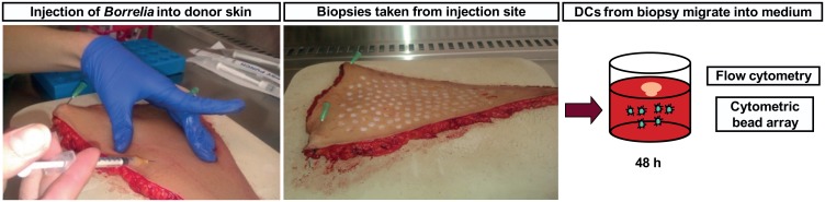
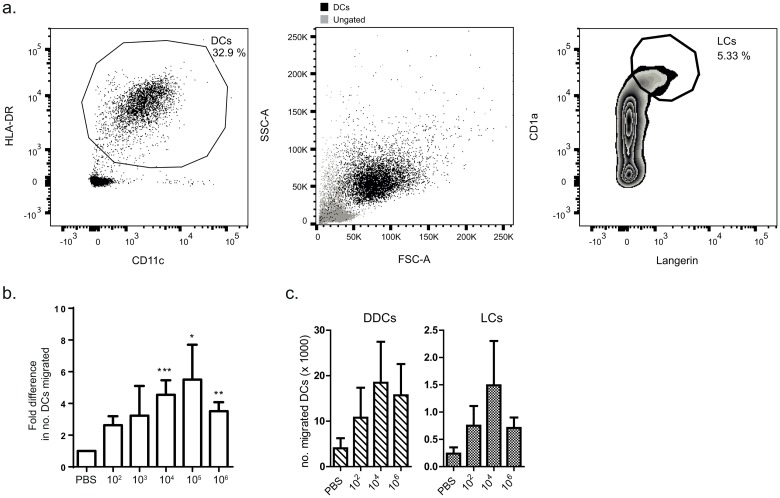
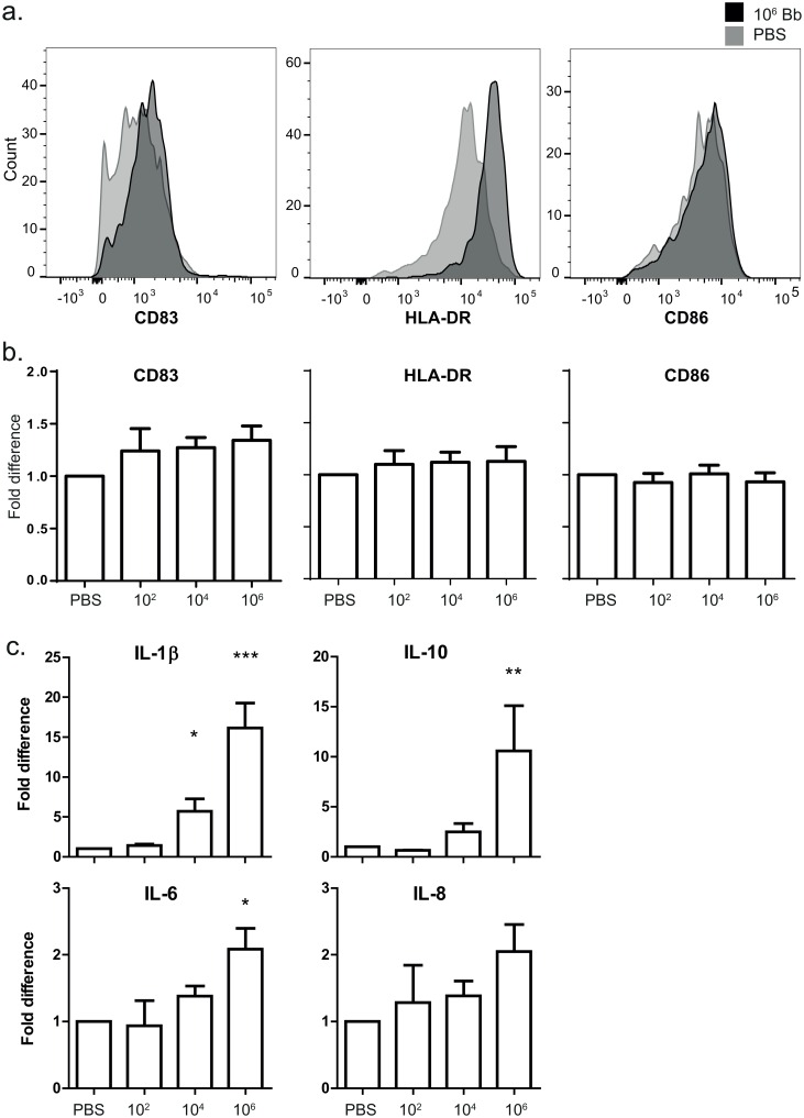
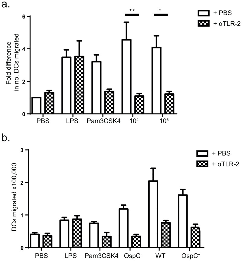
Borrelia burgdorferi is transmitted into the skin of the host where it encounters and interacts with two dendritic cell (DC) subsets; Langerhans cells (LCs) and dermal DCs (DDCs). These cells recognize pathogens via pattern recognition receptors, mature and migrate out of the skin into draining lymph nodes, where they orchestrate adaptive immune responses. In order to investigate the response of skin DCs during the early immunopathogenesis of Lyme borreliosis, we injected B. burgdorferi intradermally into full-thickness human skin and studied the migration of DCs out of the skin, the activation profile and phenotype of migrated cells. We found a significant increase in the migration of LCs and DDCs in response to B. burgdorferi. Notably, migration was prevented by blocking TLR2. DCs migrated from skin inoculated with higher numbers of spirochetes expressed significantly higher levels of CD83 and produced pro-inflammatory cytokines. No difference was observed in the expression of HLA-DR, CD86, CD38, or CCR7. To conclude, we have established an ex vivo human skin model to study DC-B. burgdorferi interactions. Using this model, we have demonstrated that B. burgdorferi-induced DC migration is mediated by TLR2. Our findings underscore the utility of this model as a valuable tool to study immunity to spirochetal infections.


Conflict of interest statement

The authors have declared that no competing interests exist.

Figures

Fig 1
Fig 1. B. burgdorferi was injected intradermally into the skin and biopsies were taken from the injection site.
Borrelia burgdorferi strain B31 in PBS was injected intradermally into full-thickness human donor skin (left panel). 8 mm biopsies were taken from around the injection site (middle panel) and then cultured in medium in a 48-wells plate for 48 hours. The migrated cells were collected and analysed using flow cytometry, and a cytometric bead array was performed to measure cytokines in the supernatant (right panel).
Fig 2
Fig 2. LCs and DDCs migrate out of the skin in response to B. burgdorferi injection.
Flow cytometry was performed on cells collected from biopsy medium. a) DCs were defined as HLA-DR+/CD11c+ cells (left panel). This population is shown in black in the forward/side scatter plot (centre panel). LCs were defined as CD1a+/Langerin+ cells and encompassed approximately 5% of the DC population (right panel). b) To calculate the number of migrated DCs per biopsy, the total number of cells retrieved from biopsy medium was counted (between 5x10³ and 2x10⁶ total cells per biopsy) and the percentage of HLA-DR+/CD11c+ cells was taken. Higher doses of B. burgdorferi induced migration of DCs that was significantly greater than PBS alone at 48 hours. The data was also significant when calculated for actual cell numbers. The graph shown depicts data pooled from 12 independent experiments, ± SEM. c) Both DDCs and LCs migrated in response to B. burgdorferi injection. This graph represents the pooled data of 4 independent experiments, ± SEM.
Fig 3
Fig 3. Migrated cells are mature and express activation markers.
a) Flow cytometry was performed on cells to measure expression of cell-surface markers CD83, HLA-DR and CD86. The geometric mean fluorescence intensity of CD83 and HLA-DR on DCs from 106 B. burgdorferi-injected biopsies (dark grey histograms) was modestly raised compared to DCs from PBS-injected biopsies (light grey histograms). CD86 expression was similar in the two conditions. These histograms are based on one donor. b) Expression of these markers varied across donors and there were no significant differences between the groups when data were pooled. These graphs depict the fold difference (MFI stimulus/MFI PBS) and are based on pooled data of 9 independent donors, ± SEM. c) A CBA was used to measure cytokine concentration in biopsy medium. Modestly higher levels of IL-1β, Il-10 and IL-6 were detected in the supernatant of biopsies inoculated with 106 B. burgdorferi compared to PBS 48 hours after inoculation. These graphs depict the fold difference (pg/ml stimulus/pg/ml PBS) and are based on pooled data of 8 independent donors, ± SEM. Mean concentrations in pg/ml (± SEM) measured in PBS-injected biopsies were as follows: IL-1β: 22.5 (7.5); IL-10: 9.9 (2.8); IL-6: 32250 (12465); IL-8: 29492 (15752). Actual concentrations of IL-1β and IL-10 were also significantly higher in the supernatant of biopsies inoculated with 106 B. burgdorferi compared to PBS.
Fig 4
Fig 4. Migration is TLR-2 mediated and OspC independent.
a) Addition of a blocking αTLR2-antibody inhibited the B. burgdorferi-induced increase in migration of DC out of the skin. The increase in DC migration induced by pam3CSK4, but not LPS, was also inhibited by blocking TLR2. The graph shown is based on the pooled data of 7 independent experiments, ± SEM. b) Inoculation of 104 B. burgdorferi lacking OspC (OspC-) induced migration of DC similarly to wildtype (WT) and OspC-complemented B. burgdorferi (OspC+). This was inhibited by blocking αTLR2 antibodies. Error bars are based on SEM of triplicate measurements from one donor, representative of two independent experiments.

References

    1. Stanek G, Wormser GP, Gray J, Strle F. Lyme borreliosis. Lancet. 2012;379(9814):461–73. 10.1016/S0140-6736(11)60103-7 . - DOI - PubMed
    1. Banchereau J, Steinman RM. Dendritic cells and the control of immunity. Nature. 1998;392(6673):245–52. 10.1038/32588 . - DOI - PubMed
    1. Mason LM, Veerman CC, Geijtenbeek TB, Hovius JW. Menage a trois: Borrelia, dendritic cells, and tick saliva interactions. Trends in parasitology. 2014;30(2):95–103. 10.1016/j.pt.2013.12.003 . - DOI - PubMed
    1. Hirschfeld M, Kirschning CJ, Schwandner R, Wesche H, Weis JH, Wooten RM, et al. Cutting edge: inflammatory signaling by Borrelia burgdorferi lipoproteins is mediated by toll-like receptor 2. Journal of immunology. 1999;163(5):2382–6. . - PubMed
    1. Shin OS, Isberg RR, Akira S, Uematsu S, Behera AK, Hu LT. Distinct roles for MyD88 and Toll-like receptors 2, 5, and 9 in phagocytosis of Borrelia burgdorferi and cytokine induction. Infection and immunity. 2008;76(6):2341–51. 10.1128/IAI.01600-07 - DOI - PMC - PubMed