

Meroxest improves the prognosis of immunocompetent C57BL/6 mice with allografts of E0771 mouse breast tumor cells
- PMID: 27695480
- PMCID: PMC5016567
- DOI: 10.5114/aoms.2014.45442
Meroxest improves the prognosis of immunocompetent C57BL/6 mice with allografts of E0771 mouse breast tumor cells
Abstract
Introduction: Recently, we have reported the antitumor properties of a new family of synthetic merosesquiterpenes, among which meroxest is highlighted, since it has high activity and specificity for ER+ breast cancer cells. In this paper, we characterize allografts of ER+ E0771 mouse breast tumor cells in immunocompetent C57BL/6 mice, and also analyze the effect of meroxest on the prognosis of the disease.
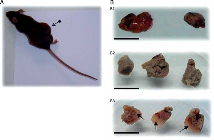
Material and methods: Twenty female C57BL/6 mice were injected with 106 E0771 cells. Once the tumors reached the appropriate size, the mice were divided into two groups, one control and another treated orally with 15 mg/kg of meroxest. After 20 days, tumor samples were taken for histopathological study and for determination of the expression of the prognostic markers Ki67 and vascular endothelial growth factor (VEGF) by immunofluorescence.
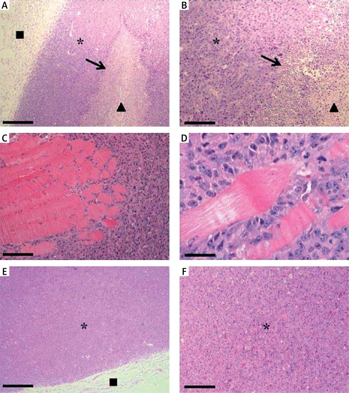
Results: In sections stained with hematoxylin-eosin, we observed that tumors have a well-defined capsule enclosing E0771 tumor cells. The central area of tumors contains necrotic regions with leukocyte infiltration. Meroxest treatment significantly reduces tumor size (68%, p < 0.05), induces changes in its structure, decreases the degree of leukocyte infiltration, and significantly reduces the expression of Ki67 (33%, p < 0.05) and VEGF (82%, p < 0.05).
Conclusions: Meroxest improves the prognosis of mice since it reduces leukocyte infiltration, and decreases the expression of Ki67 and VEGF markers. Consequently, the merosesquiterpene could become a useful antiangiogenic drug in the treatment of breast cancer. These results encourage us to deepen the study of meroxest, in order to find more evidence that supports the convenience of its evaluation in a clinical study or trial.
Keywords: Ki67; breast cancer; in vivo; merosesquiterpene; vascular endothelial growth factor.
Figures


References
-
- Piotrowski G, Gawor R, Stasiak A, Gawor Z, Potemski P, Banach M. Cardiac complications associated with trastuzumab in the setting of adjuvant chemotherapy for breast cancer overexpressing human epidermal growth factor receptor type 2 – a prospective study. Arch Med Sci. 2012;8:227–35. - PMC - PubMed
-
- Jemal A, Bray F, Center MM, Ferlay J, Ward E, Forman D. Global cancer statistics. CA Cancer J Clin. 2011;61:69–90. - PubMed
LinkOut - more resources
Full Text Sources
Other Literature Sources