

Learning neuroendoscopy with an exoscope system (video telescopic operating monitor): Early clinical results
- PMID: 27695549
- PMCID: PMC4974970
- DOI: 10.4103/1793-5482.145551
Learning neuroendoscopy with an exoscope system (video telescopic operating monitor): Early clinical results
Abstract
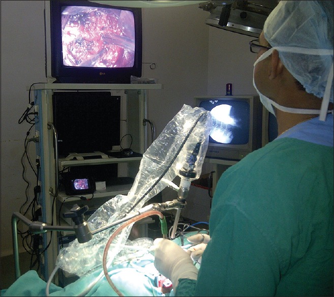
Context: Steep learning curve is found initially in pure endoscopic procedures. Video telescopic operating monitor (VITOM) is an advance in rigid-lens telescope systems provides an alternative method for learning basics of neuroendoscopy with the help of the familiar principle of microneurosurgery.
Aims: The aim was to evaluate the clinical utility of VITOM as a learning tool for neuroendoscopy.
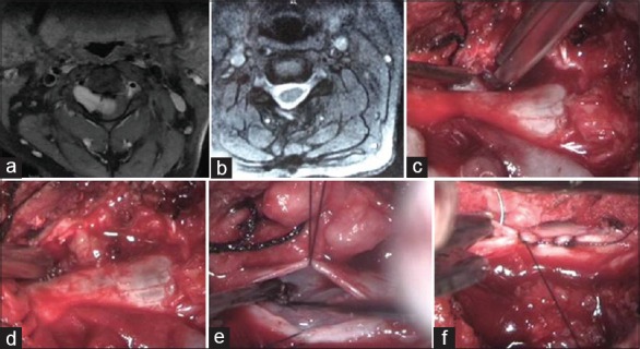
Materials and methods: Video telescopic operating monitor was used 39 cranial and spinal procedures and its utility as a tool for minimally invasive neurosurgery and neuroendoscopy for initial learning curve was studied.
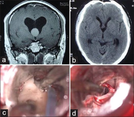
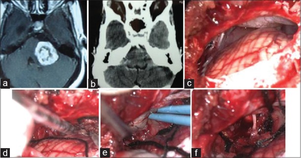
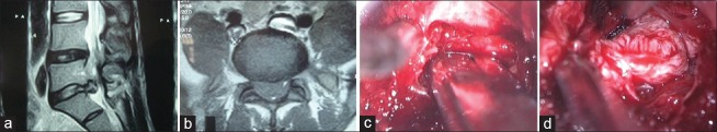
Results: Video telescopic operating monitor was used in 25 cranial and 14 spinal procedures. Image quality is comparable to endoscope and microscope. Surgeons comfort improved with VITOM. Frequent repositioning of scope holder and lack of stereopsis is initial limiting factor was compensated for with repeated procedures.
Conclusions: Video telescopic operating monitor is found useful to reduce initial learning curve of neuroendoscopy.
Keywords: Exoscope; neuroendoscopy; video telescopic operating monitor.
Conflict of interest statement
Figures
References
-
- Yadav YR, Parihar V, Namdev H, Agarwal M, Bhatele PR. Endoscopic interlaminar management of lumbar disc disease. J Neurol Surg A Cent Eur Neurosurg. 2013;74:77–81. - PubMed
-
- Yadav YR, Yadav S, Sherekar S, Parihar V. A new minimally invasive tubular brain retractor system for surgery of deep intracerebral hematoma. Neurol India. 2011;59:74–7. - PubMed
-
- Agrawal A, Kato Y, Sano H, Kanno T. The incorporation of neuroendoscopy in neurosurgical training programs. World Neurosurg. 2013;79:S15.e11–3. - PubMed
LinkOut - more resources
Full Text Sources
Other Literature Sources