

Light-curing units used in dentistry: factors associated with heat development-potential risk for patients
- PMID: 27695955
- PMCID: PMC5442227
- DOI: 10.1007/s00784-016-1962-5
Light-curing units used in dentistry: factors associated with heat development-potential risk for patients
Abstract
Objectives: To investigate how heat development in the pulp chamber and coronal surface of natural teeth with and without cusps subjected to irradiance using light-emitting diode (LED)-light-curing units (LCUs) is associated with (i) irradiance, (ii) time, (iii) distance, and (iv) radiant exposure.
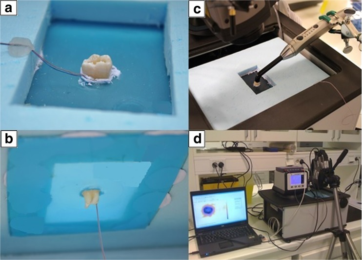
Materials and methods: Three different LED-LCUs were used. Their irradiance was measured with a calibrated spectrometer (BlueLight Analytics Inc., Halifax, Canada). An experimental rig was constructed to control the thermal environment of the teeth. The LED-LCU tip position was accurately controlled by a gantry system. Tooth surface temperature was measured by thermography (ThermaCAM S65 HS, FLIR Systems, Wilsonville, USA) and pulp chamber temperature with a thermocouple. LED-LCU tip distance and irradiation times tested were 0, 2, and 4 mm and 10, 20, and 30 s, respectively. Ethical permission was not required for the use of extracted teeth.
Results: Maximum surface and pulp chamber temperatures were recorded in tooth without cusps (58.1 °C ± 0.9 °C and 43.1 °C ± 0.9 °C, respectively). Radiant exposure explained the largest amount of variance in temperature, being more affected by time than irradiance.
Conclusions: At all combinations of variables tested, repeated measurements produced consistent results indicating the reliability of the method used. Increased exposure time seems to be the factor most likely to cause tissue damage.
Clinical relevance: Risk of superficial tissue damage at irradiances >1200 mW/cm2 is evident. There is a risk of pulp damage when only thin dentin is left at higher irradiances (>1200 mW/cm2). Clinicians should be aware of LED-LCU settings and possible high temperature generated.
Keywords: Curing lights; Dentistry; Light; Temperature; Tooth.
Conflict of interest statement
Conflict of interest
The authors declare that they have no conflict of interest.
Funding
The study was financially supported by the Norwegian Directorate of Health (14/1493).
Ethical approval
Since the experiments involved the use of human material (i.e., extracted teeth), ethical permission was asked for from the Norwegian Regional committee for Medical and Health Research Ethics (REK). The committee concluded that such permission was not required (2015/234/REK Nord).
Informed consent
For this type of study, formal consent was not required
Figures

Similar articles
-
The dark art of light curing in dentistry.J Dent. 2024 Nov;150:105375. doi: 10.1016/j.jdent.2024.105375. Epub 2024 Sep 26. J Dent. 2024. PMID: 39332516
-
Light-curing units used in dentistry: Effect of their characteristics on temperature development in teeth.Dent Mater J. 2021 Sep 30;40(5):1177-1188. doi: 10.4012/dmj.2020-305. Epub 2021 Jun 12. Dent Mater J. 2021. PMID: 34121022
-
Temperature increase at the light guide tip of 15 contemporary LED units and thermal variation at the pulpal floor of cavities: an infrared thermographic analysis.Oper Dent. 2013 May-Jun;38(3):324-33. doi: 10.2341/12-060-L. Epub 2012 Oct 23. Oper Dent. 2013. PMID: 23092145
-
Light-Curing Units: A Review of What We Need to Know.J Dent Res. 2015 Sep;94(9):1179-86. doi: 10.1177/0022034515594786. Epub 2015 Jul 8. J Dent Res. 2015. PMID: 26156516 Review.
-
High-Power LED Units Currently Available for Dental Resin-Based Materials-A Review.Polymers (Basel). 2021 Jun 30;13(13):2165. doi: 10.3390/polym13132165. Polymers (Basel). 2021. PMID: 34208978 Free PMC article. Review.
Cited by
-
How does indirect air-cooling influence pulp chamber temperature in different volume teeth and absence/presence of resin-based composite during light curing?BMC Oral Health. 2022 Nov 24;22(1):538. doi: 10.1186/s12903-022-02545-z. BMC Oral Health. 2022. PMID: 36424576 Free PMC article.
-
Effect of different curing times and distances on the microhardness of nanofilled resin-based composite restoration polymerized with high-intensity LED light curing units.Saudi Dent J. 2021 Dec;33(8):1035-1041. doi: 10.1016/j.sdentj.2021.05.007. Epub 2021 Jun 10. Saudi Dent J. 2021. PMID: 34938047 Free PMC article.
-
Pulp Temperature Rise Induced by Light-Emitting Diode Light-Curing Units Using an Ex Vivo Model.Materials (Basel). 2019 Jan 29;12(3):411. doi: 10.3390/ma12030411. Materials (Basel). 2019. PMID: 30699935 Free PMC article.
-
Assessing Dental Light-curing Units' Output Using Radiometers: A Narrative Review.J Int Soc Prev Community Dent. 2020 Jan 24;10(1):1-8. doi: 10.4103/jispcd.JISPCD_407_19. eCollection 2020 Jan-Feb. J Int Soc Prev Community Dent. 2020. PMID: 32181215 Free PMC article. Review.
-
Utilizing Light Cure Units: A Concise Narrative Review.Polymers (Basel). 2021 May 15;13(10):1596. doi: 10.3390/polym13101596. Polymers (Basel). 2021. PMID: 34063428 Free PMC article. Review.
References
-
- Nomoto R, McCabe JF, Hirano S (2004) Comparison of halogen, plasma and LED curing units. Oper Dent 29(3):287–294 - PubMed
-
- “LED”, in The American Heritage Science Dictionary (2005) Houghton Mifflin Company
MeSH terms
LinkOut - more resources
Full Text Sources
Other Literature Sources
Medical
