

Ultrasound findings of diffuse metastasis of gastric signet-ring-cell carcinoma to the thyroid gland
- PMID: 27696013
- PMCID: PMC5222898
- DOI: 10.1007/s10396-016-0746-5
Ultrasound findings of diffuse metastasis of gastric signet-ring-cell carcinoma to the thyroid gland
Abstract
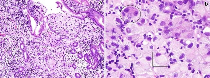
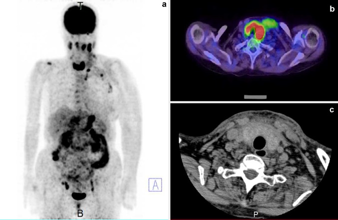
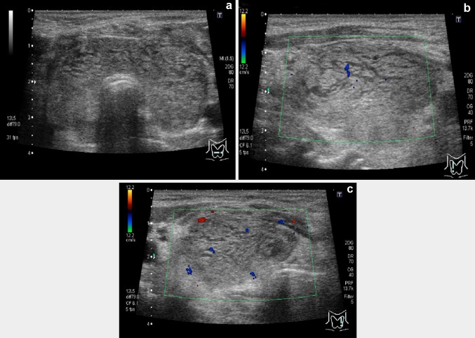
It has been shown that metastases to the thyroid from extrathyroidal malignancies occur as solitary or multiple nodules, or may involve the whole thyroid gland diffusely. However, diffuse metastasis of gastric cancer to the thyroid is extremely rare. Here, we report a case of a 74-year-old woman with diffuse infiltration of gastric adenocarcinoma (signet-ring-cell carcinoma/poorly differentiated adenocarcinoma) cells in the thyroid. The pathological diagnosis was made based on upper gastrointestinal endoscopy with biopsy and fine-needle aspiration cytology of the thyroid. An 18F-FDG PET/CT revealed multiple lesions with increased uptake, including the bilateral thyroid gland. On thyroid ultrasound examination, diffuse enlargement with internal heterogeneity and hypoechoic reticular lines was observed. On color Doppler imaging, a blood-flow signal was not detected in these hypoechoic lines. These findings were similar to those of diffuse metastases caused by other primary cancers, such as lung cancer, as reported earlier. Therefore, the presence of hypoechoic reticular lines without blood-flow signals is probably common to diffuse thyroid metastasis from any origin and an important diagnostic finding. This is the first report to show detailed ultrasound findings of diffuse gastric cancer metastasis to the thyroid gland using color Doppler.
Keywords: Color Doppler ultrasonography; Neoplasm metastasis; Signet-ring-cell carcinoma; Stomach neoplasm; Thyroid gland.
Conflict of interest statement
The authors declare that they have no conflict of interest. Human rights statements and informed consent All procedures followed were in accordance with the ethical standards of the responsible committee on human experimentation (institutional and national) and with the Helsinki Declaration of 1964 and later versions. Informed consent was obtained from the patient for being included in the report.
Figures

Similar articles
-
Diffuse metastasis to the thyroid: unique ultrasonographic finding and clinical correlation.J Korean Med Sci. 2014 Jun;29(6):818-24. doi: 10.3346/jkms.2014.29.6.818. Epub 2014 May 30. J Korean Med Sci. 2014. PMID: 24932084 Free PMC article.
-
Progression of Focal to Diffuse Thyroid Uptake Detected by 18F-FDG PET/CT: Malignant Metastatic Disease or Benign Thyroiditis?Clin Nucl Med. 2018 Sep;43(9):e310-e311. doi: 10.1097/RLU.0000000000002168. Clin Nucl Med. 2018. PMID: 29939952
-
[Malignant tumor with false negative 18F-FDG PET image].Zhonghua Zhong Liu Za Zhi. 2006 Sep;28(9):713-7. Zhonghua Zhong Liu Za Zhi. 2006. PMID: 17274383 Chinese.
-
Gastric adenocarcinoma of fundic gland type with signet-ring cell carcinoma component: A case report and review of the literature.World J Gastroenterol. 2018 Jul 14;24(26):2915-2920. doi: 10.3748/wjg.v24.i26.2915. World J Gastroenterol. 2018. PMID: 30018486 Free PMC article. Review.
-
Fine-needle aspiration of mucin-producing thyroid tumors.Acta Cytol. 2011;55(6):549-55. doi: 10.1159/000333228. Epub 2011 Dec 9. Acta Cytol. 2011. PMID: 22156465 Review.
Cited by
-
The Application Value of SMI Technology and Contrast-Enhanced Ultrasound in the Differential Diagnosis of Benign and Malignant Thyroid Nodules.Contrast Media Mol Imaging. 2022 Aug 25;2022:6157026. doi: 10.1155/2022/6157026. eCollection 2022. Contrast Media Mol Imaging. 2022. Retraction in: Contrast Media Mol Imaging. 2023 Oct 4;2023:9867806. doi: 10.1155/2023/9867806. PMID: 36082066 Free PMC article. Retracted.
-
Tumor Biology is King: Secondary Tumors of the Thyroid From 2 Medical Centers in China.Cancer Control. 2020 Jul-Aug;27(3):1073274820945984. doi: 10.1177/1073274820945984. Cancer Control. 2020. PMID: 32779493 Free PMC article.
-
Gastric Cancer Metastasis Mimicking Thyroiditis.Radiol Imaging Cancer. 2025 Jan;7(1):e240165. doi: 10.1148/rycan.240165. Radiol Imaging Cancer. 2025. PMID: 39670844 Free PMC article. No abstract available.
-
Application Value of Color Doppler Ultrasonography Combined with Thyroid Autoantibody Tests in Early Diagnosis of Thyroid Cancer.Comput Math Methods Med. 2022 Apr 27;2022:5248230. doi: 10.1155/2022/5248230. eCollection 2022. Comput Math Methods Med. 2022. Retraction in: Comput Math Methods Med. 2023 Dec 13;2023:9805780. doi: 10.1155/2023/9805780. PMID: 35529260 Free PMC article. Retracted.
-
Rare Metastasis of Gastric Cancer to the Thyroid Gland: A Case Report and Review of Literature.Front Surg. 2021 Oct 8;8:731673. doi: 10.3389/fsurg.2021.731673. eCollection 2021. Front Surg. 2021. PMID: 34692762 Free PMC article.
References
-
- Silverberg SG, Vidone RA. Metastatic tumors in the thyroid. Pacific Med Surg. 1966;74:175–180.
Publication types
MeSH terms
Substances
LinkOut - more resources
Full Text Sources
Other Literature Sources
Medical
