. 2017 Jan;103(1):67-85.
doi: 10.1111/mmi.13544. Epub 2016 Oct 27.

A novel glycan modifies the flagellar filament proteins of the oral bacterium Treponema denticola

Affiliations

A novel glycan modifies the flagellar filament proteins of the oral bacterium Treponema denticola

Kurni Kurniyati et al. Mol Microbiol. 2017 Jan.

Abstract

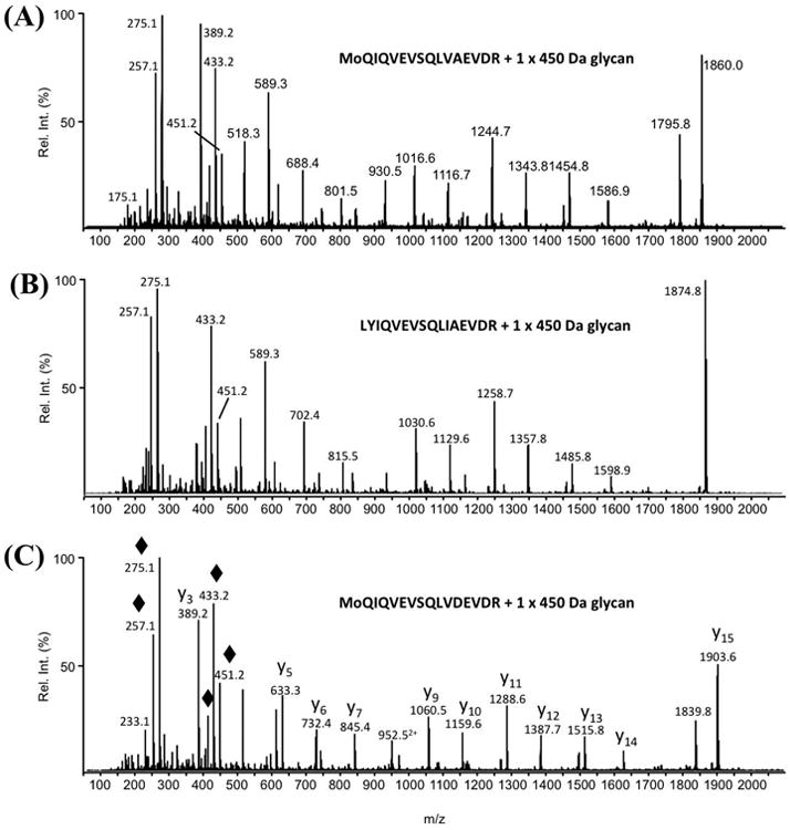
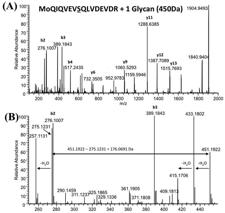
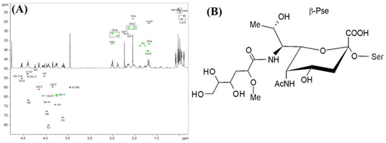
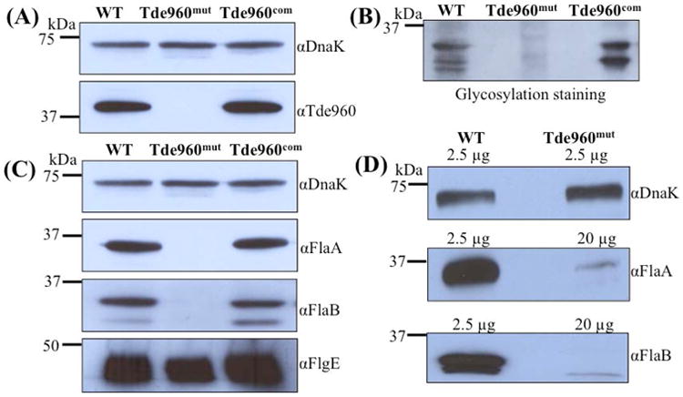
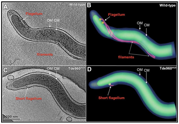
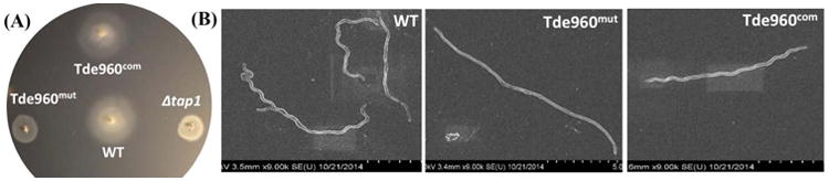
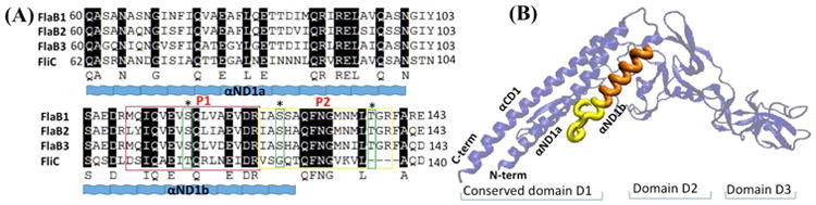
While protein glycosylation has been reported in several spirochetes including the syphilis bacterium Treponema pallidum and Lyme disease pathogen Borrelia burgdorferi, the pertinent glycan structures and their roles remain uncharacterized. Herein, a novel glycan with an unusual chemical composition and structure in the oral spirochete Treponema denticola, a keystone pathogen of periodontitis was reported. The identified glycan of mass 450.2 Da is composed of a monoacetylated nonulosonic acid (Non) with a novel extended N7 acyl modification, a 2-methoxy-4,5,6-trihydroxy-hexanoyl residue in which the Non has a pseudaminic acid configuration (L-glycero-L-manno) and is β-linked to serine or threonine residues. This novel glycan modifies the flagellin proteins (FlaBs) of T. denticola by O-linkage at multiple sites near the D1 domain, a highly conserved region of bacterial flagellins that interact with Toll-like receptor 5. Furthermore, mutagenesis studies demonstrate that the glycosylation plays an essential role in the flagellar assembly and motility of T. denticola. To our knowledge, this novel glycan and its unique modification sites have not been reported previously in any bacteria.


Conflict of interest statement

The authors declare that they have no conflict of interests.

Figures

Fig. 1
Fig. 1. The flagellin proteins of T. denticola are glycosylated
(A) SDS-PAGE (left panel) and immunoblotting (right panel) analyses of isolated PFs from the wild-type strain. 2-D electrophoresis (B) and immunoblotting (C) analyses of the isolated PFs from the wild-type strain. Purified PFs from the wild-type strain were analyzed using 2D-gel electrophoresis, followed by glycosylation staining (GS) (D) and lectin blots with ConA (E) and LFA (F). For the immunoblotting, the blots were probed with the T. pallidum FlaB antiserum (αFlaB) and T. denticola FlaA antiserum (αFlaA). For the lectin blots, the blots were probed with either ConA (Concanavalin A) or LFA (Limax Favus Agglutinin). A (FlaA); B1 (FlaB1); B2 (FlaB2); and B3 (FlaB3).
Fig. 2
Fig. 2. The three filament core proteins are glycosylated
The PFs isolated from three flaB deletion mutants (ΔflaB1, ΔflaB2, and ΔflaB3) were separated by 2D-gel electrophoresis, followed by Coomassie staining (A, B, C) and glycosylation staining (D, E, F). A (FlaA); B1 (FlaB1); B2 (FlaB2); and B3 (FlaB3).
Fig. 3
Fig. 3. Representative glycopeptide nLC-MS/MS spectra from three FlaB proteins, each modified with the 450.2 Da glycan
(A) MS/MS spectrum of MH22+ ion at m/z 1155.6 from T108-123 of FlaB1, (B) MS/MS spectrum of MH22+ ion at m/z 1163.1 from T108-123 of FlaB2, and (C) MS/MS spectrum of MH22+ ion at m/z 1177.6 from T108-123 from FlaB3. The amino acid sequences of the three glycopeptides are presented in the insets. “Mo” indicates the presence of oxidized methionine. By way of example, the major peptide fragment ions are identified in the MS/MS spectrum presented in panel c (“y” fragment ions start at the C-terminal amino acids). The mass differences between the peptide fragment ions were used to determine the amino acid sequence of this glycopeptide. The lower region of each MS/MS spectrum is dominated by the m/z 451.2 glycan oxonium ion and its related fragment ions (indicated with ◆ in panel c) that arose from multiple losses of water as well as the loss of the hexanoyl moiety.
Fig. 4
Fig. 4. High-resolution mass spectrometry analysis of the 450.2 Da glycan
nLC-HCD MS/MS analysis of the T108-123 (see sequence in Table 1) FlaB3 glycopeptide ion at m/z 1177.6: (A) full m/z range and (B) zoomed-in area showing the glycan oxonium ions plus neighboring peptide fragment ions. The best fitting elemental formulae for the m/z 451.1922 and m/z 275.1231 were C18O11H31N2, C11O6H19N2, respectively. The mass of the second glycan component was determined by subtracting the nonulosonate fragment ion from the intact glycan ion (m/z 451.1922 – m/z 275.1231 = 176.0691 Da), and the best fitting elemental composition was C7O5H12.
Fig. 5
Fig. 5. 1H-13C HSQC spectrum of the glycopeptide from T. denticola and its 1H NMR spectrum
(A) CH signals are black, and CH2 are green. Nonulosonic acid signals are labeled as N. (B) Structure of the flagellar glycan with Pse configuration (L-glycero-L-manno) of the nonulosonic acid residue β-linked to serine (NB C-8 may have the 8-epi-Pse configuration).
Fig. 6
Fig. 6. Characterizations of the TDE0960 mutant (Tde960mut) and its complemented strain (Tde960com)
(A) Immunoblotting analysis of the wild-type, Tde960mut, and Tde960com strains. Equivalent amounts of whole cell lysates were analyzed by SDS-PAGE and then probed with a specific antibody against TDE0960. DnaK was used as a loading control. (B) Glycosylation staining analysis of the whole cell lysates. The analysis was carried out as described above. (C) The levels of FlaA and FlaBs were significantly decreased in the Tde960mut strain. Equivalent amounts of the whole cell lysates were analyzed by SDS-PAGE and then probed with the antibodies of DnaK, FlaA, FlaBs, or FlgE. (D) Different amounts of whole cell lysates (as indicated on the figure) were analyzed by SDS-PAGE and then probed with the FlaA and FlaB antibodies.
Fig. 7
Fig. 7. Detection of flagellin gene mRNA by qRT-PCR and protein stability by turnover assays
(A) The levels of flaA, flaB1, flaB2, and flaB3 transcripts were measured by qRT-PCR as previously described (Bian et al., 2013). The dnaK gene transcript was used as an internal control to normalize the qRT-PCR data. The results are expressed as Tde960mut flagellin gene transcript expression relative to the wild-type levels. Asterisks indicate that the difference between the wild-type and Tde960mut transcript levels was statistically significant at a P value of < 0.01 (two-way ANOVA). (B) Protein translation was arrested by adding spectinomycin to the cultures of the wild-type and Tde960mut strains. Samples were withdrawn at the indicated time points and analyzed by immunoblotting. The whole cell lysates of the wild-type (2.5 μg) and Tde960mut strains (20 μg) were separated by SDS-PAGE, then transferred to PVDF membranes, and finally probed with antibodies against DnaK, FlaA, or FlaBs. DnaK was used as a sample loading control. SuperSignal West Femto Maximum Sensitivity substrate was used to develop the immunoblots of Tde960mut.
Fig. 8
Fig. 8. Cryo-ET analysis of the wild-type and Tde960mut strains
(A) A central slice of a typical tomographic reconstruction from a wild-type cell tip. PFs are observed in the periplasmic space between the outer membrane (OM) and the cytoplasmic membrane (CM). (B) The 3-D surface rendering of the reconstruction (A) shows two long PFs (colored in red) surrounding the cell body (green). (C) A central slice of a tomographic reconstruction from a Tde960mut cell. A short flagellum is attached to the CM. (D) The short flagellum is clearly shown in the surface rendering.
Fig. 9
Fig. 9. The Tde960mut mutant is non-motile and has an altered cell morphology
(A) Swimming plate assay of the wild-type, Tde960mut, and Tde960com strains. The assay was carried out on 0.35% agarose containing the TYGVS medium diluted 1:10 with PBS. The plates were incubated anaerobically at 37°C for 5 to 7 days to allow the cells to swim. Δtap1, a non-motile mutant, was used as a control to determine initial inoculum sizes (Limberger et al., 1999). (B) Scanning electron microscopy analysis of the wild-type, Tde960mut, and Tde960com strains. The cells were processed and observed using a Hitachi SU-70 scanning electron microscope at an acceleration voltage of 2.0 kV.
Fig. 10
Fig. 10. The first two glycopeptides of FlaBs are localized within the D1 domain of flagellin protein
(A) Sequence alignment of flagellin D1 domains. Flagellin D1 domain sequences of three FlaBs and Salmonella FliC (1IO1) (Yoon et al., 2012, Samatey et al., 2001) were aligned by Clustal X. The secondary structures of α-helices (αND1a and αND1b) are shown as waves; color-boxed P1 (orange) and P2 (yellow) sequences are two glycopeptides identified in all FlaB proteins; and green color-coded residues represent three glycosylation residues. (B) Homology mapping of the glycopeptides. Based on the results from pairwise sequence alignment shown on (A), P1 and P2 were directly mapped to the corresponding sequences on the crystal structure of 1IO1 using homology mapping (Beaver et al., 2007). Both P1 and P2 are located within αND1b located within interface-B between FliC and TLR5 (Yoon et al., 2012).

Similar articles

Cited by

References

    1. Agulleiro JI, Fernandez JJ. Tomo3D 2.0--exploitation of advanced vector extensions (AVX) for 3D reconstruction. J Struct Biol. 2015;189:147–152. - PubMed
    1. Anand A, Luthra A, Edmond ME, Ledoyt M, Caimano MJ, Radolf JD. The major outer sheath protein (Msp) of Treponema denticola has a bipartite domain architecture and exists as periplasmic and outer membrane-spanning conformers. J Bacteriol. 2013;195:2060–2071. - PMC - PubMed
    1. Beaver JE, Bourne PE, Ponomarenko JV. EpitopeViewer: a Java application for the visualization and analysis of immune epitopes in the Immune Epitope Database and Analysis Resource (IEDB) Immunome Res. 2007;3:3. - PMC - PubMed
    1. Berg HC, Anderson RA. Bacteria swim by rotating their flagellar filaments. Nature. 1973;245:380–382. - PubMed
    1. Bian J, Fenno JC, Li C. Development of a modified gentamicin resistance cassette for genetic manipulation of the oral spirochete Treponema denticola. Appl Environ Microbiol. 2012;78:2059–2062. - PMC - PubMed

LinkOut - more resources